Anterior Abdominal Wall
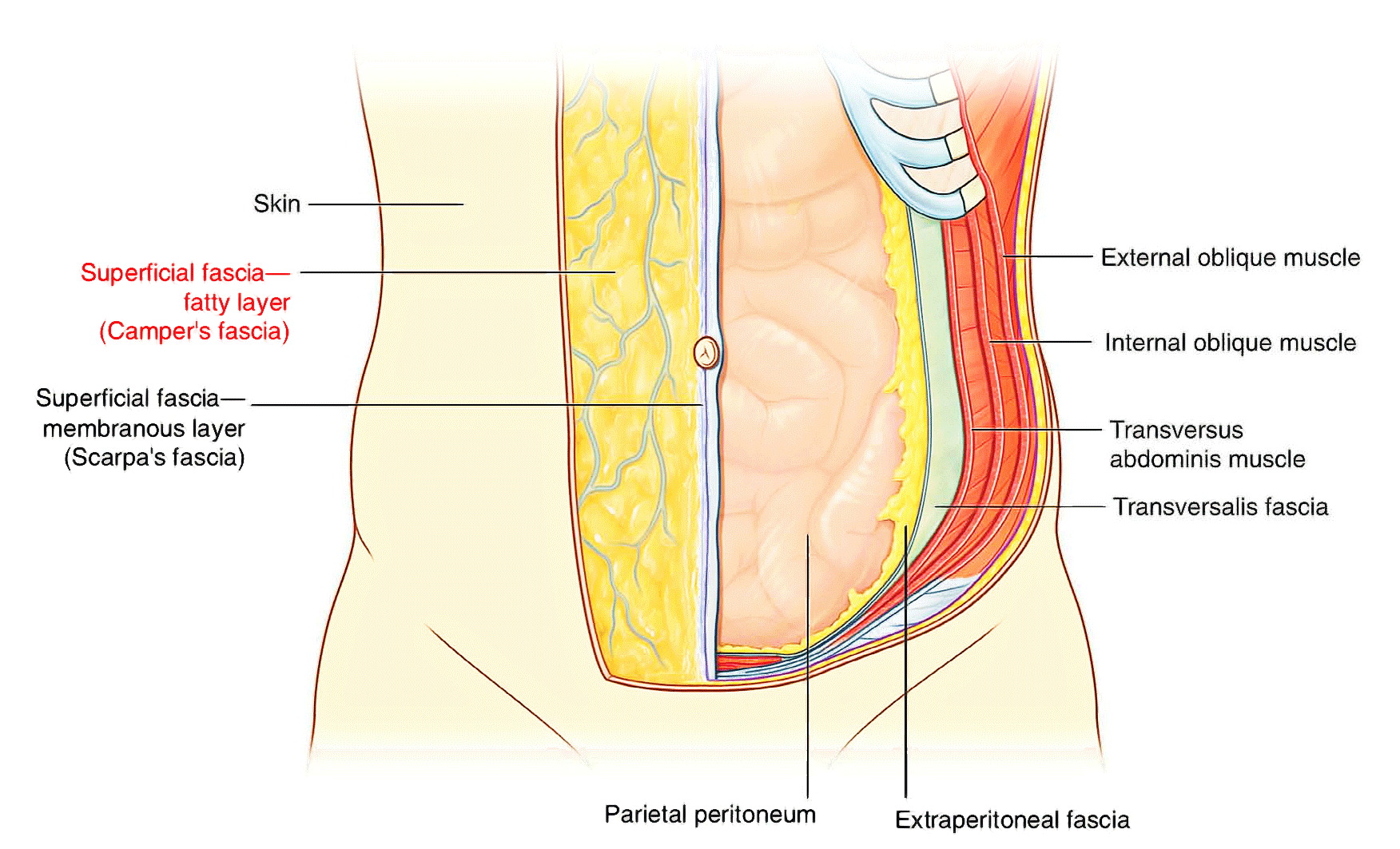
Layers of Anterior Abdominal Wall
- Skin
- Superficial fascia
a. Camper’s fatty layer
b. Scarpa’s membranous layer
- External oblique muscle
- Internal oblique muscle
- Transversus abdominis muscle
- Rectus abdominis muscle
- Fascia transversalis
- Extraperitoneal fat
- Parietal peritoneum
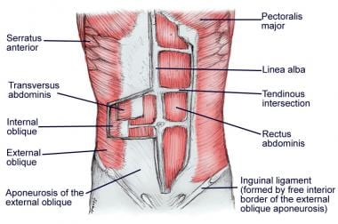
Muscles of the Anterior Abdominal Wall
- External oblique muscle
- Internal oblique muscle
- Transversus abdominis muscle
- Rectus abdominis muscle
- Pyramidalis muscle
Note: The inguinal (lowest) fibers of the internal oblique and transversus abdominis muscles are inserted by the conjoint tendon into the pubic crest and pectineal line of pubis. It is innervated by the ilioinguinal nerve (L1)
External Oblique Muscle
%%Origin%%: fleshy digitations from the lower 8 ribs
%%Insertion%%:
- fleshy fibers: outer lip of the anterior 1/2 of the iliac crest
- aponeurosis:
- medial part: linea alba from xiphoid process to symphysis pubis
- lateral part: folded upwards and backwards upon itself to ==form the inguinal ligament== (ASIS to pubic tubercle)
%%Direction of fibers%%: downwards, forwards, medially
%%Nerve supply%%: intercostal nerves (T7 - T11) & subcostal nerve (T12)
Internal Oblique Muscle
%%Origin%%:
- anterior 2/3 of intermediate line of iliac crest
- lateral 2/3 of the inguinal ligament
- lumbar fascia
%%Insertion%%:
- lower 6 costal cartilages
- linea alba
- pubic crest and pectineal line via the conjoint tendon
%%Direction of fibers%%: upwards, forwards, medially
%%Nerve supply%%: T7 - T12, iliohypogastric nerve, ilioinguinal nerve
Transversus Abdominis Muscle
%%Origin%%:
- lower 6 costal cartilages
- lumbar fascia
- anterior 2/3 of inner lip of iliac crest
- lateral 1/3 of inguinal ligament
%%Insertion%%:
- linea alba
- pubic crest and pectineal line via the conjoint tendon
%%Direction of fibers%%: horizontally
%%Nerve supply%%: T7 - T12, iliohypogastric nerve, ilioinguinal nerve
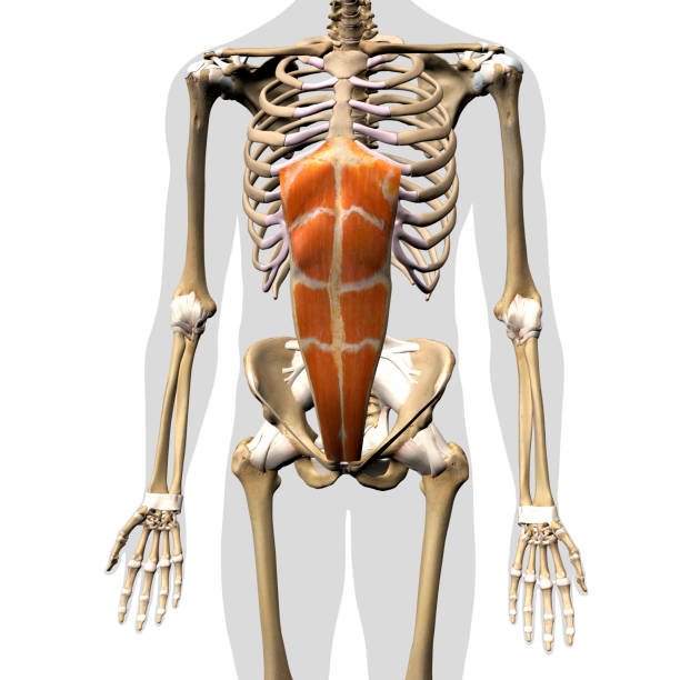
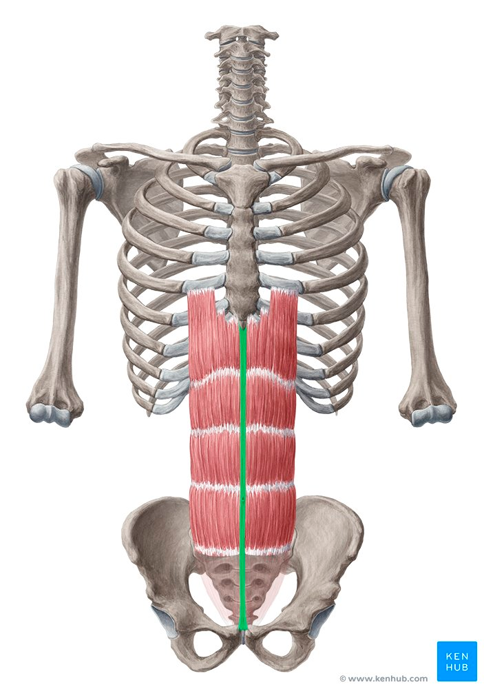
Rectus Abdominis Muscle
%%Origin%%: from the pubic crest, pubic symphysis
%%Insertion%%: 5th, 6th, 7th costal cartilages, xiphoid process
%%Nerve supply%%: intercostal nerves (T7 - T11) & subcostal nerve (T12)
Note: the muscle is divided into segments by tendinous intersections, which indicate that the muscle arises from a number of myotomes fused together
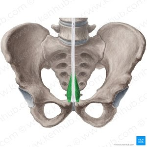
Pyramidalis Muscle
- small triangular muscle
- in front of the lower part of the rectus abdominis within the rectus sheath
%%Origin%%: from the front of the pubis and pubic symphysis
%%Insertion%%: linea alba 4cm above its orign
%%Nerve supply%%: subcostal nerve (T12)
Note: between the 2 recti all the aponeurosis fuse to form the ==linea alba==, a strong fibrous structure which is firmly attached to the xiphoid process above and the symphysis pubis below
Actions of Anterior Abdominal Wall Muscle
- assist in raising the intra-abdominal pressure (vomiting, cough, delivery)
- keep the abdominal viscera in position
- movements of the trunk
- flexion of the truck by rectus abdominis
- lateral flexion by on sided contraction of 2 oblique muscles
- rotation of the trunk by a combined action of the external oblique with the opposite internal oblique muscles
- act as accessory expiratory muscles
Rectus Sheath
Formation of Anterior Wall of Rectus Sheath
| Above Costal Margin: | aponeurosis of External oblique |
|---|---|
| Below Costal Margin and Arcuate Line: | aponeurosis of External oblique and anterior lamella of aponeurosis of Internal oblique |
| Below Arcuate Line: | aponeurosis of External oblique, Internal oblique, and Transversus abdominis |
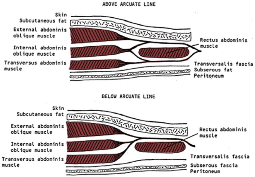
Formation of Posterior Wall of Rectus Sheath
| Above Costal Margin: | 5th, 6th, 7th costal cartilages |
|---|---|
| Below Costal Margin and Arcuate Line: | posterior lamella of aponeurosis of Internal oblique and Transversus abdominis |
| Below Arcuate Line: | fascia transveralis |
Content of the Rectus Sheath
| Muscles | Vessels | Nerves |
|---|---|---|
| Rectus abdominis muscle | superior epigastric vessel | terminal parts of T7-T11 nerves |
| pyramidalis muscle | inferior epigastric vessel | terminal parts of T12 nerve |
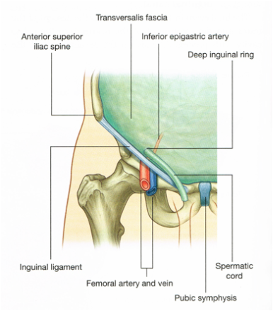
Deep Inguinal Ring
%%Origin%%: fascia transversalis
- oval opening
- in ==fascia transversalis==
- lies about 1/2 inch (1.25 cm) above the inguinal ligament midway between the anterosuperior iliac spine and the symphysis pubis (midinguinal point)
Superficial Inguinal Ring
%%Origin%%: aponeurosis of external oblique
- triangular in shape
- lies in ==aponeurosis of the external oblique muscle==
- lies above and lateral to the pubic tubercle
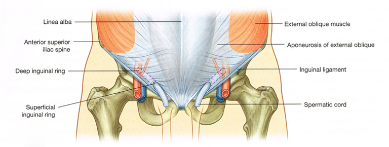
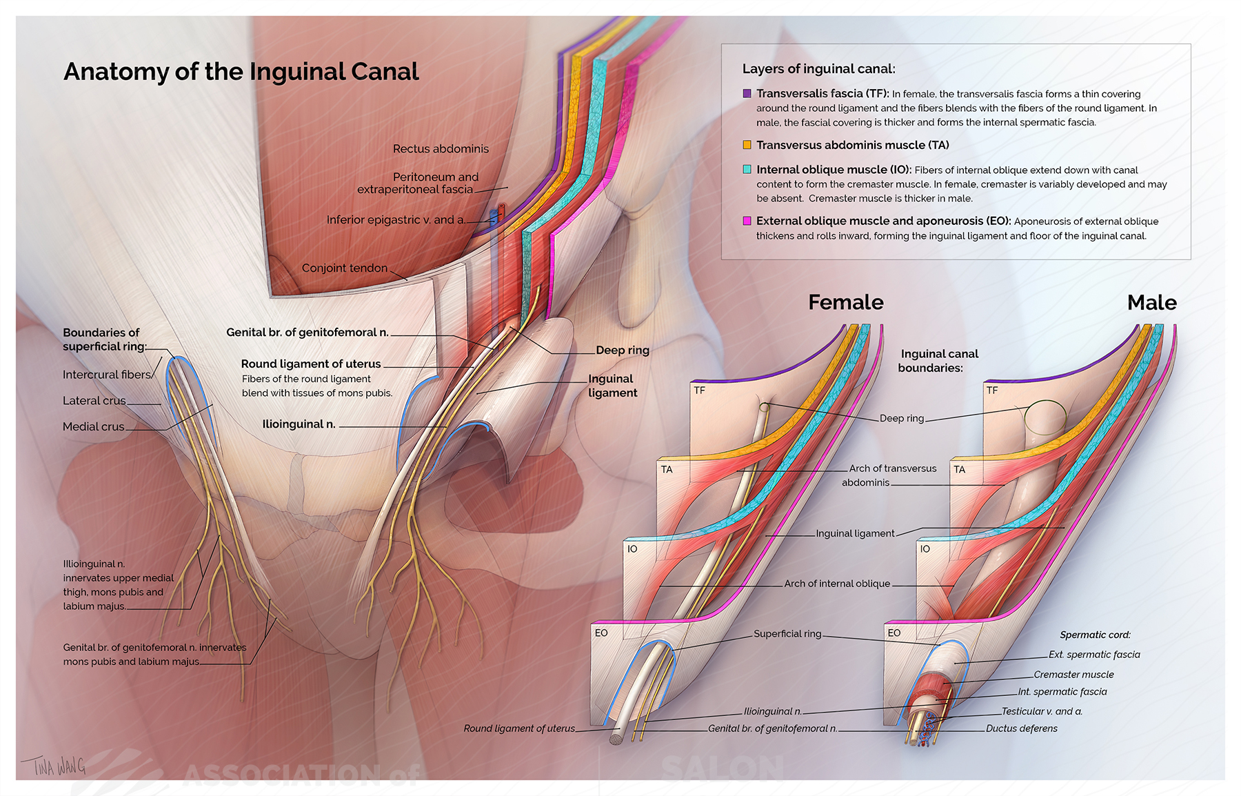
Inguinal Canal
%%Nature%%: an oblique space in the lower part of anterior abdominal wall extending from the deep inguinal ring to the superficial inguinal ring.
%%Length%%: 4 cm long
%%Direction%%: downwards, forwards, medially
%%Structures passing through%%:
- ilioinguinal nerve
- spermatic cord in males
- round ligament of the uterus in females
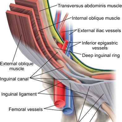
%%Boundaries:%%
| Anterior wall | external abdominal oblique aponeurosis | lateral third: internal abdominal oblique |
|---|---|---|
| Posterior wall | fascia transversalis | conjoint tendon (medial half) and reflected part of the inguinal ligament |
| Roof | arched fibers of the internal oblique and transversus abdominis | |
| Floor | concave upper surface of the inguinal ligament and lacunar ligament |