Week 7 & 8: Haematology
Blood Components:
Plasma-
- Contains:
- Low [K+] & [Mg2+]
- High [Na+] & [Cl-]
- High protein concentration (7% of plasma conc, 91.5% water), e.g.
- albumin- maintains osmotic pressure and transports insol. molecules
- globulins- transport ions, hormones & lipids → assist in immune function
- fibrinogen- involved in blood coagulation - formation of blood clot to prevent leakages
- No cellular components - just ions and nutrients
Leukocytes (WBC)
3 types:
Granulocytes:
- Neutrophils (50-72%)- defence against bacteria and some fungi- they are active fungi, engulfing bacteria & destroy them using enzymes with lysosomes- produce reactive O2 species e.g. h2o2 to degrade the pathogen.
- Eosinophil (2-4%) - lead attack against parasitic worms that are too large to be phagocytosed, where they surround the worm and release their cytoplasmic granules onto parasites surface, digesting it. (stain red with acid dyes)
- Basophil (0.5-1%)- filled with histamine that acts as a vasodilator (stains purple/black with basic dyes).
Monocytes & Macrophages (3-8%)- monocytes circulate in the blood for 1-3 days where they are the 1st line of defence against viruses, then mature into macrophages when in tissues, have a role in inflammatory response.
Mast cells (<0.2%)- generated in the bone marrow, role in allergic reactions and inflammation - release of histamine. Important in defence against parasites, as they recruit other types of leukocytes. Involved in angiogenesis.
Lymphocytes:
- B-Lymphocytes (B-cells)- destroy bacteria and inactivate their toxins, differentiate into plasma cells that secrete antibodies to act against antigens through agglutination & opsonisation.
- T-Lymphocytes (T-cells)- recognise specific foreign proteins, attack cells that have been infected by viruses & tumour cells. Stimulate B-cells to produce antibodies.
- Natural Killer cells (NK cells)- attack infected body cells, detect and control early signs of cancer.
Platelets (thrombocytes)- promote blood clotting when blood vessels are damaged, and are generated in bone marrow.
Erythrocytes (RBC)
- Erythropoiesis- process of generating mature RBCs, regulated in feedback process by hormone EPO.
- Cytokines, growth factors and hormones influence erythroid proliferation (GROWTH).
- Transports O2 and CO2, facilitated by haemoglobin. Lifespan 120 days
- Erythrocyte breakdown:
- proteases degrade haemoglobin using macrophages.
- Globin broken down into amino acids & re-used.
- Haeme degraded into bilirubin- metabolised & bile acids are used to make it soluble.
- Fe2+ recycled- it binds to transferrin & transported to bone marrow for new erythrocyte synthesis.
- Haematocrit (HCT)/ Packed cell volume (PCV): proportion of blood made up by cells (RBC & WBC).
ABO Blood grouping
Type A= A antigen
Type B= B antigen
Type AB= Both A&B antigens
Type O= Neither A nor B antigens
Rhesus (Rh) system
- Two proteins are responsible for different serotypes: D & C/E
- D is very antigenic and Rh+/- relates to D
- C & E are 2 diff regions of the same protein
- In the Rh system, individuals whose erythrocytes have Rh antigens are classified as Rh+; those who lack the antigen are Rh−.
- Haemolytic disease of the newborn (HDN) can occur when an Rh− mother is pregnant with an Rh+ foetus, but the mother has previously been exposed to RhD positive blood and has developed an immune response to it.
Other systems include:
- Kell system→ anti-kell antibodies are usually antibody class IgG
- Kidd system
- Duffy system
The Lymphatic system
Functions:
- Removal interstitial fluid
- Transport system for WBCs
- Recognition of bacteria in lymph → lymphocyte proliferation in the lymph nodes cause swelling and tenderness
- Lymph nodes act as filters→ macrophages remove and destroy microorganisms & debris
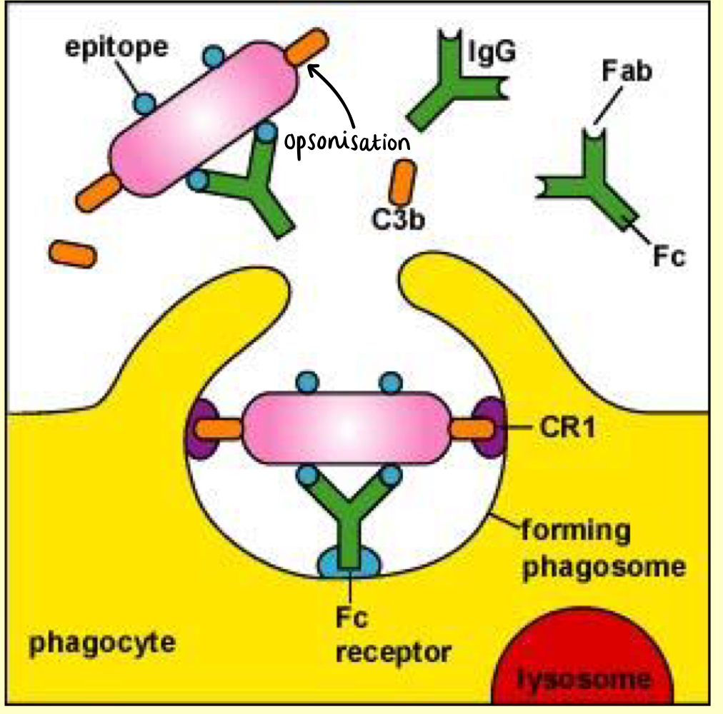
Opsonisation
Process of marking a pathogen for destruction
- Opsonins coat the outside of the cell/microorganism to be attacked
- Opsonins→ complement proteins or antibodies
- Both provide ‘handles’ for the phagocyte receptors to bind
Complement Cascade
Cascade of proteases→ hydrolysis of protein causes them to become more reactive
Functions:
- Removal of microbes or damaged cells
- Promotes inflammation • Attacks cell membrane of microbe/damaged cell
Downstream effects:
- Chemotaxis→ attracting macrophages and neutrophils
- Opsonisation→ enhancing phagocytosis of antigens
- Cell Lysis→ rupturing membranes of foreign cells (e.g. bacteria)
- Activation and migration of leukocytes
- Agglutination→ clustering and binding of pathogens together
Stimulates:
- Degranulation of mast cells & basophils
- Release of inflammatory mediators e.g. histamine
Achieved through activation of the Cell-killing membrane complex
Haemostasis
- Blood coagulation is initiated and terminated, together w/ removal (fibrinolysis) of the clot as part of vascular remodelling.
- Haemostatic system→ mosaic of activating or inhibitory pathways that integrates 5 major components:
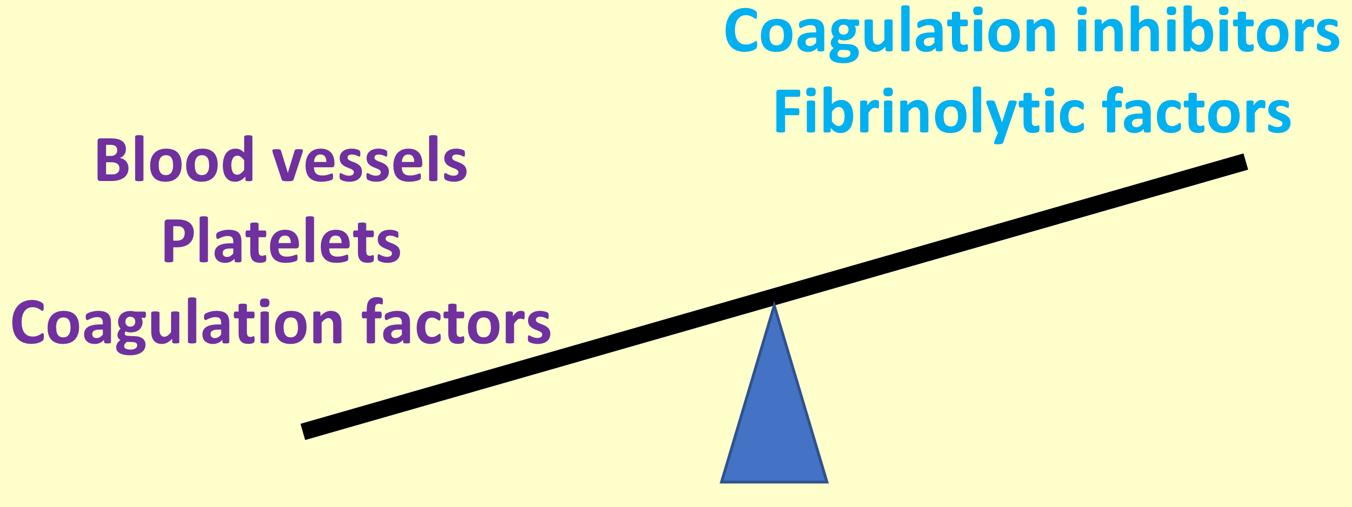
It is sensitive, rapid, localised, stable, coordinated, controlled. Upsetting the balance results in inappropriate bleeding or clotting.
Preserves integrity of the vascular system in response to injury
Prevents microbes from entering the body
The body is high pressure vascular system ∴ response must be rapid. Regulated to prevent inappropriate clot formation and localised to prevent loss of blood flow through the blood vessels.
Inappropriate blood coagulation may block vessels through formation of a thrombus, which
- Restricts blood flow
- Starves tissues of oxygen
- Leads to cell death
Failure to achieve blood coagulation (haemophilia) is life threatening
Steps