Capillary Exchange & Lymphatic System
A) Exchange at Capillaries
Plasma: a liquid component of the blood, found within blood vessels.
Contains formed elements, dissolved proteins, and other molecules.
Interstitial fluid: the fluid found in the space that makes up tissues
Identical to the plasma, except not have plasma proteins.

C6H12O6 + 6O2 → 6CO2 + 6H2O + 38 ATP
Plasma supplies O2 and nutrients to cells, and removes CO2 and wastes from cells.
This exchange occurs in the capillaries.
Cells are continually low in O2 and nutrients because they are used by the cells.
→ Diffusion of these substances will occur from the blood to the interstitial fluid, and then from the interstitial fluid into the cells’ cytoplasm.Cells are high in CO2 and waste (due to constant production).
→ These substances will naturally diffuse from the cells to the interstitial fluid, and then from the interstitial fluid to the plasma.Not only are dissolved solutes exchanged between the blood and the interstitial fluid at the capillaries, but so are the fluids.
B) Mechanisms of Tissue Fluid Exchange at Capillaries

Plasma is always hypertonic to the interstitial fluid due to the presence of plasma proteins that are too large to leave the capillaries with the plasma through the gaps in the capillary walls.
This creates a natural osmotic pressure of ~ 25 mmHg, which will drive water from the interstitial fluid to the plasma.
At the beginning of the capillary bed, the blood pressure is ~ 40 mmHg.
This exceeds the osmotic pressure (25 mmHg), and the fluid will always travel in the direction of greater pressure.
Therefore, fluids filter through the capillary walls and become interstitial fluid.
Large plasma proteins cannot leave the capillary walls because the gaps are too small for them to fit.
Blood pressure dissipates as the fluid within the blood vessels reduces.
The “new” interstitial fluid will be high in O2 and nutrients, and low in CO2 and waste.
The cells have opposite concentration conditions.
Thus, diffusion of gases and nutrients and active transport of nutrients will occur between the cells and the interstitial fluid.
The interstitial fluid becomes high in waste and lower in dissolved oxygen and nutrients.
At the end of the capillary bed, blood pressure has dropped to ~ 10 mmHg due to the loss of blood velocity and volume.
The osmotic pressure is still ~ 25 mmHg.
Therefore, the osmotic pressure exceeds the blood pressure, resulting in the interstitial fluid being driven into the capillaries.
The waste is also carried away from the tissues into the blood, as this occurs.
The reabsorption of fluid back into the capillaries is not 100%. Some fluid remains left in the tissues and is not returned to the plasma.
→ This is where the lymphatic system becomes critical.
C) Lymphatic System
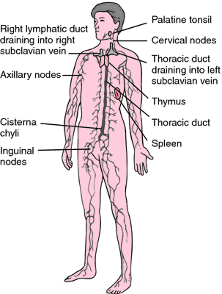
Lymph capillaries wind through the capillary bed and reabsorb any remaining tissue fluid.
Lymph: interstitial fluid inside the lymph vessel
Lymph capillaries join to form larger lymph veins.
Lymph veins have valves and skeletal muscle contraction to move lymph through the vessels (just like regular veins).
Lymph rejoins the plasma at 2 locations in the body where the lymphatic veins join the regular circulatory system.
The right subclavian vein joins the right lymphatic duct to drain the upper right portion of the body.
The left subclavian vein joins the thoracic duct to drain the rest of the body.
The lymphatic system has 3 functions:
Drain excess tissue fluid to prevent edema (swelling) of tissues.
Absorb lipoprotein (lipids) from the intestinal villi.
Help fight infection: within the lymph, there are white blood cells, which perform 2 functions.
Produce antibodies to fight infection.
Perform phagocytosis to destroy any foreign cells or cellular debris to clean the lymph.
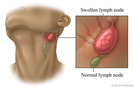
These white blood cells are stored within lymph nodes: small, round structures located in key areas.
When you are sick, the white blood cells reproduce rapidly, and your lymph nodes swell.