Pain and Nociception
What is pain?
An unpleasant sensory and emotional experience associated with actual or potential tissue damage. Pain is highly subjective, influenced by past experiences, expectations, and emotional states. It serves as a crucial protective mechanism, alerting an individual to danger or injury.
The feeling, or perception, of something unpleasant – sore, stinging, irritating, aching, throbbing, miserable or unbearable sensations arising from a part of the body
Hyperalgesia is an increased sensitivity to pain, where a normally painful stimulus causes an intensified pain response. Hypoalgesia is the opposite: a diminished sensitivity to pain, where a normally painful stimulus results in a decreased pain response
Nociception
The sensory process that provides the signals that trigger pain. It is the detection of noxious (damaging) stimuli by specialized sensory neurons called nociceptors.
The perception of a noxious stimulus (damaging)
Painful stimuli (causing tissue damage) activate nociceptors (sensory neurons)
Sex differences and pain
Physiological
testosterone treatment increases pain threshold (Pednekar & Mulgaonker, 1995)
Endogenous opioids and sex hormones like estrogen and testosterone play a role in modulating pain sensitivity, with higher testosterone levels generally associated with higher pain tolerance.
Psychological
Sex role beliefs can influence how pain is experienced and expressed. For instance, societal expectations about stoicism in men versus emotional expression in women can affect pain reporting.
Lower pain tolerance in women linked to self-efficacy beliefs (Jackson et al, 2002). Beliefs about one's ability to cope with pain significantly impact the pain experience.
Pain coping strategies differ, with some studies suggesting women are more likely to use emotion-focused coping and men more problem-focused coping, although this is variable.
Pain-related expectancies: Anticipation of pain can amplify or diminish its perception (e.g., placebo/nocebo effects).
Social
“No pain, no gain” – mentality that ability to withstand pain is something to value
“Boys don’t cry” - social pressure on men
Cultural factors and upbringing also shape how individuals interpret and react to painful experiences.
Why do we feel pain?
Insensitivity → the painful stimulus is not even perceived: a patient cannot describe the intensity or type of pain
This can be due to genetic conditions (e.g., Congenital Insensitivity to Pain), severe neuropathy, or pharmacological blockade.
Indifference → the patient can perceive the stimulus, but lacks an appropriate response: they will not flinch or withdraw when exposed to pain
This often involves higher-level cognitive or emotional processing deficits, such as observed in some psychiatric conditions or frontal lobe damage.
How do we feel pain?
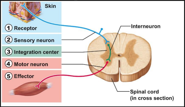
Nociceptors
Two types:
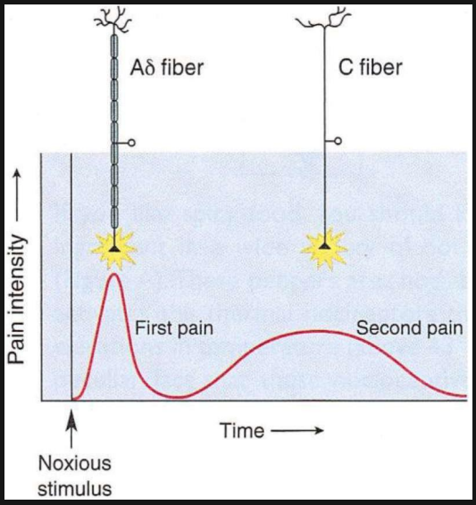
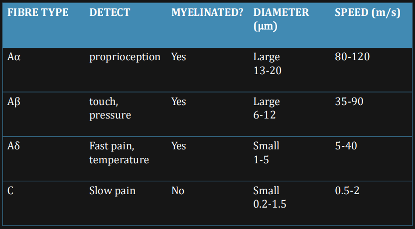
Aδ fibres (fast and specific)
Myelinated: Possess a myelin sheath, allowing for rapid conduction of nerve impulses.
‘Fast’ pain sensation: Transmit sharp, acute, well-localized pain. This initial, immediate pain allows for rapid withdrawal from the noxious stimulus.
Unimodal: Typically respond to intense mechanical or thermal stimuli.
C fibres (slow and flexible)
Unmyelinated: Lack a myelin sheath, resulting in slower conduction speeds.
‘Slow’ pain sensation: Transmit dull, aching, throbbing, or burning pain that is poorly localized and often persistent. This second, prolonged pain signal contributes to guarding behavior and promoting healing.
Polymodal: Respond to a wide range of noxious stimuli, including mechanical, thermal, and chemical agents.
Inflammation
Inflammation often accompanies injury or infection. It is a complex biological response of vascular tissues to harmful stimuli, such as pathogens, damaged cells, or irritants, aimed at removing the initial cause of cell injury, clearing out necrotic cells and tissues damaged from the original insult and the inflammatory process, and initiating tissue repair. The inflammatory process leads to increased blood flow to the area, promoting healing.
Many inflammatory mediators
Histamine: Released by mast cells, causes vasodilation and increased vascular permeability.
Prostaglandins: Synthesized from arachidonic acid by cyclo-oxygenase (COX) enzymes, contribute to vasodilation, fever, and sensitize nociceptors.
Substance P: A neuropeptide released by nociceptor terminals, promotes vasodilation and mast cell degranulation, and facilitates pain transmission.
Bradykinin: Formed during tissue injury, directly activates nociceptors and increases vascular permeability.
These mediators sensitise nociceptors, lowering their activation threshold, meaning they fire with less intense stimuli.
Hyperalgesic states arise from these processes, where a normally painful stimulus is perceived as more painful than usual, or allodynia, where a non-painful stimulus becomes painful. This sensitization can also contribute to chronic pain.
NSAIDs (Non-Steroidal Anti-Inflammatory Drugs) block synthesis of prostaglandins by inhibiting COX enzyme (cyclo-oxygenase). By reducing prostaglandin production, NSAIDs reduce inflammation, pain, and fever.
Pain - Ascending pathway
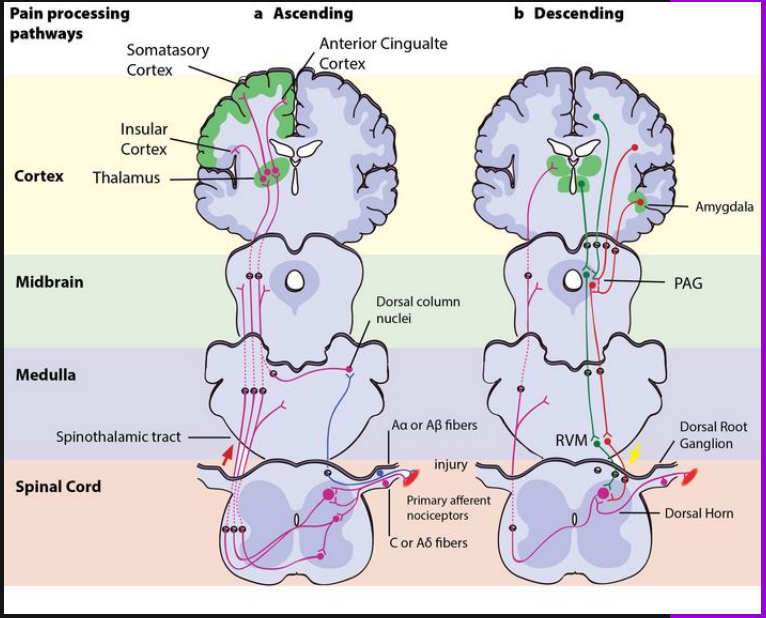
Axons from nociceptors (first-order neurons) enter the dorsal horn of the spinal cord (specifically lamina I, II, and V of Rexed) and ascend or descend a few segments in ‘Lissauer’s tract’ before synapsing.
Enter the substantia gelatinosa (lamina II) or nucleus proprius (lamina I & V) and synapse with ‘second-order’ nociceptors/projection neurons. These interneurons process and relay the information.
These second-order neurons then cross the midline (decussate) at the level of entry in the spinal cord via the anterior white commissure and ascend contra-laterally as the lateral spinothalamic tract. This tract carries pain and temperature information.
They synapse in the VPL (ventral posterolateral) nucleus of the thalamus for body pain and VPM (ventral posteromedial) for facial pain. The thalamus acts as a crucial relay station.
From the thalamus, third-order neurons project to various cortical areas for higher-level processing.
Pain - Descending pathway
Modulate ascending sensory pathways, providing an endogenous system for pain control, known as antinociception. This system can either inhibit or facilitate pain transmission.
Projections originate from several brainstem nuclei:
PAG (Periaqueductal Gray) – Opioidergic: A key midbrain structure. It receives inputs from the cortex and limbic system and projects to other brainstem nuclei. Its activation leads to the release of endogenous opioids (e.g., enkephalins) that inhibit pain transmission in the spinal cord.
Locus coeruleus - Noradrenergic: Located in the pons, it primarily releases norepinephrine (noradrenaline), which has antinociceptive effects in the spinal cord by inhibiting dorsal horn neurons.
Nucelus raphe magnus (NRM) - Serotonergic: Located in the medulla, it releases serotonin (5-HT), which can both inhibit and facilitate pain at the spinal cord level, often through activating inhibitory interneurons.
Reticular formation: A diffuse network of nuclei spanning the brainstem, involved in various functions including pain modulation. It contributes to both pain inhibition and facilitation.
These descending pathways can act directly on dorsal horn neurons or indirectly via inhibitory interneurons, affecting the release of neurotransmitters and hyperpolarizing pain-signaling neurons.
Pain in the brain
Pain is not solely processed in one brain area but involves a complex network, often referred to as the 'pain matrix'.
Thalamus acts as a sensory relay station to cortex: All sensory information (except olfaction) to the cortex passes through the thalamus. It broadly processes pain intensity and basic characteristics.
Insula and cingulate cortex → make pain ‘unpleasant’
These limbic structures are crucial for the emotional and affective components of pain. The anterior cingulate cortex (ACC) is involved in pain's unpleasantness and emotional response, while the insula integrates sensory and emotional information, contributing to the subjective experience of pain intensity and distress.
Primary somatosensory cortex
Located in the postcentral gyrus, it contains a sensory homunculus, a topographical map of the body. S1 is primarily involved in locating painful stimuli, discriminating pain intensity, and identifying the type of pain. It provides sensory-discriminative aspects.
Damage to S1 does not get rid of pain: While S1 is involved in localization, the distributed nature of the pain matrix means that other areas (like the insula and ACC) can still process the emotional and unpleasant aspects of pain, leading to continued experience of pain even with S1 lesions.
Types of pain
Acute
Less than 6 months
Pain often related to tissue damage (not always)
Medication usually helpful
Usually curable
Chronic
More than 6 months
Pain that lasts after healing process has occurred
Medication can be unhelpful
No known cure
Nociceptive
Due to stimulation of peripheral nociceptors during damage to body tissues
Neuropathic (due to neuronal damage)
Pain persisting after injury has healed or with no obvious injury
Fibromyalgia pain fits this pattern, and some studies suggest that fibromyalgia pain may be neuropathic
Psychogenic
Physical pain resulting from emotional factors
Tension headache, back pain, stomach pain
Nociceptive pain
Response to an actual bodily injury
Provide a warning signal
Fast pain and slow pain
Aδ-fibres & C-fibres
Stabbing, throbbing, aching, stiffness
Treatment of pain is usually achieved by the treatment of the underlying condition
Usually acute pain
Neuropathic pain
No immediately apparent cause
Alleviating pain more difficult
Shooting, electric shock-like, burning, tingling or numbness
Inflammation contributes to pain: Sensitization of nociceptors by inflammatory mediators
Often chronic pain – not always (e.g. arthritis)
Phantom limb pain
Limb removed, but still feel it’s there
Common – 80% amputees
Why? Still mapping for that limb
Mirror box therapy
Review of phantom limb theory see link: https://www.nature.com/articles/nrn1991
Fear/ expectation of pain
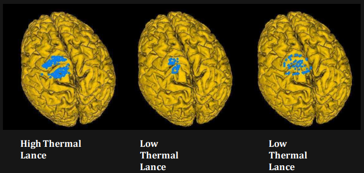
Research using PET scan - Shows electrical activity in the brain
Pain as a complex experience
Not a simple relationship between tissue damage and pain
Pain magnitude does not reflect the extent of tissue damage
Lesions of the pathway do not stop pain
Biopsychosocial model of pain (Campbell and Edwards 2009)
Preventing pain
Nociceptors like all neurons conduct signals via action potentials
shut these down, you shut down pain
Block voltage gated sodium channels
Local anaesthetics like lidocaine/lignocaine do this
Shut down pain (and everything else)
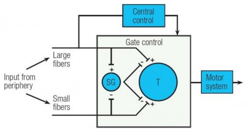
Gate Theory
Melzack and Wall, 1965
Tried to explain a range of pain phenomena
Pain is not simply related to extent of tissue damage
How anti-nociception works
Role of large diameter neurons
Role of descending pathways from the brain
Neurotransmitter system involved in gating mechanism = opioid system
Opioids are natural anti-nociceptive substances – enkephalins
Pain-Gate Open | Pain-Gate Closed |
Focus on pain (pain watching) | Distraction (but see later on) |
Negative emotions such as: Depression, Anxiety, Anger, Frustration, Guilt | Happiness and laughter |
Anxiety and panic | Relaxation and feeling calm |
Sedentary lifestyle / being unfit | Aerobic exercise / being fit |
Fear of pain | Reassurance |
Preventing pain
Activity in Aβ fibres inhibits nociceptive transmission
Activates inhibitory interneurons in spinal cord
Rubbing injured body part
Adrenaline playa a role too
Stress induced analgesia
Locus coeruleus (blue area) descending projections provide endogenous analgesia
Value of Gate Theory
Integrative biological psychology – how cognitive or psychological factors can affect pain
Good to know this to design pain control interventions
Transcutaneous electrical nerve stimulation (TENS) - stimulates low-threshold large-diameter neurons to close the gate
Acupuncture – stimulates opioid release in the PAG to increase descending inhibition
Pain Management
Also called pain medicine or algiatry
More than just pills and bandages
Multiple methods:
Pharmacological
Procedural (stimulation)
Physical (physiotherapy)
Psychological
Biological | Psychological | Social |
Stretching | Habits | Communication |
Exercise | Depression | Relationships |
Pacing | Stress | Having fun |
Relaxation | Irritability | Friends |
Medication | Understanding | Work |
Gate theory | Sleep |
Link between physical and emotional pain
DeWall et al (2008) – subjects took paracetamol (for several weeks) and felt less emotional pain (link to social pain)
Depressed people show higher levels of substance P (primary NT at 1st-2nd synapse)
Anterior cingulate cortex implicated in depression as well as pain