Week 6: Inflammatory and Structural Heart Disorders
Inflammatory & Structural Heart Disorders
Overview of Cardiac Disorders
Key focus on inflammation and decreased cardiac output, leading to altered tissue perfusion.
Part 1: Infective Endocarditis
Definition
Infective Endocarditis is an infection of the endocardium, which lines the heart and forms the heart valves.
Commonly caused by bacteria (e.g., staphylococci, enterococci) but can also be due to fungal infections.
Structure of the Heart
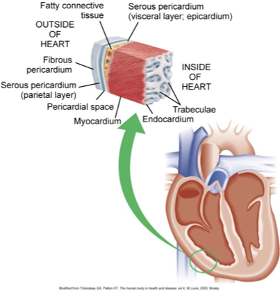
Pericardium (sac surrounding the heart):
Fibrous Pericardium (outer layer)
Serous Pericardium:
Visceral layer (epicardium)
Parietal layer
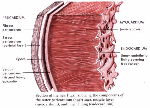
Heart Wall Components:
Myocardium (muscle layer; part of the muscle dies due to infarction/death due to lack of oxygen)
Endocardium (inner lining covering trabeculae; they form the valves)
Causes of Infective Endocarditis
Modes of Infection:
Use of shared needles
Direct contamination
Surgical or dental procedures
Development from a systemic infection.
Pathophysiology
Colonization of microbes occurs at sites of abnormalities or injuries (e.g., prolapsing valves, injured valves, or artificial valves).
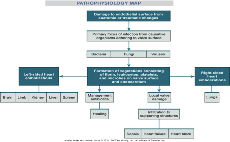
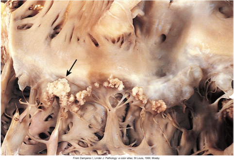
The inflammatory response leads to endothelial damage, promoting clustering of platelets, fibrin, white blood cells (WBCs), and bacteria (a vegetation). This forms around the valves. Can be a viral or fungal infection.
Vegetations form on the endocardium:
Fragile and easily crumbled, can break off, causing emboli.
Can damage valve leaflets and mechanical valves, leading to abscess formation.
Risk Factors for Infective Endocarditis
Pre-existing heart disease.
Intravenous (IV) drug use.
Cardiac valvular diseases.
Rheumatic heart disease.
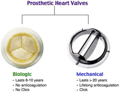
Presence of prosthetic valves.
Congenital and structural abnormalities (e.g., mitral valve prolapse).
Common valve replacements: aortic valve (blood from the left ventricles to the rest of the body) and mitral (between the left atrium and left ventricle).
Mechanical replacements, the client needs to be on anticoagulation therapy, and you’ll hear a click upon auscultation.
Biological ones, which come from animals, deteriorate more quickly.
Long-term intravenous catheters.
Recent body piercings or similar procedures.
Clinical Manifestations
Symptoms:
Rapid onset with fever and chills.
Subacute presentations with gradual onset, low-grade fever, and constitutional symptoms.
Notable signs include heart murmurs, worsening with vegetation damage.
General symptoms may include myalgias and joint pain. Fever is not mandatory.
Specific Signs:
Splinter Hemorrhages and Petechiae (occurring in ~40% of cases due to hemorrhage from distal capillaries).
Petechiae: common in those with low platelets/thrombocytopenia.
Osler Nodes: Painful, red nodules in fingers/toes due to microemboli.
Janeway Lesions: Painless red or purple macules on palms or soles.
Embolization Risks
Left-sided heart embolizations can impact major organs (e.g., brain, kidneys, liver, spleen).
Right-sided heart embolizations primarily affect the lungs.
Management
Antibiotics for treating infections.
Obtain a blood culture to diagnose and determine how to treat the infection.
Monitoring for local valve damage and potential complications: sepsis, heart failure, and heart block.
Part 2: Acute Pericarditis
Definition
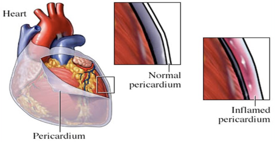
Inflammation of the pericardium, the protective sac around the heart.
Holds the heart in place.
Baroreceptors control heart rate and BP.
Structure of the Pericardium
Composed of:
Fibrous Pericardium (outer layer)
Serous Pericardium:
Parietal layer (outer)
Visceral layer (inner; epicardium)
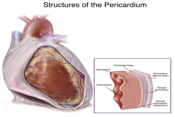
Contains 15-50 ml of pericardial fluid to reduce friction.
Causes of Acute Pericarditis
Can be caused by infections (bacterial, viral, fungal) or secondary to inflammation from:
Myocardial infarction (MI)
Cardiac surgery
Malignancy
Lupus
80% of cases are idiopathic, often thought to be viral.
Risk of pericardial effusion leading to cardiac tamponade (pressure on the heart). Constrictive pericarditis can occur, restricting heart movement and filling, leading to decreased cardiac output.
It smoothers the heart with too much fluid.
Tamponade: a medical emergency characterized by the accumulation of fluid in the pericardial space, which can significantly impair the heart's ability to pump effectively and requires immediate intervention.
Clinical Manifestations
Symptoms range from mild to severe.
Common symptom: Chest pain (similar to MI) characterized as:
Sharp
Aggravated by deep breaths, coughing, and lying down (indicative sign!).
Persistent nature, with relief upon sitting or leaning forward.
Retrosternal pain that can radiate.
Signs include:
Friction Rub: A distinctive heart sound arising from the rubbing of inflamed pericardial layers.
Commonly heard at the left lower sternal border (4th intercostal space) using the diaphragm of the stethoscope, loudest when the patient sits forward.
Other symptoms:
Fever
Increased white blood cell (WBC) count
Elevated sedimentation rate/C-reactive protein
Exertion-independent dyspnea
Widespread ST-segment elevation on 12-lead EKG
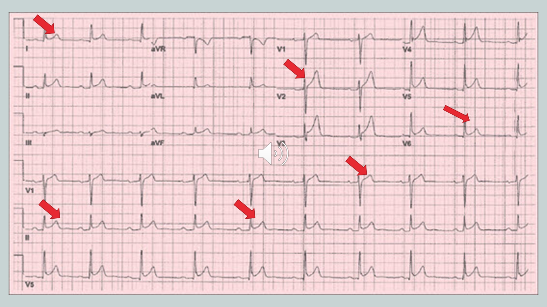
Possible pericardial effusion on echocardiogram.
Nursing Care for Acute Pericarditis
Aim to relieve pain and manage symptoms.
Assess for cardiac tamponade signs.
First-line treatments include NSAIDs (e.g., Ibuprofen, ASA, Indomethacin).
Given anti-inflammatories.
Corticosteroids are used for patients intolerant to NSAIDs, and colchicine for recurrent cases.
Positioning is key for comfort, providing reassurance that the symptoms are not indicative of a heart attack.
Relieved with sitting and leaning forward to reduce pressure from the parietal pericardium.
Part 3: Valve Disorders and Cardiomyopathy
Overview of Valvular Diseases
It occurs when heart valves do not function properly, affecting blood flow.
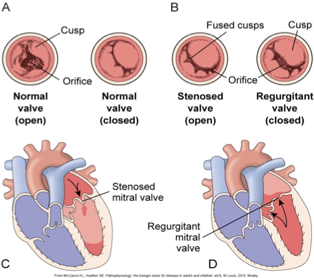
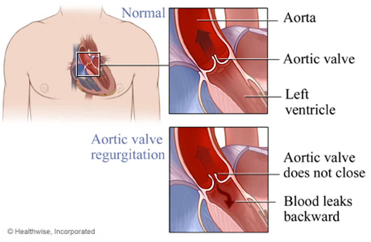
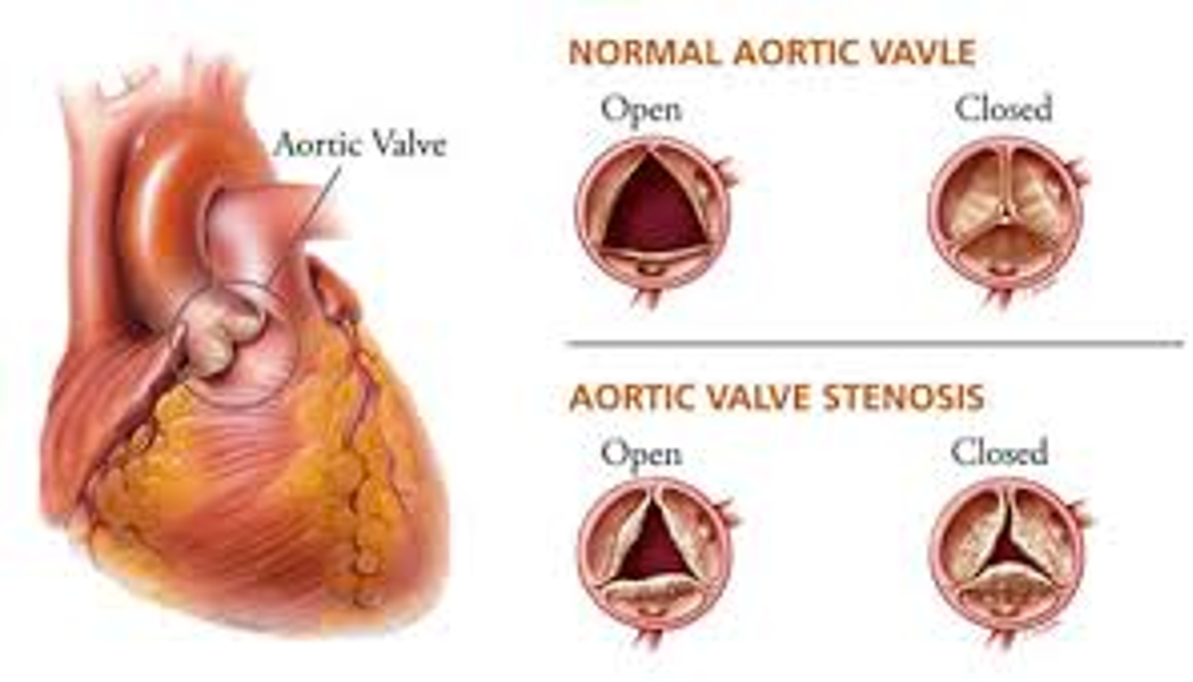
Stenosis: Narrowing of valve openings, reducing blood flow through the valve.
It doesn’t fully open or close all the way.
Regurgitation: Incomplete closure of valves, allowing backward blood flow.
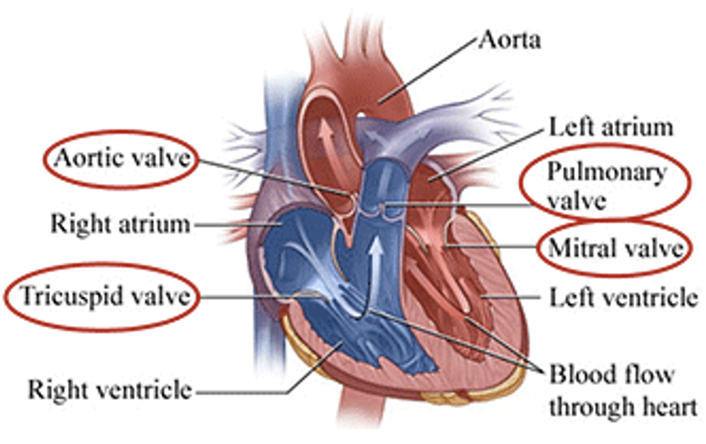
Visual Representation of Blood Flow Through the Heart
Aorta: Origin point of blood after leaving the left ventricle.
Valves involved:
Mitral valve (left atrioventricular; has 2 leaflets)
Tricuspid valve (right atrioventricular; has 3 leaflets)
Pulmonary valve (out to lungs)
Aortic valve (out to body).
Specific Valve Disorders
Mitral Valve Stenosis: Constriction of the mitral valve.
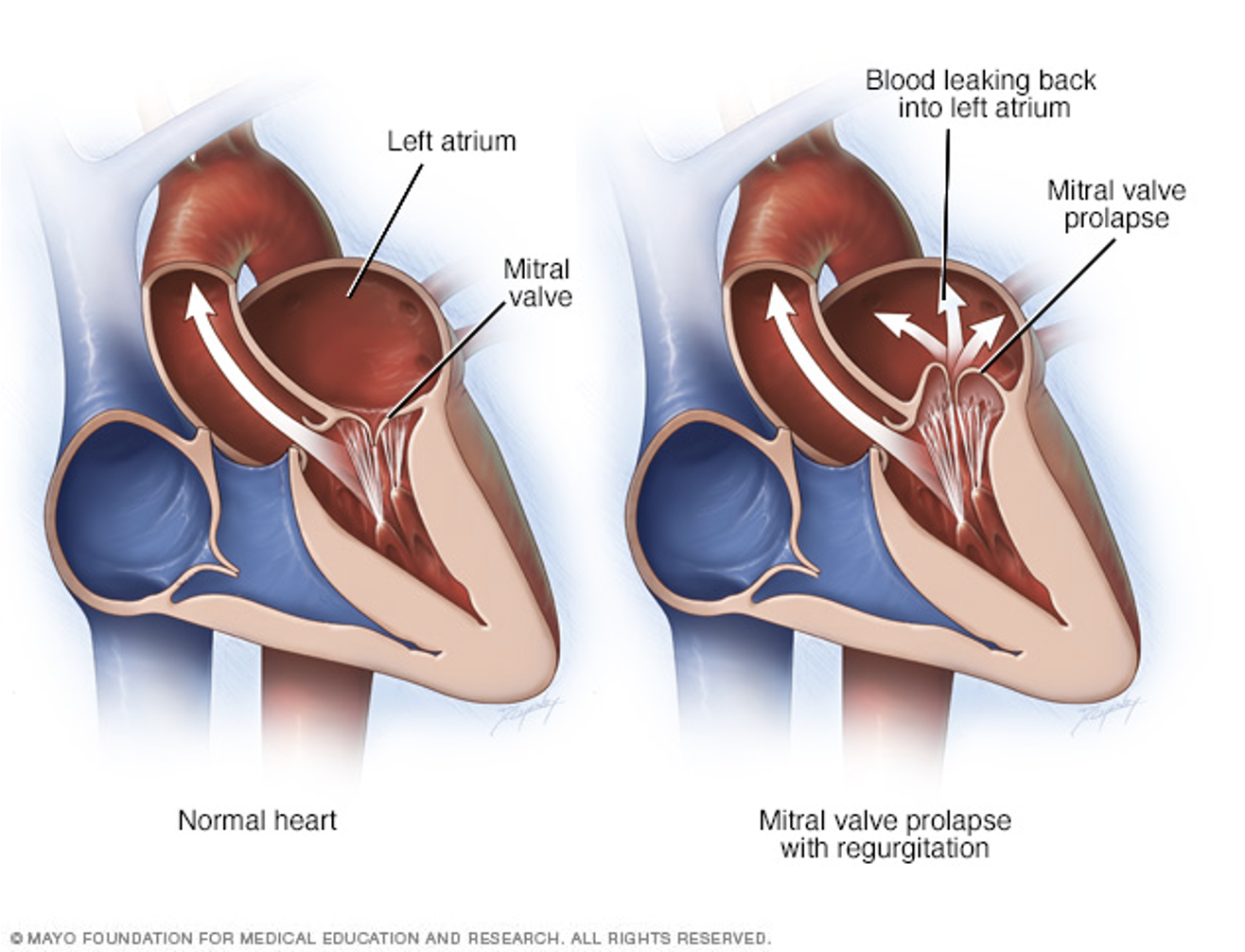
Mitral Valve Regurgitation: Backflow of blood into the left atrium due to improper valve closure.
Mitral Valve Prolapse: The mitral leaflets bulge backward into the left atrium, causing regurgitation.
Limited to the mitral valve.
Common in women.
The mitral valve can be replaced if this issue cannot be fixed.
The leaflet folds downward/dips (hooding) into the left atrium during systole, which can lead to mitral regurgitation if the valve does not close properly.
Aortic Stenosis and Regurgitation: Aortic stenosis involves narrowed openings; regurgitation features leakage back into the left ventricle when the valve fails to close completely.
Regurgitation usually follows stenosis or prolapse, as it is present in relation to issues of the valve.
Diagnostic Studies for Valvular Heart Disease
Includes:
Electrocardiogram (EKG)
Chest X-ray
Echocardiogram (including Transesophageal Echocardiogram/TEE)
3D Doppler echocardiography
Cardiac catheterization
Exercise stress test.
Management of Valvular Heart Disease
Conservative Treatments:
Medications and rest.
Percutaneous trans-luminal balloon valvuloplasty (replacement/repair of the valve): a minimally invasive procedure (percutaneously or surgically) used to open narrowed heart valves by inserting a balloon through a catheter and inflating it to widen the valve opening.
Annuloplasty: repair of the outer ring around the valve.
Surgical Treatments:
Valve repair and replacement.
Cardiac transplant in severe cases.
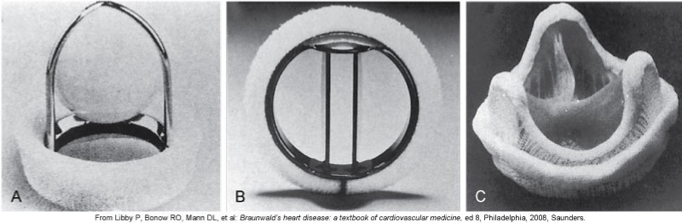
Prosthetic Valve Types:
Mechanical (e.g., bi-leaflet, caged-ball)
Tissue valves (e.g., homograft, pericardial, xenograft).
Postoperative Care for Valve Repair/Replacement
Monitoring for signs/symptoms of emboli and heart failure.
Care aligns with postoperative protocols for patients recovering from coronary artery bypass surgery.
High risk of post-op complications (thromboembolism, infection, arrhythmias, hemolysis).
Part 4: Cardiomyopathy
Definition
A group of disorders involving the myocardium, leading to mechanical and/or electrical dysfunction.
Results in impaired cardiac output.
Types of Cardiomyopathy
Dilated Cardiomyopathy (DCM): Weakened and enlarged left ventricle.
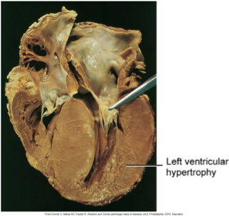
Hypertrophic Cardiomyopathy (HCM): A genetic condition resulting in a thickened left ventricular wall.
Can be seen in children.
Restrictive Cardiomyopathy (RCM): Rigid heart muscle restricting filling.
Arrhythmogenic Right Ventricular Cardiomyopathy (ARVC): Replacement of heart muscle cells with fat and scar tissue.
Temporary, broken heart as the muscles are weakened for a while.
Stress-Induced Cardiomyopathy: Typically resolves in weeks.
Dilated Cardiomyopathy Etiology
Results in thinning of the left ventricular wall and increased chamber size and volume.
An over-stretched rubber band that doesn’t fling back the way it normally does.
Hypertrophic Cardiomyopathy Etiology
Characterized by left ventricular hypertrophy, thickening, impacting heart's ability to pump efficiently.
The muscle is overworked, causing this growth/hypertrophy.