Untitled Flashcards Set
HHP: 4300 Sensorimotor Neurophysiology Unit 1 Learning Objectives
Lecture 1
1. Sensory System vs Motor System vs Sensorimotor System (top-down vs bottom up/outside in)
a. Sensory system: bottom up or outside in (afferent)
i. Receives info from environment through senses and sends info to the brain
ii. Environment body/PNS Brain/SC/CNS
iii. M1 = primary motor cortex
iv. S1 = somatosensory cortex
v. Senses going to spinal cord through thalamus and gets integrated
vi. Afferent = sensory = towards NS = peripheral in
b. Motor system: top down or inside out (efferent)
i. Sends signals from the brain to muscles to produce movement
ii. Brain/SC/CNS body/PNS environment
iii. M1 = precentral gyrus
iv. S1 = postcentral gyrus
v. Efferent = motor = away from CNS = internal out
c. Sensorimotor system: integrates sensory info and motor output
i. Brain is a prediction engine and injuries are typically incorrect predictions (such as picking up box and hurting back)
2. List the different senses
a. Proprioception – body sensing where you are in space
b. Smell
c. Taste
d. Hearing
e. Vision
f. Touch
g. Vestibular – balance
h. Interoception – sense and interpret bodily sensations
i. Hunger, pain, heart burn, etc
3. Integration and the sensorimotor system
a. Sensory inputs are processed and integrated with motor commands to produce coordinated movement
i. Sensory input brain processes motor output sensory feedback adjustments
b. Integration of sensory information to inform and regulate motor function
c. Involves the inputs from afferent and efferent nerves in order to produce coordinated movement
4. Sensorimotor Feedback Loop
a. Sensorimotor system is constantly predicting and adjusting
b. Continuous cycle of sensory input guiding motor output
5. CNS vs PNS Terminology Differences
a. CNS = brain and spinal cord
i. Axon = tract
ii. Cell body = nuclei
b. PNS = everything else, nerves outside the CNS including sensory and motor pathways
i. Axon = nerve
ii. Cell body = ganglia/ganglion
6. Coronal vs Sagittal vs Transverse Planes
a. Coronal: divides anterior vs posterior
b. Transverse: divides superior vs inferior
c. Sagittal = divides medial and lateral
Lecture 2
1. CNS and PNS components
a. CNS
i. Spinal cord: all the nerves going to the rest of the body comes here
1. Essentially an extension of the brain
2. Grey vs White Matter – CNS
a. Brain
i. Gray matter = superficial
1. Nuclei, dendrites, contains both myelinated and unmyelinated axons, glia cells, and blood capillaries
ii. White matter = deep
1. Myelinated, made up of fat, long-range myelinated axons
b. Spinal cord – gray matter = deep and white matter = superficial
3. List the 3 meninges and the order of the layering
a. Protective membranes that enclose the brain and spinal cord
i. Dura mater = outermost layer
ii. Arachnoid mater = middle layer
iii. Pia mater = innermost layer
4. CSF
a. Primary Roles of the CSF?
i. Beneath the subarachnoid layer in the subarachnoid space
ii. Part of blood brain barrier for protection
iii. Reduces friction
iv. Lessons flow of impact from injuries
v. Makes brain lighter - buoyancy
5. Define Meningitis and the common sources
a. Meningitis: inflammation of the meninges due to bacterial or viral infection
b. Can come from injuries, cancer, certain drugs and other types of infection
6. Glia Cells
a. Types, CNS vs PNS
i. Oligodendrocytes = produces myelin and wrap around neuron in CNS
ii. Schwann cells = produces myelin and wrap around neuron in PNS
iii. Microglia = NS macrophages in CNS
7. Myelin
a. Purpose, composition
i. Insulating layer / sheath that forms around nerves in the brain and SC, made up of protein and fatty substances
ii. Allows electrical impulses to transmit quickly and efficiently along the axon
8. MS
a. Pathophysiology, common symptoms
i. Autoimmune disease where immune system decides myelination is bad and affects CNS axonal myelin
ii. Tcells infiltrate the BBB and wrongly recognizes myelin as an invasive substance
iii. Tcells signal other immune cells, like plasma cells, to tag myelin with antibodies
iv. Macrophages destroy the tagged myelin and the oligodendrocytes that product/maintain myelin
b. Define Clinico-radiological paradox
i. Patients clinical symptoms not aligning with the severity of abnormality seen on an MRI
c. Types of MS (3 most common)
i. Relapsing-remitting: episodes of relapsing (worsening) for day-month and then go through remissions (recovery)
ii. Secondary progressive: starts as RRMS but worsens over time with no relapses
iii. Primary progressive: progressive worsening of symptoms from onset without relapses
9. Autonomic Nervous System
a. PNS vs SNS
i. Effect on HR, BP, blood vessel diameter (i.e., constriction/dilation)
1. PNS: decrease HR, decrease BP, vasodilation
2. SNS: increase HR, increase BP, vasoconstriction
b. Enteric NS
i. Primary role
1. Controls GI function, nerves in the GI
10. Draw a flow chart of the naming convention of the different branches of the nervous system
a. CNS Brain/SC
b. PNS Somatic (sensory and motor divisions) and Autonomic (parasympathetic, sympathetic and enteric divisions)
Lecture 3
1. Define signal transduction
a. Conversion of sensory stimuli into electrical signals
2. Importance of the thalamus
a. Relay station for sensory processing
3. Tonic vs Phasic adaptation
a. Tonic: slow adapting receptors that respond for the duration of a stimulus (pain receptors)
b. Phasic: fast adapting to a constant stimulus and turn off. They fire once more when the stimulus turns off (touch receptors)
4. Rapid vs slow adapting mechanoreceptors
a.
5. Tase buds are what kind of receptor?
a. Chemoreceptors
6. Vision Blind Spot
a. Where the optic nerve connects to the eye and brain guesses to fill info in
7. Specialized photoreceptors in the eye
a. Rods = gray
b. Cones = color
8. Olfactory receptors are what kind of receptor?
a. Chemoreceptors
9. Explain Photic Sneeze reflex (do not need to know names of specific cranial nerves)
a. V1 and V2 are close together and when there’s a random influx of light it gets overstimulated and causes you to sneeze
b. V1 hops to V2
10. Briefly (i.e., 2 sentences or less) explain auditory transduction
a. Sound travels through auditory canal and goes to tympanic membrane where the vibrations bounce back and forth. The sound then goes to the 3 ear bones and then travels to the cochlea where it creates waves and activates signaling based on the waves.
11. Semicircular canals vs Otolith organs
a. Roles, motions for each
i. Vestibular system
1. Semicircular canals: rotational movement
2. Otolith organs: head position and linear movement
12. Importance of going to spine and cerebellum with vestibular reflexes
a. Important for balance posture, spatial orientation
b. Would want the reflexes to act first before the brain interprets what it is
13. Explain why dizziness occurs
a. Occurs due to mismatches vestibular and visual input
b. Visual input knows that spinning has stopped but the fluid in the ears is still moving
14. Proprioceptors – provide essential information about body position and movement
a. Skin, joints, muscle, tendons – what is being sensed at each level
i. Skin: sensing touch and pressure
ii. Joints: sensing angle, direction and speed
iii. Muscles: sensing length and stretch
iv. Tendons: sensing tension
b. Define and differentiate Golgi tendon organs vs muscle spindle fibers
i. Muscle spindle fibers sense muscle length and velocity of change of muscle length
ii. Golgi tendon organ senses active muscle tension
c. Example: Walking
i. Taking a step involves the muscle spindle fibers in your leg to detect the length and velocity of change of that muscle length
ii. Golgi tendon organs sense the tension as you land on the ground to maintain balance and prevent injury
iii. Joint receptors: detect angle and movement of the joints as you take that step and room for adjustment
iv. Skin receptors: soles of feet detect the pressure and texture as you step foot on the ground which sends information to the brain to be processed
15. Explain the phenomena of light touch with regards to balance
a. Light touch provides additional sensory input to stabilize posture and provide better balance
Lecture 4
1. M1 vs S1
a. M1 = primary motor cortex (voluntary movement)
b. S1 = primary somatosensory cortex (sensory processing)
c. Homunculus?
i. Map of body parts in the brain
2. Cerebrum
a. Function
i. Higher cognitive functions, sensory processing, motor control
b. Regions not considered cerebrum
i. Cerebellum, spinal cord, and brain stem
3. List Primary Motor Control Regions and their relay hub
a. M1, premotor cortex, and supplementary motor area
b. Relay hub = thalamus
4. Other regions near M1 that help with planning and coordinating movement
a. Premotor cortex and supplementary motor area
5. Cerebellum
a. Primary Roles
i. Balance, coordination, fine motor control
b. Role in coordination?
i. Compares intended vs actual movement
6. Basal Ganglia
a. Structures & function
i. Initiation and modulation of voluntary movement. Suppresses unwanted movement
ii. Structures:
1. Caudate nucleus
2. Putamen
3. Globus pallidus
4. Subthalmic nucleus
5. Substantia niagra
7. Draw the Motor Control “Workflow”
a. Motor areas sensory areas cerebellum basal ganglia
Lecture 5
1. Primary Function of the spinal cord
a. Central relay hub between brain and body
2. Functional organization
a. Segments
i. Cervical thoracic, lumbar, and sacral
b. White/Grey Matter, Horns, Nerves
i. White matter = superficial
ii. Gray matter = deep
1. Cell bodies (dorsal = sensory and ventral = motor)
c. Be able to draw a spinal cord cross section
3. Sensory Processing & Motor Commands
a. Horns, roots, tracts associated with each
i. Dorsal horns: receiving sensory input from periphery
1. Processing occurs
ii. Ventral horns: contain cell body of motor neurons that send signals to muscles
4. Describe spinal cord Role in autonomic regulation
a. Controls sympathetic and parasympathetic outflow
5. Spinal Reflex Arcs
a. Mono vs Polysynaptic
i. Patellar reflex = mono = direct sensory-motor connection
ii. Withdrawal reflex = poly = interneurons involved
b. Reciprocal Inhibition
i. Agonist contracts and antagonist relaxes (gets inhibited)
c. Describe Patellar, nociceptive withdrawal, crossed extensor
i. Patellar = Reflex arc - ipsilateral
1. Monosynaptic sensory and motor neurons, no interneurons (except inhibitory)
2. Rapid, involuntary responses to stimuli
3. Help protect the body from harm and maintain balance tests of neurological damage
4. RECIPROCAL INHIBITION quad extensors are activated from this flex but the interneurons send signals to inhibit the quad extensor
ii. Nociceptive withdrawal - ipsilateral
1. Polysynaptic sensory, motor, and interneurons
2. Rapidly remove body from noxious/painful stimuli
3. Feel
4. Feel pain first and then hot
iii. Crossed extensor – withdrawal reflex - ipsilateral
1. Withdrawal reflex when one limb is pulled away from a painful stimulus while the opposite limb extends to support the body and maintain balance
2. Crossed = involves both sides of the spinal cord (ipsilateral and contralateral)
6. Define Central Pattern Generator
a. Rhythmic activity that can occur without the brain and cognitive input
b. Imagine cycles of reciprocal inhibition and crossed extensor reflexes
c. Polysynaptic
i. Common activity examples
1. Walking, biking, swimming
7. Kyphosis vs Lordosis vs Scoliosis
a. Kyphosis = ant and posterior curvature
i. Abnormal structure of thoracic spine, rounded or hunchback appearance
b. Lordosis = anterior and Posterior
i. Anterior deviation of lumbar or cervical spine
c. Scoliosis = lateral. And medial
i. Lateral curvature or twisting of spine
8. Incomplete vs Complete Spinal Cord Injuries
a. Incomplete = partial cord intact
b. Complete = entire cord damaged
9. Autonomic Dysreflexia
a. Vasodilation above level of injury
b. Vasoconstriction below level of injury
/
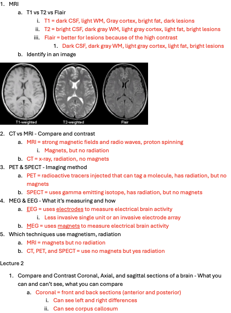
1. MRI
a. T1 vs T2 vs Flair
i. T1 = dark CSF, light WM, Gray cortex, bright fat, dark lesions
ii. T2 = bright CSF, dark gray WM, light gray cortex, light fat, bright lesions
iii. Flair = better for lesions because of the high contrast
1. Dark CSF, dark gray WM, light gray cortex, light fat, bright lesions
b. Identify in an image
2. CT vs MRI - Compare and contrast
a. MRI = strong magnetic fields and radio waves, proton spinning
i. Magnets, but no radiation
b. CT = x-ray, radiation, no magnets
3. PET & SPECT - Imaging method
a. PET = radioactive tracers injected that can tag a molecule, has radiation, but no magnets
b. SPECT = uses gamma emitting isotope, has radiation, but no magnets
4. MEG & EEG - What it’s measuring and how
a. EEG = uses electrodes to measure electrical brain activity
i. Less invasive single unit or an invasive electrode array
b. MEG = uses magnets to measure electrical brain activity
5. Which techniques use magnetism, radiation
a. MRI = magnets but no radiation
b. CT, PET, and SPECT = use no magnets but yes radiation
Lecture 2
1. Compare and Contrast Coronal, Axial, and sagittal sections of a brain - What you can and can’t see, what you can compare
a. Coronal = front and back sections (anterior and posterior)
i. Can see left and right differences
ii. Can see corpus callosum
iii. Can see medial and lateral white matter tracts
iv. Can see the lobes and thalamus deep nuclei and brainstem
b. Axial (transverse/horizontal) = top and bottom sections (superior and inferior)
i. Can see left and right hemisphere differences
ii. Can see ventricles and basal ganglia
iii. Can see anterior and posterior white matter tracts
c. Mid-sagittal = left and right hemispheres
i. Viewing rostro-caudal (anterior-posterior) size
ii. Can see anterior and posterior structures
iii. Can see white matter tracts
iv. Cerebellar structure and brainstem structure
v. NOT helpful for left and right differences
2. Number of Cortical layers – within superficial GM of cerebral cortex
a. 6 layers in total – 3 are focused on in class
i. Molecular layer = superficial (axons, dendrites, and crossover)
ii. External pyramidal layer = middle (pyramidal cells)
iii. Multiform cell layer = deep (multitude of cell types)
3. Ventricles
a. Identify the ventricles
i. Produces, transports, and excretes CSF
b. Function of the choroid plexus
i. Within the 4th ventricle and makes CSF
4. Action Potential
a. Know all the basic defining characteristics
i. -70mv = resting membrane potential
ii. Neurotransmitters interacting with receptors causes depolarization of the neuron
iii. -55mv = threshold potential Voltage-gated Na+ channels open for Na+ to enter the cell causes massive depolarization of the neuron (becomes positive like +10mv) creating action potential
iv. Ca2+ channels open and rushes into the cell vesicle with NS fuse with membrane and dump NT into synaptic space to bind to the gate / channel
v. At the peak positive mv = Na+ close and K+ channels open where K+ flows out of the cell Repolarization membrane potential going back to negative
vi. Hyperpolarization occurs where membrane potential falls below -70mv
vii. K+ channels close membrane potential reached
b. Refractory Periods
i. Where the membrane potential is below -70mv
ii. Difficult for the neuron to fire again
c. IPSP and EPSP
i. IPSP = inhibitory postsynaptic potential = less likely to fire AP
ii. EPSP = excitatory postsynaptic potential = more likely to fire AP
d. Glutamate and GABA
i. Glutamate = primary excitatory NS
ii. GABA = primary inhibitory NS
5. Basal Ganglia
a. Input vs output vs intrinsic nuclei
i. Input = striatum (caudate nucleus and putamen)
ii. Intrinsic nuclei = external globus pallidus (GPe), subthalamic nucleus, and pars compacta of the substantia nigra (SNc)
iii. Output = internal globus pallidus (GPi) and pars reticulata of the substantia nigra (SNr)
b. Caudate Nucleus – major input for basal ganglia, movement planning, learning, memory, reward, motivation, emotion, romantic interaction
i. Role, afferent and efferent connections
1. Afferent = ipsilateral frontal lobe
2. Efferent = hippocampus, globus pallidus, and thalamus
ii. Striatum (with putamen) – largest subcortical structure
c. Putamen – Learning, motor control, speech articulation, language function, reward, cognitive function, addiction, sensory and motor aspects of pain
i. Role, afferent and efferent connections
1. Afferent = cortex
2. Efferent = cortex via thalamus
ii. Lentiform Nucleus vs striatum
1. Striatum with CN
2. Lentiform nucleus with GP
d. Globus Pallidus – conscious and proprioceptive movements
i. Externus vs Internus
1. Externus = intrinsic = GPe
a. Inhibitory tone, indirect pathway
2. Internus = output = GPi
a. Excitatory tone, direct pathway
ii. Role, afferent and efferent connections
1. Input from multiple structures
2. Intrinsic nucleus = relay for info in basal ganglia
3. Output nucleus = sends info to the thalamus for movement
iii. Lenticular Nucleus – with putamen
e. Substantia Nigra – compart part and net-like part, “black substance”
i. Pars compacta vs Pars reticulata
1. Pars compacta = compact part
2. Pars reticulata = net-like part
ii. Role, afferent and efferent connections
iii. SNc and dopamine
1. SNc = produces and releases dopamine
2. Dopaminergic projections to the striatum
a. Synapse on DA1 or DA2 receptors
i. DA1 = excitatory
ii. DA2 = inhibitory
6. Subthalamic Nucleus
a. Is it Basal Ganglia?
i. not “officially” BG
b. Inputs, outputs, function
i. Input from GPe
ii. Outputs to GPi
iii. Primarily glutamate
iv. Target of deep brain stimulation in Parkinson’s
7. Excitatory Vs Inhibitory Movement Control
a. Direct pathway = excitatory
i. Cortex sends glutamate signals to striatum
ii. D1 receptors are activated and send GABA signals to GPi
iii. GPi normally inhibits thalamus
iv. Inhibition of inhibitor at GPi thalamus less inhibited and facilitates movement
b. Indirect pathway = inhibitory
i. Cortex sends glutamate signals to striatum
ii. D2 receptors are activated and send GABA signals to GPe
iii. GPe normally inhibits STN inhibition of the inhibitor
iv. STN is active which sends glutamate signals to GPi
v. GPi inhibiting thalamus suppresses movement
Lecture 3
1. Thalamus – allows selected info to pass to cerebral cortex, regulation of consciousness, alertness, sleep, and wakefulness
a. Anterior Nuclei
i. Inputs = mammillary body, hippocampus, and limbic
ii. Output = cingulate gyrus
iii. Function = learning, memory, emotion
b. Ventral Posteromedial Nuclei
i. Inputs = trigeminothalamic tract and rostral solitary nucleus
ii. Outputs = primary somatosensory cortex and gustatory cortex
iii. Function = touch, position, pain, and temperature from the face as well as gustatory information
c. Ventral Posterolateral Nuclei
i. Inputs = body and limbs dorsal column medial lemniscus (pressure, vibration, fine touch, and proprioception), and spinothalamic tract
ii. Outputs = somatosensory cortex
iii. Function = relays somatosensory spinal inputs to the somatosensory cortex
d. Ventral Anterior and Lateral Nuclei
i. Ventral anterior nucleus
1. Input = basal ganglia
2. Outputs = motor, premotor, and supplementary motor cortices (frontal)
3. Function = relays basal ganglia inputs to motor cortices for motor planning
ii. Ventral lateral nucleus
1. Input = cerebellum
2. Outputs = motor, premotor, and supplementary motor cortices (frontal)
3. Function = relays cerebellar inputs to motor cortices for motor planning and control
e. Dorsomedial Nuclei
i. Inputs = amygdala, olfactory, limbic, and basal ganglia
ii. Outputs = frontal cortex, prefrontal association areas
iii. Functions = emotions, cognition, and learning
f. Pulvinar
i. Inputs = visual inputs from the superior colliculus and extrastriate visual areas, auditory and other sensory pathways
ii. Outputs = parietotemporal-occipital association areas
iii. Functions = precise function unknown, might be involved in orientation toward visual, auditory, and other sensory stimuli
g. Medial and Lateral Geniculate Bodies
i. Medial geniculate body
1. Input = inferior colliculus
2. Output = auditory cortex of the temporal lobe
3. Function = relays auditory input to the cortex
ii. Lateral geniculate body
1. Input = retina/optic nerve
2. Outputs = primary visual cortex
3. Function = relays visual inputs to the primary visual cortex
h. Intralaminar/Midline Nuclei
i. Inputs = ascending reticular formation, spinal cord, and hypothalamus
ii. Outputs = limbic cortex and basal ganglia
iii. Functions = arousal, motivation, affect (emotion / feelings), pain
2. Limbic System
a. Overall function
i. Process and regulate emotion, memory, learning, motivation, and social processing, sexual stimulation
b. Brain regions (and how it is a fugazi)
c. Hypothalamus
i. Role
1. Coordinates the endocrine system, temperature, autonomic, and appetite regulation
2. Input = various brain regions
3. Output = pituitary thyroid, adrenal, and reproductive organs
d. Hippocampus – learning and memory
i. Role, input, output
1. Learning and memory processing
2. Inputs = olfactory, visual, auditory, and somatosensory
3. Outputs = thalamus, hypothalamus, and other limbic structures
ii. 3 zones & 4 regions
e. Amygdala
i. Role, functional connection
1. Regulates anxiety, aggression, fear conditioning, emotional memory, and social cognition
2. Involved in conditioning for food, sex, and drugs
3. Functionally connected to hypothalamus and ventral pallidum (GP) and ventral striatum
f. Parahippocampal Gyrus
i. Role
1. Memory encoding and retrieval
2. Environmental scene and not faces
g. Entorhinal Cortex
i. Entorhinal vs parahippocampal gyrus
ii. Role
1. Memory formation and consolidation – sleep optimization
2. Declarative memory
3. Input = hippocampus
4. Output = dentate and hippocampus
Lecture 4
1. Cerebellum – motor regulation and balance control, posture, muscle tone, voluntary muscle activity, UNABLE to initiate muscle contraction
i. Input from cortex and other cerebellar tracts
b. Primary and secondary functions
c. Anatomy
i. Lobes - 3
1. Anterior
2. Posterior
3. Flocculondular
ii. Deep nuclei
1. Fastigial nucleus
2. Interposed nucleus
3. Dentate nucleus = largest and most important
iii. Molecular, Purkinje, granular layers – within GM
1. Molecular = superficial
2. Purkinje = intermediate, has dendrites and axons
3. Granular = deep
iv. Vermis – role
1. Control area of cerebellum
2. Coordinates trunk movement = neck, shoulders, thorax, abdomen, and hips
2. Number of cranial nerves – 12 pairs, 24 cranial nerves
a. General functions cranial nerves control not specifics for each nerve
i. Control sensory and motor functions in the head and neck
3. White Matter Tracts – communication between brain areas
a. Categories
i. Projection tracts = connections of cortex
ii. Association tracts = connect cortex to ipsilateral cortex
iii. Commissural tracts = connect left and right hemispheres
b. Functions of key tracts
i. corticospinal, stria terminalis, thalamocortical
1. corticospinal = motor activity, controls voluntary movement
a. terminate in spinal grey matter
2. thalamocortical = relays sensory and motor from thalamus to cortex
3. limbic tracts = cingulum = interconnect cingulate gyrus with medial lobes role in emotion and motivation
a. fornix = connected to hippocampus and thalamus via mamillary bodies
b. stria terminalis = limbic system, emotional stress response, start at amygdala
4. Brain blood supply
a. Internal carotid and vertebral arteries
i. Two internal carotid and two vertebral arteries
b. Circle of willis
i. Don’t need specifics or diagram, just know about basics
1. Primary blood supply for brain
2. Supplied by internal carotids and vertebral arteries
Lecture 5
1. Brain Stem – unconscious control such as breathing, HR, BP
i. Has 10/12 cranial nerves coming off brainstem
b. Midbrain
i. Location on brainstem
1. Connects cerebrum to pons
ii. Colliculi functions
1. Superior colliculi = visual reflexes
a. Output = lateral geniculate body (thalamus) and optic tract
2. Inferior colliculi = auditory processing
a. Output to medial geniculate body (thalamus
iii. Substantia Nigra
1. Base of the midbrain and is part of the basal ganglia
iv. Ventral Tegumental Area
1. Reward pathway
v. Red Nucleus
1. Motor control and muscle tone
c. Pons
i. Location on brainstem
1. Superior to inferior = midbrain, pons, and medulla oblongata
2. Connects to cerebellum via middle cerebellar peduncle
3. Medulla junction contains cranial nerves
ii. Locus coeruleus – synthesizes NE
1. RAS
a. Helps regulate arousal and attention
iii. Pontine nuclei = assists in coordinating movement
d. Medulla Oblongata
i. Location on brain stem
1. Most inferior of brainstem, pons to spinal cord
2. Meets spinal cord at foramen magnum
3. Connects to cerebellum via inferior cerebellar peduncle
ii. Pyramids
1. Motor fibers from M1 spinal GM
2. Anterior fissure that extends to spinal cord
iii. Olives = lateral to pyramids
1. Aid in motor coordination via connection with cerebellum
iv. Other functions
1. Respiratory center with pons controls respiration
2. Vasomotor center where it controls arterial BP
/1. Action potentials
a. Resting membrane potential, threshold potential,
i. RMP = -70mv
ii. Threshold potential = -55mv
iii. Negative to less negative = depolarization till about +10mv
1. Na+ rushing into the cell
iv. Peak = K+ channels opening and slowly leaving the cell
1. Becoming more negative = repolarization
v. Overshoot = more negative than the RMP due to the slow closing of those K+ channels
b. Role of NA/K Pump
i. Actively transporting 3 Na+ out of the cell and 2 K+ into the cell using energy from ATP
ii. Creates an electrochemical gradient
c. Glutamate and GABA
i. Glutamate = primary excitatory NS
ii. GABA = primary inhibitory NS
2. Sensory Receptors
a. List all the receptor types and what stimuli they respond to
i. Chemoreceptors: transduces chemical substances into biological signals, detect changes in normal environment
1. Internal peripheral: detect chemical changes in blood
a. Carotid bodies detect an increase in CO2 in blood
b. Chemical action potential
2. Distance: sense of smell where you aren’t directly interacting with the substance
a. Detect chemicals in gaseous states (odors and pheromones)
3. Direct: requires direct interaction with the chemical’s source – tastebuds
a. Taste doesn’t adapt as quickly whereas smell does
b. Dorsal respiratory group = inspiration
c. Ventral respiratory group = stimulates DRG for non-normal respiration
d. Pneumotaxic = turns off DRG
4. Peripheral chemoreceptors = internal
a. Carotid and aortic bodies
i. Sense partial CO2 and O2 pressure
ii. Sense pH in blood
5. Central chemoreceptors
a. Within ventral medulla
b. Sense pH in CSF
ii. Photoreceptors: transduce light into biological signals (best transduction = vision)
1. Rods: sensitive to light, scotopic (dim) vision
2. Cones: rapid responses to variations in light intensity, photopic (bright) vision, color vision and visual acuity
a. Less cones than there are rods, which shows how good they are at producing imaging
b. Concentrated in the macula and fovea
3. Intrinsically photosensitive retinal ganglion cells
a. Play a role in circadian rhythm and the pupillary reflex
4. Destroyed distally via phagocytosis
5. Produced proximally via morphogenesis
iii. Thermoreceptors
iv. Mechanoreceptors
v. Proprioceptors
vi. Nociceptors
b. Common receptors responsible for regulation of homeostatic variables such as BP, blood plasma chemical concentrations
i. Chemoreceptor’s cardiac function
1. Cardiac center medulla controls HR
2. Baroreceptors detects pressure from decrease O2, increase CO2, and decrease pH
3. Heart rate
4. Vasodilation/vasoconstriction
Lecture 2
1. Vision
a. name of receptors, specific proteins involved
i. Rods
1. Has both retinal and opsin primary proteins
2. Retinal + opsin = Rhodopsin proteins
ii. Cones
1. Different type of opsin proteins = Photopsin proteins
iii. which are involved in low light situations and which are involved with color vision?
1. Rods = low light conditions
2. Cones = color vision
b. what makes the vision sensory transduction process unique from other sensory transduction processes?
i. Phototransduction cascade
1. Opsin in disk membrane absorbs photon causes change of configuration of the 3D structure unstable intermediates activate transducin protein
ii. Stimulus = light photons
c. What colors of cones do humans possess?
i. S = Blue, M = green, L = red, and black
d. What determines what color we see when we only have 3 “color” cones?
i. Receptor output signal is proportional to the number of photons absorbed
1. Estimation of wavelength color vision
2. Specific to a wavelength
2. Temperature
a. Temperature change vs absolute temperature
i. “non-specialized” receptor that codes relative changes in temp
1. We’re used to relative changes whereas not good at absolute temperature
b. Receptors involved
i. Warm receptors
ii. Cold receptors
c. Receptors involved in nociception of temperature
d. Temperature change transduction pathway
i. Warm receptors
1. Increase in warming increase in AP firing rate, decrease in warming decrease in AP firing rate
ii. Cold receptors
1. Increase in cooling increase in AP firing rate, decrease in cooling decrease in AP firing rate
Lecture 3
1. Major categories of mechanoreceptors
a. Merkel’s disk
b. Meissner’s corpuscle
c. Ruffini endings
d. Pacinian corpuscles
i. Stimuli for each type
1. Merkel’s disk = pressure and texture
2. Meissner’s corpuscle = light touch and vibration
3. Ruffini endings = stretch and pressure
4. Pacinian corpuscles = deep pressure and vibration
ii. Slow vs Fast adapting
1. Slow adapting
a. “tonic”, fire continuously when a stimulus is present, crucial for providing info about constantly present stimuli, skin stretch, muscle tension, Ruffini endings and GTOs
2. Fast adapting
a. “phasic”, rapid initial response to stimulus, firing rate quickly decreases and eventually stops even if stimulus persists, sensitive to changes in stimulus, fine touch, movement, vibration, Meissner’s corpuscles, and Pacinian corpuscles
2. Minor categories
a. Hair nerve fibers
b. Free nerve endings
3. Types of skin and unique characteristics important for sensation
a. Glabrous
i. Mostly meissner’s corpuscles, but also has Pacinian corpuscles, Merkel’s disks, and Ruffini endings
ii. Concentrated in fine touch areas fingers, palms, soles
iii. More sensitive than hairy and is involved in detailed, tactile sensation
b. Hairy
i. Less specialized for detailed tactile perception
ii. Fewer touch receptors
iii. Hair nerve, free nerve endings, and ruffini endings
4. Transduction pathway for Meissner Corpuscles
a. Involves light touch or vibrations converting into electrical signals
i. External force physical deformation of lamellae bending of nerve axon terminals action potential nerve dorsal root ganglion dorsal column of spinal cord medulla (decussation) thalamus S1 FEEL
1. What nuclei in the thalamus?
a. VPL – receives sensory input related to the body
2. What white matter tract from the Thalamus to S1?
Lecture 4
1. Definitions of Proprioception and kinesthesia
a. Proprioception is the sense of position and movement of the body including forces and loads applied to the body
i. Not in isolation but they integrate other senses at the same time including touch, vision, and vestibular
1. Active joint position: you are moving your own arm
2. Passive joint position: someone else moving it
ii. Two basic categories (all get info from joint capsule, ligaments, muscles, tendons, skin and get sent to the brain) = redundancy
1. Sense of force effort, heaviness, tension
a. Reproduce and match desired level of force
2. Sense of change in velocity
a. Detects vibration of oscillating objects
b. Kinesthesia is active but during movement
i. Awareness of motion
1. Duration, direction, amplitude or size, speed, acceleration
2. Muscle Spindles vs. Golgi Tendon Organs (GTO)
a. Similarities and differences
i. Muscle spindles = spindle like
1. Intrafusal fibers = not contractile, no tension
a. Nuclear bag = absolute center of muscle spindle
i. type of intrafusal fiber
ii. many nuclei in bags
iii. cause excitation of sensory fibers
1. dynamic nuclear bag = fast contraction speed
2. static nuclear bag = slow contraction speed
iv. sensitive to length changes
b. Nuclear chain = nuclei aligned in chain, wrapped around intrafusal fiber
i. Type of intrafusal fiber
ii. Excites secondary nerve
iii. Half the size of bag fibers
1. Measures stress and strain on muscle
2. Spring “stretch of spring”
2. Extrafusal fibers = contractive, tension
ii. Golgi Tendon Organs
b. Functions
c. Roles in proprioception and kinesthesia
i. Muscle spindles = muscle length
ii. Golgi tendon organ = tension
3. Muscle Spindle Specifics
a. Intrafusal vs extrafusal fibers
1. Intrafusal fibers = not contractile, no tension
a. Nuclear bag = absolute center of muscle spindle
i. type of intrafusal fiber
ii. many nuclei in bags
iii. cause excitation of sensory fibers
1. dynamic nuclear bag = fast contraction speed
2. static nuclear bag = slow contraction speed
iv. sensitive to length changes
b. Nuclear chain = nuclei aligned in chain, wrapped around intrafusal fiber
i. Type of intrafusal fiber
ii. Excites secondary nerve
iii. Half the size of bag fibers
1. Measures stress and strain on muscle
2. Spring “stretch of spring”
2. Extrafusal fibers = contractive, tension
b. α and γ motor neurons
i. y gamma motor neurons
1. don’t “contract” the muscle
2. adjusting stretch sensitivity by contracting intrafusal fibers
3. keeps the spindle tight
4. allows a motor neuron to continually fire and contract muscle
5. release acetylcholine intrafusal fibers open Na2+ influx depolarization increased stretch sensitivity of spindle afferents
6. do not contribute to force of muscle/extrafusal contraction
ii. a alpha motor neurons
1. cause muscle contraction
c. sensory components (group 1 vs 2 afferents)
i. 1a afferent = large diameter, spiral around intrafusal fibers, middle of spindle
ii. II afferent = medium diameter, lateral on chain fibers
1. Afferent stretch info via mechanically gated on channels
2. Bag2 and chain fibers increase firing rate on these two at a given muscle length
3. Bag1 increase stretch sensitivity of 1a afferents
d. α - γ coactivation hypothesis
i. during voluntary muscle contractions, both a and y motor neurons are activated simultaneously to maintain the sensitivity of the muscle spindle
ii. ensures that the muscle spindle remains sensitive to changes in muscle length, even during contraction
4. GTO specifics
a. Spindles have y motor neurons, no motor innervation
b. Mechanically activated by muscle-tendon unit
i. Group 1b afferents
1. Thinner than 1a, myelinated, still high conduction velocity
2. Reflexive control with spinal
3. Reciprocal innervation when too much force
4. Relaxation of contracting muscle and contraction of antagonist
5. Protects against overstraining the muscle / tendon
ii. how does it get activated?
1. High tension from voluntary contractions or external forces acting on the muscle
iii. GT reflex afferent and efferent pathways
1. Muscle generates force sensory terminals compress stretch sensitive channels open action potential spinal cord reflex and brain
2. Golgi tendon senses excessive tension sensory neuron conducts action potential sensory neurons synapse with interneurons a motor neurons to quads is inhibited while the hamstring is stimulated hamstring contracts and the quads relax
3. Muscle contraction (afferent pathway) activation of GTO sensory signal sent to SC spinal cord integration inhibition of agonist muscle (efferent pathway) activation of antagonist muscle reflex
a. What is the purpose?
i. Protection