Genetics - lecture 16 - Cancer Genetics
Cancer Overview
1 in 2 individuals will be diagnosed with cancer in their lifetime.
Cancer survival rates have improved significantly since the 1970s.
Learning Outcomes
Define the hallmarks of cancer.
Identify gene types linked to tumor formation.
Understand the clonal origin of cancer.
Cancer Definition and Types
Definition:
A group of disorders characterized by uncontrolled cell growth and the invasion and spread of cells from the site of origin to other sites.
Malignant tumors invade other tissues.
Types of Cancer:
Carcinomas (85%):
Arise from epithelial cells (lung, mouth, intestines).
Sarcomas:
Derived from mesoderm (bone, muscle).
Adenocarcinomas:
Derived from glandular tissues (breast).
Benign Tumors:
Locally growing, encapsulated in fibrous tissue, and non-invasive.
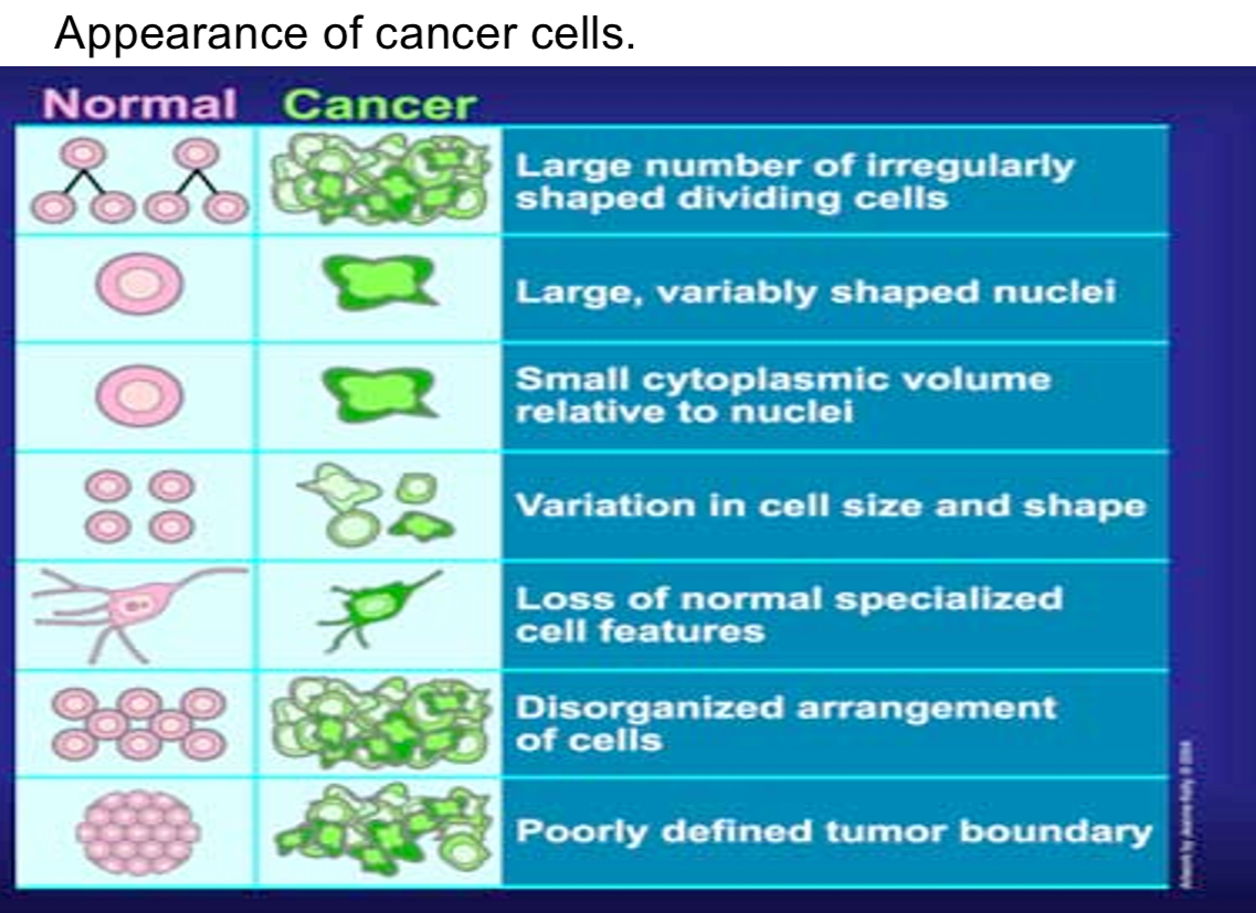
Appearance of Cancer Cells
Normal vs. Cancer Cells:
Cancer cells show irregular shapes, large variably shaped nuclei, smaller cytoplasmic volume, and disorganized arrangements.
Metastasis
Example: Breast cancer often spreads to the brain.
Metastasis can be observed in studies of melanoma in mice.
Development and Progression of Cancer
Multi-step Process:
Cancer is not caused by a single mutation.
Accumulation of approximately six mutations required for certain cancers.
Process affected by aging and environmental factors.
Evidence of Multi-step Development:
Single mutation is not enough to cause cancer and incidence is a function of age.
Time lag exists between carcinogen exposure and cancer development (e.g., smoking and lung cancer).
Histopathological studies show progressive changes in intestinal epithelia leading to malignancy.
Large amount of cell division so more likely to develop cancer because more quickly accumulating mutations.
Clonal Origin of Cancer
Tumors originate from a single ancestral cell (clonal hypothesis).
Evidence:
Tumor cells often show the same X chromosome inactivated when tested for X-linked markers.
Mtelomas produce a single type of Ig.
Hallmarks of Cancer Cells
Growth Signal Autonomy:
Cancer cells bypass normal growth factor pathways.
Insensitivity to Anti-growth Signals:
Respond to inhibitory pathways but do not stop growth.
Evading Apoptosis:
Resistance to programmed cell death despite DNA damage.
Limitless Replicative Potential:
Cancer cells maintain telomere length, allowing continuous division.
Angiogenesis:
Induce formation of new blood vessels for nutrient access.
Invasion and Metastasis:
Mutations reduce cell adhesion, enabling tissue invasion.
Genetic Mutations in Cancer
Proto-oncogenes:
Encode proteins to promote cell division; mutations lead to oncogenes (dominant).
Dominant mutations.
Tumor Suppressor Genes:
Proteins limit cell proliferation, prevent cell cycle pogression, or activate apoptosis; mutations require both alleles to be inactive (recessive).
Classes of Oncogenes
Five Main Classes:
Secreted growth factors (e.g., PDGF, IGF2).
Relatively small proteins mediating intracellular communication.
Platelet derived GF (PDGF): wound healing
Insulin-like GF (IGF2): promote growth during gestation.
Cell surface receptors (overexpression examples, like Her2).
Signal transduction components (e.g., Ras mutations).
DNA binding proteins (e.g., Myc).
Cell cycle regulators (e.g., cyclin D2).
Activation of Proto-oncogenes to Oncogenes
Point Mutation:
Mutations that abolish GTPase activity.
Usually affects Ras genes.
Amplification:
Multiple copies of oncogenes (e.g., HER2 in breast cancer).
Duplication of a limited region of a chromosome will display a repeating binding pattern.
Chromosomal rergion may break away and replicate as an independent particle, forming double.
Chromosomal Translocation:
Results in novel fusion genes (e.g., bcr-Abl in CML).
An autoinhibitory domain of N terminal domai of Abl is lost in the bcr-ABl fusion, and the kinase domain is permanently active.
De-regulated kinase activity leads to at least two consequences that contribute to tumourgenesis: uncontrolled cell division and a defect in DNA repair
Fusion Gene Formation:
Example: MYC from Burkitt’s lymphoma.
myc now placed under the control of a promoter that drives synthese of Igs.
myc now a potent oncogene driving relentless proliferation of lymphoid cells.
Tumor Suppressor Genes
Corresponding proteins limit cell cycle entry, limit normal cell proliferationm and can activate apoptosis (e.g., Rb).
Recessive
Examples:
Rb (Retinoblastoma):
Regulates cell cycle at restriction point R.
p53:
Mutated in 50% of tumors; halts cell cycle for DNA repair or triggers apoptosis.
APC gene:
Associated with familial adenomatous polyposis; affects colon cancer development.
Causes of Cancer
Multi-step process driven by mutation accumulation.
Environmental Factors:
e.g., tobacco exposure, UV radiation, HPV infection.
Mutation Rates:
Approximately three mutations per cell division; overall can exceed 2,000 mutations in a lifetime.