Lab 16 Muscles of the Head and Neck
Section 1: Overview of Structural and Functional Characteristics of Skeletal Muscle
Before we get into specific muscle groupings, it is important to recognize skeletal muscle not just as individual tissues but as a highly organized and dynamic organ system. Composed of muscle fibers, connective tissue, blood vessels, and nerves, skeletal muscle plays a critical role in voluntary movement, posture, thermoregulation, and metabolic homeostasis. Understanding the structural and functional characteristics of skeletal muscle at the organ level provides essential context for studying how specific muscle groups work together to generate force and support movement.
Structural Characteristics of Skeletal Muscle
Structural organization of skeletal muscle as an organ can be understood by studying the connective tissue components (Figure 16.1) from smallest (thinnest) to largest (thickest) and from deep to superficial.
The smallest and deepest connective tissue component is a thin sheet of loose connective tissue that surrounds each individual muscle fiber called endomysium. Endomysium allows room for nerves to stimulate and blood capillaries to nourish muscle cells.
The perimysium is the next layer of connective tissue with larger nerves and blood vessels. The perimysium is thicker and wraps about 20 to 60 muscle fibers together into bundles called fascicles. The next layer of connective tissue is an even thicker fibrous sheath that surrounds the entire muscle called the epimysium. The deep side (inner surface) of the epimysium extends between fascicles to help form the perimysium, whereas the superficial (outer surface) side grades into what is called fascia.
Fascia is the most superficial and thickest sheet of connective tissue that separates muscle groups from each other and subcutaneous tissue. The collagenous fibers of the connective tissue components wrapping the muscles emerge or extend past the muscle and merge with the tendon or other bone attachment.
The tendon is a straplike cord of connective tissue that attaches muscle to bone as the collagenous fibers that emerged from muscle to the tendon now merges with the periosteum and matrix of the bone as an attachment site.
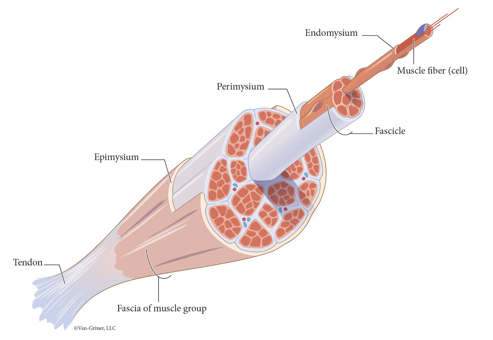
Figure 16.1Connective tissue components of skeletal muscle
When studying the structural organization of skeletal muscle, we must also consider the orientation of the fascicles as defined by the perimysium. This is important because the fascicles can be arranged in such a way that will determine the strength of a muscle as well as the direction that a muscle will pull. Muscles can be classified according to fascicle arrangement as follows (see Figure 16.2):
Parallel muscles have fascicles that are arranged in parallel bundles of uniform width.
Example: rectus abdominusFusiform muscles are thickest in the middle and fascicles taper on each end.
Example: biceps brachiiCircular muscles (sphincters) have fascicles arranged in rings or circles around certain body openings and passages.
Example: orbicularis oculi of the eyelidTriangular (convergent) muscles are broad or wide at one end of the muscle, and the fascicles converge to a narrower end of the muscle. Example: Trapezius
Pennate muscles are feather shaped with three types of pennate muscles.
Unipennate muscles have fascicles that all approach the tendon from one side.
Example: semimembranosus of thighBipennate muscles have fascicles that approach the tendon from both sides.
Example: rectus femoris of thighMultipennate muscles have fascicles arranged in multiple rows, with each row converging on one or more tendons, resembling the structure of multiple feathers placed side by side with quills converging on a single point.
Example: deltoid muscle of the shoulderMuscles of the head and neck
Functional Characteristics of Skeletal Muscle
When considering the functional characteristics of skeletal muscle, let's first study the intrinsic-extrinsic muscle distinction. A muscle that acts upon and is contained entirely in the body region of interest is an intrinsic muscle. A muscle that acts upon a body region of interest and is not completely contained in that body region of interest is an extrinsic muscle.
For example, we will study the extrinsic muscles of the eye later in this lab lesson. The muscles of the eye that we will study are responsible for movement of the eye but are not located in the eye. Thus, these are considered extrinsic muscles of the eye. In a later lab, we will discuss intrinsic muscles of the eye as it pertains to the autonomic nervous system and the fight or flight—sympathetic nervous system response or the rest and digest—parasympathetic nervous system response. These muscles are contained or located in the eye and are responsible for pupil dilation (fight or flight—sympathetic nervous system response) and pupil constriction (rest and digest—parasympathetic nervous system response).
Notice that we just used terms like “act upon,” “movement,” “dilation,” and “constriction.” These terms refer to the effect produced by the muscle of interest called the action of the muscle. The action, or effect produced by a muscle can produce or prevent a certain type of movement. Actions of skeletal muscles can include but are not limited to movements such as flexion, extension, rotation, abduction, adduction, etc.
Skeletal muscles are often arranged in opposing pairs at a joint. The muscle that is most responsible for producing a movement is called the agonist, or prime mover. A muscle that opposes the agonist is usually found on the opposite side of the bone or body and is called an antagonist. A synergist aids the agonist.
Additional Information
As you learn the names of the labeled muscles, keep in mind that the name of the muscle is often related to its location (example: the occipitalis muscle overlies the occipital bone). Muscles may also be named according to shape (example: the trapezius is a diamond-shaped muscle), size (example: gluteus maximus), or action (example: levator labii superioris).
Also, when studying the figures of the muscles, recognize that muscles are layered. Muscles that are located closest to the skin are superficial muscles. They can be removed to reveal deep muscles (muscles underneath other muscles) that are closer to internal body organs. Many of the figures used will give a representation of the superficial and deep muscles of interest by having on side of the figure contain the superficial muscles and the other side depicting the removal or reflection of the superficial muscle(s) by showing only the deep muscles of interest. In Figure 16.3, both the anterior and posterior views show superficial muscles on one side and deep muscles on the other.
Muscles of Facial Expression
The muscles we will study in this section are primarily superficial muscles termed as the Muscles of Facial Expression. These muscles are distinctive compared to the other muscle groups we will study because they originate in fascia or bone and insert in the dermis and subcutaneous tissues of the head and neck. When these muscles tense and pull, they produce facial expressions and can contribute to speech, chewing, and other oral functions.
We start our study of the muscles of facial expression at the scalp with the two muscles and aponeurosis that come together to form the epicranius, also referred to as the occipitofrontalis muscle (Figure 16.4).
The frontalis and occipitalis muscles (Figure 16.4) of the epicranius are named after the cranial bones that lie underneath each. They are mechanically connected by a broad, flat, tendon-like structure known as the cranial aponeurosis (Figure 16.4) once termed the galea aponeurosis.
In understanding the action(s) of these muscles, we consider the origin (stationary attachment site) and insertion (movable attachment site) of the muscle. For example, the frontalis muscle originates (stationary attachment site) at the anterior border of the cranial aponeurosis and inserts onto the subcutaneous tissue of the eyebrow. When this muscle contracts or tenses, the insertion is pulled in the direction of the origin. The result of this action is that the eyebrows are raised as in the universal facial expression of surprise or horror.
In addition, the occipitalis muscle originates at the occipital bone and inserts at the posterior border of the cranial aponeurosis. Again, when this muscle contracts, the insertion is pulled in the direction of the origin. The result of this action is that the scalp is retracted or pulled back towards the occipital bone.
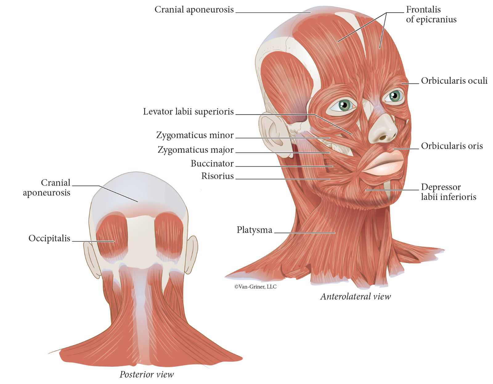
Figure 16.4Muscles of facial expression
In continuation of our study of the muscles of facial expression, two muscles, the orbicularis oculi and orbicularis oris (Figure 16.4), form circles around facial openings. Their names should help you locate them in an image or your own body (oculus refers to the eye, and oris refers to the mouth).
The zygomaticus major and zygomaticus minor (Figure 16.4) muscles both originate on the zygomatic bone and have slightly different insertion sites at the corner of the mouth. The risorius muscle draws the mouth laterally in facial expressions such as laughing. All three of these muscles are used when smiling.
Recall that muscles are sometimes named for their action—this is the case for the levator labii superioris (Figure 16.4)—which elevates the upper lip. The depressor labii inferioris (Figure 16.4) depresses the lower lip.
The buccinator (Figure 16.4) muscle forms part of the lateral wall of the mouth. It is responsible for compressing the cheek against the teeth. Sucking through a straw or producing a good tone with a musical instrument requires use of this muscle.
Anteriorly, the most superficial neck muscle is the platysma (Figure 16.4). This broad muscle extends from the sternum and clavicle and inserts on the mandible. It is primarily responsible for depressing the lower lip.
Muscles of Facial Expression—Actions
To study the attachment sites and actions of all the indicated muscles of facial expression, see Table 16.1. Use Table 16.1 as a guide to learn the required muscles of facial expression and their action(s). Although the muscle site attachments are listed in the table, muscle site attachments are used only for the purpose of understanding the muscle actions and will not be used for questions on the lab exam.
Table 16.1
Muscles of Facial Expression and Their Actions
Muscle
Origin
Insertion
Action
Frontalis of epicranius
Aponeurosis
Skin of eyebrow
Raises eyebrows
Occipitalis of epicranius
Occipital bone
Aponeurosis
Retracts scalp
Orbicularis oris
Mandible and Maxilla
Skin of lips
Purses lips
Orbicularis oculi
Orbital bones
Skin around eye
Closes eyelids
Buccinator
Mandible and Maxilla
Orbicularis oris
Compresses cheeks
Zygomaticus major
Zygomatic bone
Corner of mouth
Pulls corners of mouth upward
Zygomaticus minor
Zygomatic bone
Corner of mouth
Pulls corners of mouth upward
Risorius
Zygomatic arch and fascia near the ear
Corner of mouth
Pulls corners of mouth laterally
Levator labii superioris
Zygomatic and maxilla bones
Skin of upper lip
Elevates upper lip
Depressor labii inferioris
Mental protuberance
Skin of lower lip
Depresses lower lip
Platsyma
Fascia of chest muscles
Mandible
Tenses neck
Depresses mandibleMuscles of Mastication
The second group of muscles that we are studying is used in the process of mastication, or the chewing of food. Mastication involves elevating and depressing the mandible (or opening and closing the jaw). Closing the jaw means working against the force of gravity; therefore, there are three muscles to elevate the mandible and only one to depress it on each side of the head. The strongest of the muscles to elevate the mandible is the masseter (Figure 16.5), which extends from the zygomatic arch to the mandible.
The large fan-shaped temporalis (Figure 16.5) originates on the parietal bone and overlies the temporal bone. It inserts onto the coronoid process of the mandible, and it also functions to elevate the mandible (i.e., close the mouth).
The pterygoid muscles are deep (Figure 16.5), and both move the mandible side-to-side to help grind our food. The medial pterygoid also elevates the mandible, and the lateral pterygoid depresses it.
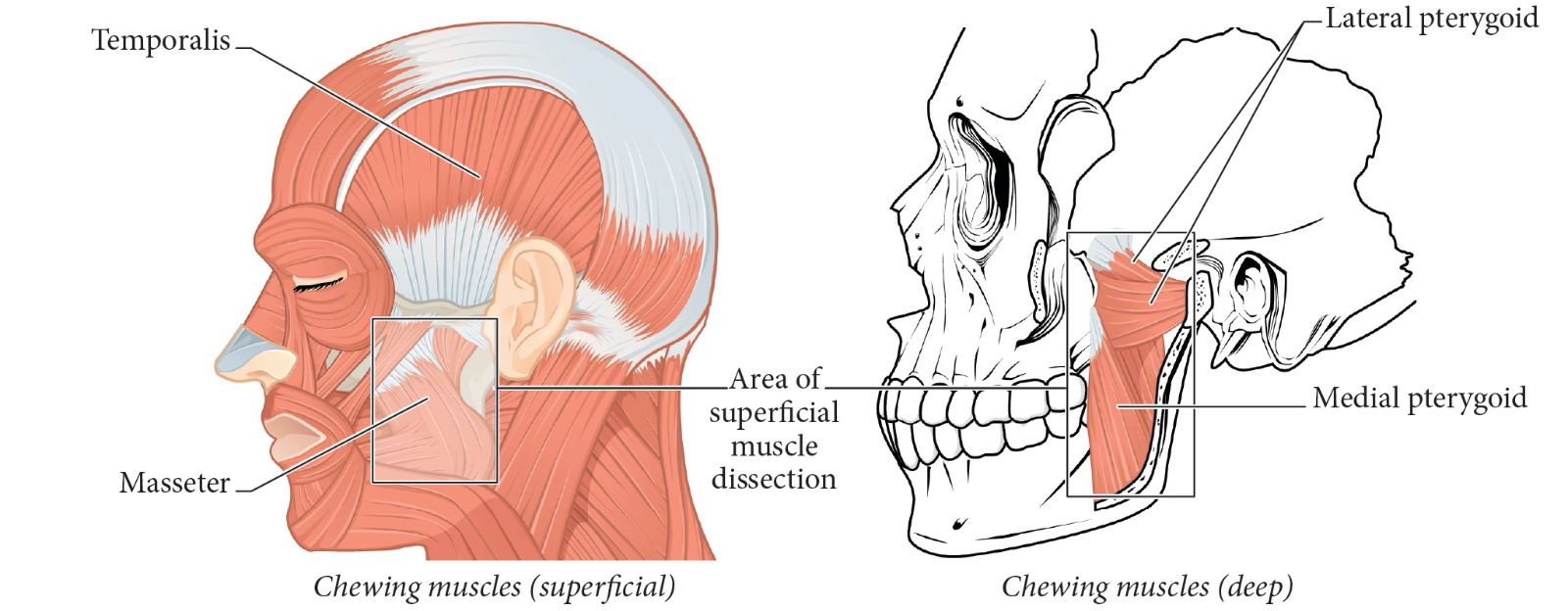
Figure 16.5Muscles of mastification (chewing muscles)
Access for free at Openstax.Muscles of Mastication—Actions
To study the attachment sites and actions of all the indicated muscles of mastication, see Table 16.2. Although the muscle site attachments are listed in Table 16.2, they are used only for the purpose of understanding and mastering the muscle actions and will not be used for exam questions. However, you should be able to name and locate each indicated muscle and identify the action(s) of each.
Table 16.2
Muscles of Mastication (Chewing Muscles) and Their Actions
Muscle
Origin
Insertion
Action
Masseter
Zygomatic arch
Lateral ramus of mandible
Elevates mandible
Temporalis
Temporal bone
Coronoid process of mandible
Elevates mandible
Medial pterygoid
Sphenoid, palatine, maxilla bones
Medial surface of mandible
Elevates mandible; moves mandible side to side
Lateral pterytoid
Sphenoid bone
Mandibular condyle
Depresses mandible; moves mandible side to side
Muscles of the Neck that Move the Head
Our next group of muscles we are studying move the head and are found in the neck area. In anatomical position, the neck is extended with the head facing forward. Movements of the neck and head include flexion (chin to chest), extension, hyperextension (tilting the head back to look at the stars, for example), and rotation. We will consider two superficial muscles, one in the anterior neck, and the other in the posterior neck.
The large, four-sided trapezius (Figure 16.6 and Figure 16.7) is a muscle with multiple fiber directions. This means it will have multiple actions, and not all fibers are active at the same time. The superior fibers of the trapezius attach to the occipital bone and the spine of the scapula. If the head is in a flexed position (chin to chest), bilateral contraction (both muscles of the pair of muscles contract simultaneously) trapezius of the superior fibers of the trapezius will extend the head. With unilateral contraction (only one muscle of the pair contracts), the head will rotate.
When the platysma is removed as in Figure 16.6 and Figure 16.7 the sternocleidomastoid muscle can be viewed on the lateral aspect of the neck. This muscle is named for its attachment sites—the sternum, clavicle (cleido- means clavicle), and mastoid process of the temporal bone. When both sternocleidomastoid muscles contract, they will flex the neck causing the head to bow. However, when only one sternocleidomastoid muscle contracts, it rotates the head by turning it to the opposite side.
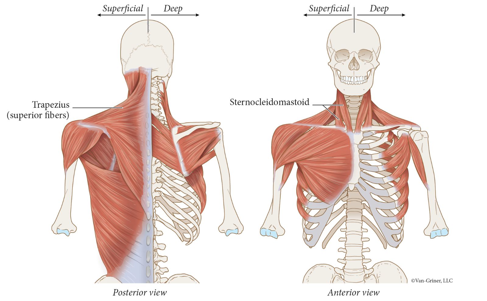
Figure 16.6Superficial muscles of the neck that move the head
When a muscle contracts, one attachment tends to remain relatively stationary, and the other attachment moves. But with both the superior fibers of the trapezius and the sternocleidomastoid, the movable attachment can be switched. With the sternocleidomastoid muscle, if the attachments to the mastoid processes move, the action is flexion of the neck/head. But if those attachments remain stationary, and the sternal and clavicular attachments move, the action is to raise the rib cage. This will facilitate inspiration (breathing in). For this reason, sternocleidomastoid is an "accessory muscle of inspiration."
There are two deeper muscles, one in the anterior neck and the other in the posterior neck. The scalenes (Figure 16.7) are three paired muscles (anterior, middle and posterior) located in the anterolateral aspect of the neck, and like the sternocleidomastoid, they flex the neck and can elevate the rib cage when acting as accessory muscles of inspiration. Posteriorly, deep to the superior fibers of the trapezius are the splenius capitis muscles (Figure 16.7) which extend the neck.
.jpg)
Figure 16.7Deep muscles of the neck that move the head
Muscles of the Neck that Move the Head—Actions
To study the attachment sites and actions of all the indicated muscles of the neck that move the head, see Table 16.3. As a reminder, although the muscle site attachments are listed in Table 16.3, they are used only for the purpose of understanding and mastering muscle actions and will not be used for exam questions. However, you should be able to name and locate each indicated muscle and identify the action(s) of each.
Table 16.3
Muscles of the Neck that Move the Head and Their Actions
Muscle | Origin | Insertion | Action |
|---|---|---|---|
Superior fibers of trapezius | Clavicle and spine of scapula | Occipital bone | Bilateral contraction: Extends the head (or elevates the scapula) |
Sternocleidomastoid | Sternum and clavicle | Mastoid process | Neck/head flexion (or elevation of rib cage) |
Scalenes | Ribs 1 and 2 | Transverse process of cervical vertebrae | Synergist for neck flexion and rotation (or elevation of rib cage) |
Splenius capitis | C7–T6 spinous processes | Occipital bone; mastoid process | Extends the head |
Extrinsic Muscles that Move the Eye
As we conclude our study of the muscles of the head and neck, recall that if the entirety of a muscle is within a particular body area of interest, it is called an intrinsic muscle. In contrast to intrinsic muscles, extrinsic muscles are not located in the body area of interest yet play a role in movement of that body area. There are six extrinsic muscles of the eye that insert on the eye itself. However, they originate externally to the eye, specifically from the back wall of the orbital cavity. These extrinsic eyes muscles are the focus of our study for this last section of the lab.
Four of the muscles are known as "rectus muscles". Rectus means "straight," and these muscles approach their attachment site in a straight line from the origin. The muscles are named for where they attach to the eye and their direction of movement—superior rectus, inferior rectus, lateral rectus, and medial rectus (Figure 16.8). When the medial and lateral rectus muscles contract, the eye moves medially and laterally, respectively. The superior and inferior rectus muscles produce the action their names suggest, however they both have an additional action of medial rotation.
The superior oblique and inferior oblique muscles (Figure 16.8) attach to the eye at an angle, which causes their action to be the opposite of their name (think "o" for opposite). In addition, they both have additional actions of lateral rotation. Notice that the superior oblique muscle is anchored to the medial orbital cavity wall by a ring of connective tissue called the trochlea. You can use this structure as a marker to determine if you are viewing the right or left eyeball and whether you a looking at a medial or lateral view of the eye.
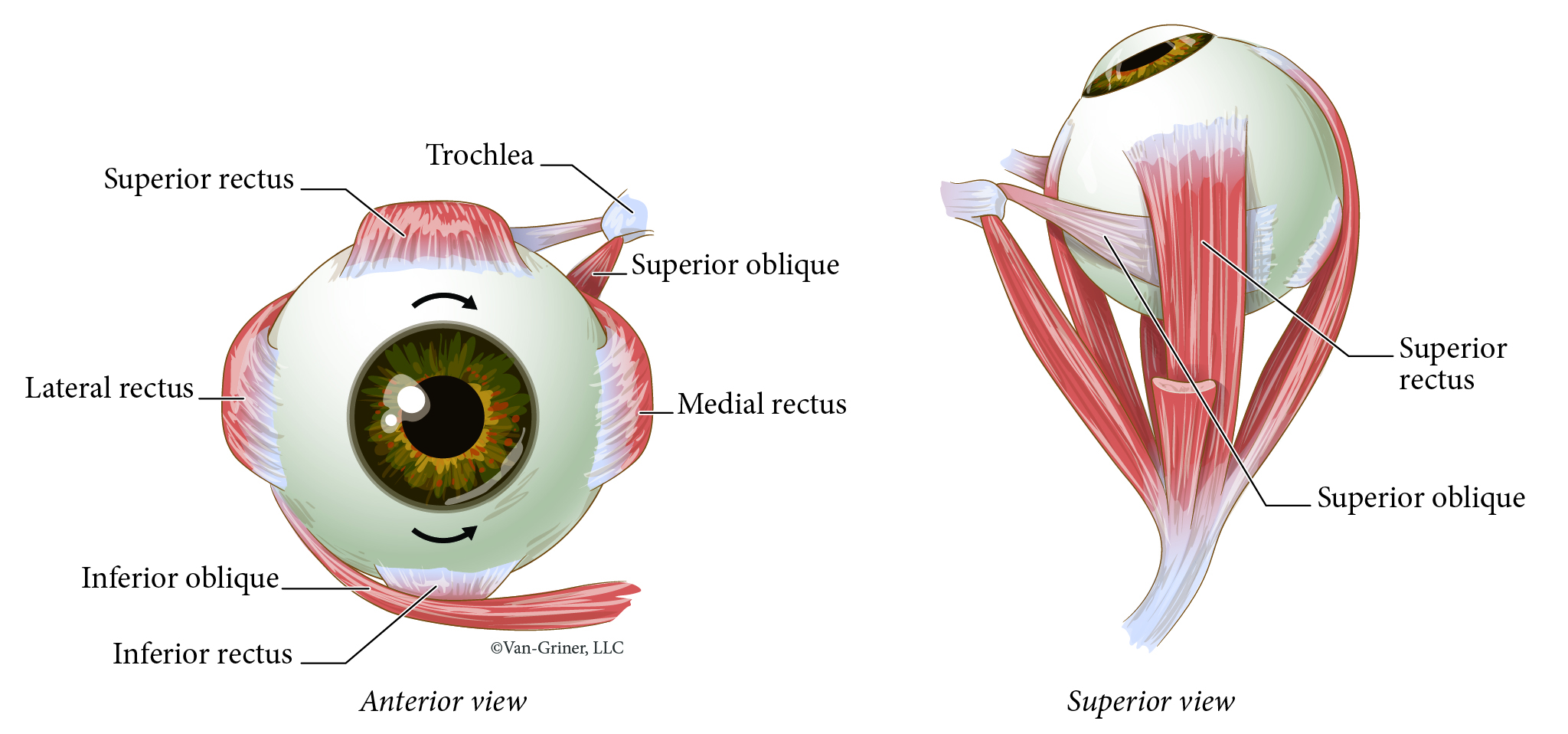
Figure 16.8Extrinsic Muscles that Move the Eye
Extrinsic Muscles that Move the Eye—Actions
To study the actions of all the indicated extrinsic muscles that move the eye, see Table 16.4. Note that there are no muscle site attachments for the extrinsic eye muscles. We will only focus on the actions or movements of the eye. Again, you should be able to name and locate each indicated muscle and identify the action(s) of each.
Table 16.4
Extrinsic Muscles that Move the Eye and Their Actions
Muscle | Action |
|---|---|
Lateral rectus | Lateral rotation |
Medial rectus | Medial rotation |
Superior rectus | Superior and medial rotation |
Inferior rectus | Inferior and medial rotation |
Superior oblique | Inferior and lateral rotation |
Inferior oblique | Superior and lateral rotation |