Physiological Psychology 1
Physiological psychology The study of biological basis of behaviors
The relationship between the nervous system and behavior
Nervous System and Nerve Cells
Central Nervous System
Brain and spinal cord
Peripheral nervous system
Nerve and sensory organs
Two kinds of cells in the nervous system:
Neurons: they process and transmit information in the nervous system
Glial cells: they support, nourish, and protect neurons, playing a crucial role in maintaining homeostasis and facilitating communication within the nervous system.
Only 10% of cells in the nervous system are neurons
Neurons
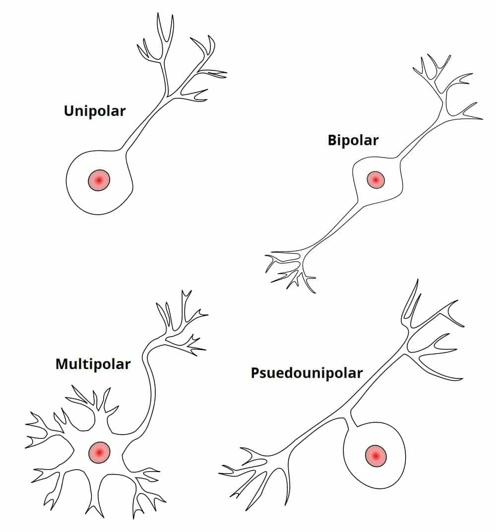
Types of Neurons (Bipolar, Multipolar, Unipolar, Anaxonic)
Bipolar neurons:
two extensions off of the cell body, one axon and one dendrite
allow for the integration of information from multiple sources, making them essential in sensory pathways such as vision and olfaction.
Multipolar neurons: most common type of neuron
characterized by multiple dendrites extending from the cell body and a single axon
allowing for the integration of a large amount of information from other neurons.
Unipolar neurons: primarily found in sensory neurons,
characterized by a single elongated process that extends from the cell body, which splits into two branches – one serving as a dendrite and the other as an axon,
facilitating the transmission of sensory information to the central nervous system.
Psuedounipolar neurons:
these neurons also have a single elongated process that bifurcates into two branches; however, they are predominantly found in the peripheral nervous system and are involved in transmitting sensory information from the periphery to the central nervous system.
Anaxonic neurons:
these are a type of interneuron characterized by their lack of axons, which allows them to integrate and process information locally within the central nervous system, playing a crucial role in modulating the activity of other neurons.
Neurons are limited to one axon but can have an unlimited amount of dendrites
Afferent, efferent, and interneurons:
Sensory neurons (Afferent neurons) (in)
Have afferent fibers
Receptors that pick up information located in the periphery
bring external information from the sensory organs to the central nervous system
Ex. Eye, (light) ear, (sound) nose, tongue, (taste) skin (pain)
Convert information to nerve impulses and send them to interneurons
Structure
Unipolar neuron
Interneurons
Receive information from sensory neurons and send it to motor neurons
Allow sensory and motor neurons to communicate with each other
Receive and send information from/to other interneurons
Essential for reflexes and complex processing of sensory information
Motor Neurons (Efferent neurons) (away)
Receive information from interneurons
Carry information away from the CNS to the muscle
Control movement
Structure
Multipolar neuron
Example If you touch a hot stove with your hand, sensory neurons will pick it up, send it to the interneurons in the CNS, who send it to the information to the motor neurons, causing you to move your hand away.
Components of a neuron:
All kinds of neurons have four major parts — dendrites, cell body, axon, and synaptic terminals, which work together to transmit signals efficiently throughout the nervous system.
Dendrites: Branch-like structures that receive messages from other neurons.
Presynaptic neuron: The neuron that sends the signal to the synapse
Post-synaptic neuron: The neuron that receives the signal from the presynaptic neuron at the synapse
Information travels from neuron to neuron amongst synapses (gaps)
Cell body: The main part of the neuron that contains the nucleus, ribosomes, mitochondria, and organelles, responsible for maintaining the cell's health.
Axon: A long, thin projection that transmits electrical impulses away from the cell body to other neurons or muscles.
Conducts information, an electrical signal (Nerve impulse, or Action Potential)
Transmission of signals is an electrical process
Synaptic (Axon) terminals: The endpoints of the axon where neurotransmitters are released to communicate with neighboring neurons.
When AP reaches the synaptic terminals, neurotransmitters are released and attach to receptors in the dendrites which make the postsynaptic cell more or less likely to fire an action potential, thereby facilitating or inhibiting the transmission of signals across the synapse.
This is a chemical process
Myelin: rich membrane that covers the axons of many neurons, facilitating faster transmission of electrical impulses.
Glia
Astrocytes: Most abundant glial cells in the CNS
Star-shaped glial cells
Provide neurons with nutrients: blood vessels → astrocytes → neurons
provide structural support, surrounding and isolating synapses, protect synapses from circulating chemicals
modulate synaptic communication
maintain the firing rate of neurons
help build and destroy synapses
hold neurons together
clean away debris from dead neurons through phagocytosis
support the overall health of the nervous system by providing nutrients and maintaining homeostasis.
Microglia: primary immune cells of the CNS
very small cells that remove waste material as well as viruses,
fungi, and other microorganisms
destroy infectious agents through phagocytosis
associated with brain disease — may overreact and attack healthy brain cells in older brains
Radio glia:
Guide neuronal migration during CNS development
Oligodendrocytes and Schwann cells
provides neutrons for axons
produce myelin which protects and facilitates the transmission of nerve impulses in axons
The Nerve Impulse or Action Potential
Neuronal Membrane
Made of two layers of lipid molecules
Semipermeable: allows some substances to pass through but disallows others to pass
Membrane potential/electrical gradient the difference in electrical charges between the inside and outside of neurons
Maintained by the electrical gradient, the concentration gradient, and the sodium-potassium pump
Resting membrane potential: neurally suppressed
A neuron is at rest when it is not receiving or sending messages. It is polarized, a difference in charge of electrons
Inside of the neuron is more negative than the outside of the neuron
The resting potential is measured by very thin microelectrodes.
A typical resting membrane potential is -70 millivolts (mV). This may vary from one neuron to another.
A change in membrane potential will indicate whether an Action Potential has been fired
Depends on the permeability of the neuronal membrane to different ions and the difference in electrical charges
Relative concentration of charges
Sodium-potassium pump: This pump is essential for maintaining the resting membrane potential by actively transporting sodium ions out of the cell and potassium ions into the cell, thus creating an electrochemical gradient.
When the membrane is at rest, the inside is more negatively charged than the outside, and two forces work on sodium ions:
The electrical gradient/force of electrostatic pressure:
opposite electrical charges attract, so sodium (which is positively charged) is attracted to the negative charge inside the cell.
The concentration gradient/force of diffusion (difference in distribution of ions between
the inside and the outside of the membrane):
Determines which directions ions will move
Sodium is more concentrated outside the membrane than inside and is thus more likely to enter the cell than to leave it.
Given that both the electrical and concentration gradients tend to move
sodium into the cell, sodium would be expected to quickly enter the cell.
However, when the membrane is at rest, sodium channels are closed.
Potassium ions are subject to the same two forces; however, the forces are in opposition to each other. Potassium ions are positively charged, so the electrical gradient tends to move potassium in, but since potassium is concentrated on the inside of the cell, the concentration gradient causes
potassium to flow out of the cell.
The potassium takes positive charges with it making the inside of the cell more negative than the outside
There is a greater number of positive sodium ions than negative chloride ions making the outside of the cell more positive compared to the inside, which contributes to the overall resting potential and influences the excitability of the neuron.
Relative permeability
Membrane is impermeable to negatively charged ions, they cannot leave the cell
Sodium and Chloride can diffuse through the membrane through specialized protein channels: leak and voltage gated channels
At rest, leak channels are open-gated and voltage gated channels are closed
When the membrane is at rest, sodium can transfer from one membrane to another only through leak channels
Leak channels are ALWAYS open
Voltage gated channels are caused by electrical changes or changes in the resting membrane potential
When the neuron is at rest, voltage gated channels are closed
There are more potassium channels than sodium
Therefore when the neuron is at rest it is more permeable to potassium than sodium
The potassium takes positive charges when they leave through the leak channel making the inside of the cell more negative than the outside
The inside of the neuron is more negative than the outside due to a greater number of negatively charged proteins than positively charged ions inside of the cell, and because when the neuron is at rest, positively charged potassium ions leave the cell through leak channels, making the inside more negative.
The force of diffusion when there are no forces preventing them from doing so, molecules will diffuse from areas of higher concentration to areas of lower concentration, (caused by the sodium-potassium pump) which contributes to the overall resting membrane potential by allowing potassium ions to exit the neuron more freely than sodium ions can enter.
Ex: when neuron is at rest, potassium, which is at higher concentration inside the neuron, will move to a region of lower concentration through a leak potassium channel
The force of electrostatic pressure counteracts the force of diffusion, ions of the same charge repel each other and ions of different charges are attracted to each other
Ions will be attracted to the side of the membrane with the opposite charge, therefore forcing potassium ions to the negatively charged inside of the membrane
whys potassium so highly concentrated inside if its constantly leaving?
TO DUMB IT DOWN. THE POTASSIUM WHICH IS IN HIGH CONCENTRATION INSIDE OF THE CELL IS PUSHED OUT DUE TO THE FORCE OF DIFFUSION BUT BECAUSE POTASSIUM IS POSITIVELY CHARGED IT IS PUSHED BACK INSIDE BECAUSE THE OUTSIDE OF THE CELL IS MORE POSITIVELY CHARGED BUT THE TWO FORCES ARE ALMOST IN BALANCE THEREFORE POTASSIUM TENDS TO REMAIN INSIDE OF THE CELL AND ONLY A SMALL AMOUNT OF SODIUM ENTERS THROUGH THE LEAK CHANNELS
sodium remains highly concentrated outside because
THE MEMBRANE IS MORE PERMEABLE TO POTASSIUM, ONLY A SMALL AMOUNT OF SODIUM ENTERS THE NEURON THROUGH THE LEAK CHANNELS WHEN THE NEURONS ARE AT REST BECAUSE OF THE VOLTAGE CHANNELS B EING CLOSED
The sodium-potassium pump forces the sodium and potassium ions to move against their
concentration gradients.
Requires a large amount of energy, about 40% of a neurons metabolic resources
Forces the ions to move from regions of low concentration to regions of higher concentration
Basically exchanges sodium for potassium
3 sodium ions out of the cell for every 2 potassium ions into the cell
Helps maintain higher concentration of sodium outside of the neuron and higher concentration of potassium inside the neuron
Action potential results from changes in the resting membrane potential, resting membrane potential changes when the neuron is stimulated (receives or sends messages to other neurons)
When neurotransmitters bind to receptors of the post-synaptic neuron, they produce changes in the resting membrane potential of the post-synaptic neuron that may increase or decrease the probability that an action potential will occur in the post-synaptic neuron
Inhibitory neurotransmitters produce hyperpolarization,
which makes the resting membrane potential more negative
(the inside of the neuron becomes more negative)
and decreases the likelihood of an action potential firing in the post-synaptic neuron.
Excitatory neurotransmitters produce depolarization,
leading to a more positive membrane potential, which increases the likelihood of an action potential firing.
Enough excitation (if the threshold is reached) will result in the generation of an action potential, allowing the signal to be transmitted along the neuron.
Stages of Action Potential
resting membrane potential
if neuron is depolarized leading to the opening of voltage-gated sodium channels.
(3 and 4) Voltage-gated K+ channels open later than the voltage-gated Na+ channels.
The outflow of K+ produces hyperpolarization. Voltage-gated K+ channels begin closing.
5. Voltage-gated K+ channels close and the membrane returns to the resting potential.
Absolute refractory period
During the absolute refractory
period another AP can not be
generated.
• The absolute refractory period
limits the maximum number of
AP generated in a second .
• During the relative refractory
period, a stronger stimulus may
cause an AP.
The image above shows the stages of the action potential
All-or- none law
•An AP either occurs or does not occur.
•An AP is always the same size.
• An AP does not die
The Rate Law
• Stimulus Intensity is represented by
the rate of firing.
• As the stimulus intensity increases,
the firing rate increases.
Records showing APs in a neuron that responds
to light entering the eye.