Agarose Gel electrophoresis
This technique is used to separate DNA fragments by their size
Principle of Agarose Gel Electrophoresis
DNA loaded into one end of the gel
electric current applied to pull them through the gel
Due to the negative charge of the DNA fragments, they move towards the positive electrode
smaller fragments move through the gel faster than the larger ones
when the gel is stained with the DNA binding dye, the DNA fragments can be seen as bands
each band represents a group of same sized DNA fragments
materials required
An electrophoresis chamber and power supply
gel casting tray and comb
agarose gel
a gelatinous substance derived from a polysaccharide that accumulates in the cell walls of red algae
melts - 85ºC , solidifies - 35ºC
Agarose polymer in solidified gel forms a porous network
pore size can be adjusted by varying the percentage of the agarose in the gel, in order to efficiently resolve nucleic acids of different sizes
lower agarose concentration (large pore sizes) used to separate larger DNA fragments
higher agarose concentration (smaller pore size) used to separate smaller DNA fragments - hence better separation and resolution of smaller DNA fragments
DNA staining agent is added into gel before it solidifies
electrophoresis buffer e.g. Tris-borate-EDTA (TBE)
molten gel is loaded into gel casting tray to set (the comb creates the wells
solidified gel is submerged in electrophoresis buffer
electrophoresis buffer is an ionic solution with buffering capacity
used in gel runs to allow current flow while impeding pH changes that may occur
Loading dye
contains a dense solution (e.g. glycerol)
mixed with DNA sample to allow the sample to “sink” into the wells
DNA staining agent e.g. SYBR Green, Ethidium bromide
inserts in between the bases of DNA stranf and fluoresce orange under UV → carcinogen
UV Transilluminator
how the machine works
electrophoresis chamber is connected to power pac
electric current causes DNA to move through gel
steps to conduct gel electrophoresis
melt the agarose powder in TBE buffer
add SYBR Green dye
Pour the molten agar into a gel casting tray
Use a comb to create the wells
Allow the gel to set
Submerge the gel in TBE buffer in an electrophoresis chamber
Mix DNA sample with loading dye
Load the DNA sample into the wells
Connect to power supply
View the gel with UV light
components and funcitons
power supply
provide the electric current for DNA fragments to move from negative to positive electrode
gel casting comb
make wells in the gel for loading DNA sample
agarose gel
forms a gel matrix with pore sizes for separation of DNA fragments
TBE buffer
allow current flow while impeding pH changes that may occur
Loading dye
contains glycerol which allow DNA sample to sink into wells
contains dye (bromophenol blue) which provides colours for easy monitoring of sample loading and progress of the electrophoretic run
DNA staining agent
allow for visualisation of DNA samples after the run under UV light
UV transilluminatior
to visualise and capture the gel image
analysing results
DNA marker / ladder
short fragments of DNA of known sizes (bp)
loaded together with the unknown DNA samples as reference
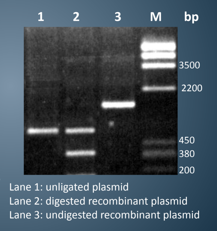
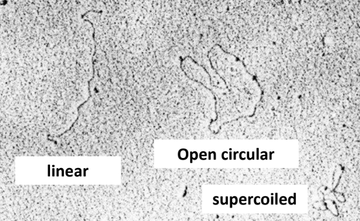
plasmids exhibit 3 conformations
when both strands are intact → supercoiled
when one strand is nicked → open circular
when both strands are cut → linear
more !!
different conformations of the same plasmid (linear, open circular, supercoiled) migrate at different speeds during agarose gel electrophoresis
both shape and molecular weight affect travelling distance
determination of DNA molecular weight
Measure the distances travelled by 5 DNA marker bands around the DNA band and the DNA band
Convert MW (molecular weight) of DNA marker bands into logarithm format (just log the MW) this is to make the graph linear
plot graph of travel distance (mm) [y axis] vs logMW [x axis]
use graph to determine the MW of the DNA of interest (rmb to un-log)