Hippocampal Dependent Neurogenesis and Behaviours
Hippocampal Dependent Neurogenesis
Adult hippocampal neurogenesis (AHN) is the process of generating new neurons in the adult brain that contribute to normal brain function.
It affects brain structure/plasticity and contributes to normal brain function and spatial memory.
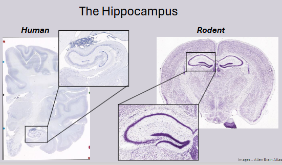
AHN occurs in the subventricular zone (SVZ) of the lateral ventricles and the dentate gyrus (DG) of the hippocampus.
AHN declines with age and can be affected by exercise, environmental enrichment, and dietary manipulation.
Why Investigate Neurogenesis?
Increasing aging population.
Increase in neurological disease/disorders that affect memory function.
Examples:
Alzheimer’s: 900,000 people with dementia in the UK.
Parkinson’s: 153,000 people currently living with PD in the UK.
Epilepsy: 630,000 people in the UK.
Depression: 1 in 6 people (16% of the UK population).
Anxiety: 8.2 million cases in the UK (2013).
Adult Neurogenesis
The process of generating new neurons in the adult brain that contribute to normal brain function
First discovered by Joseph Altman in 1965.
Altman used autoradiographic evidence of post-natal neuronal cell differentiation.
Experiment:
Long Evans rats treated with H3-Thymidine (various ages) and terminated after 2 weeks.
Labelled cells found in the basal area of the granule layer of the dentate gyrus (DG).
Results:
10-day-old rats: 35 labelled cells.
30 to 40 days old: ~5 cells.
4 months or older (adults): ~2 labelled cells or less.
A decline of 19% to 3% labelled cells.
Stages of Hippocampal Neurogenesis and Cell Markers

BrdU - Bromodeoxyuridine
5-bromo-2′-deoxyuridine.
Thymidine analogue that incorporates into the DNA of dividing cells (S-phase of cell cycle).
Used for birth-dating and monitoring cell proliferation – labels new-born cells.
Can be administered orally (drinking water) or via i.p. injection for in vivo labelling.
The timing of BrdU administration is important for interpreting the outcome of an experiment/treatment.
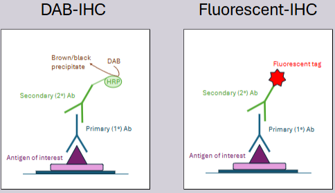
Immunohistochemistry (IHC)
Process of detecting the presence of proteins and antigens on tissue sections using antibodies.
Antibody-antigen interactions are visualised either chromogenically (coloured enzyme substrate) or fluorescently (fluorescent probe).
Provides information on the location of proteins in a whole tissue context.
Tissue processing (fixation – perfusion vs. immersion) can impact staining outcomes.
Types of IHC
DAB-IHC:
Primary antibody (1° Ab) binds to the antigen of interest.
Secondary antibody (2° Ab) with HRP (horseradish peroxidase) binds to the primary antibody.
DAB (diaminobenzidine) substrate is used, resulting in a brown/black precipitate.
Fluorescent-IHC:
Primary antibody (1° Ab) binds to the antigen of interest.
Secondary antibody (2° Ab) with a fluorescent tag binds to the primary antibody.
Assessing Hippocampal and Neurogenesis-Dependent Behaviours
Contextual Fear Conditioning:
Learned fear memory (survival response).
Requires inputs from the hippocampus and CA3 region.
Rodents learn to associate a neutral context with an aversive stimulus, e.g., foot shock.
Display fear response, e.g., freezing behaviour.
Spontaneous Location Recognition (SLR) Task
Pattern separation memory – DG dependent
Highly sensitive to changes in hippocampal neuroplasticity.
SLR manipulates the similarity of spatially landmarked locations during the sample phase when memory is encoded, and pattern separation processes are active.
Takes less than 1 month to run this task.
Touchscreen Tasks – Location Discrimination
Operant box task, same concept as SLR hand task.
Rodents are required to discriminate between 2 white squares on the screen.
The distance between squares can be varied.
Neural systems used:
Hippocampus.
Neurogenesis.
Behavioral Tasks - Anxiety
Open Field Test.
Elevated Plus Maze.
Elevated Zero Maze.
Theory in Practice
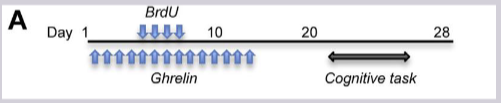
Experiment:
Day 1: BrdU and Ghrelin administration.
Days 10-20: Cognitive task.
Day 28: Analysis.
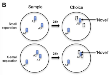
SLR Task:
Sample phase with small separation (A2, A3) and X-small separation (A1, A4, A5) between landmarks.
Choice phase 24 hours later. 'Novel' location.
Discrimination ratio:
Results:
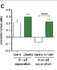
Ghrelin improves discrimination ratio in small separation condition.
DCX staining:
Ghrelin increases DCX+ cells in the dentate gyrus.
BrdU/NeuN staining:
Ghrelin increases the number of BrdU+ cells and BrdU/NeuN+ cells in the dentate gyrus.