Psychology Yr11 ATAR
Everything deleted - redoing notes. [soon]
Nervous System
Structure
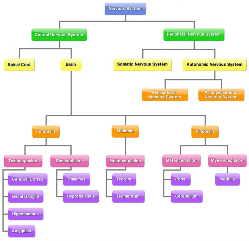
The nervous system: system of networks of neurons that connect different parts of the body to each other and the brain via electrochemical signals.
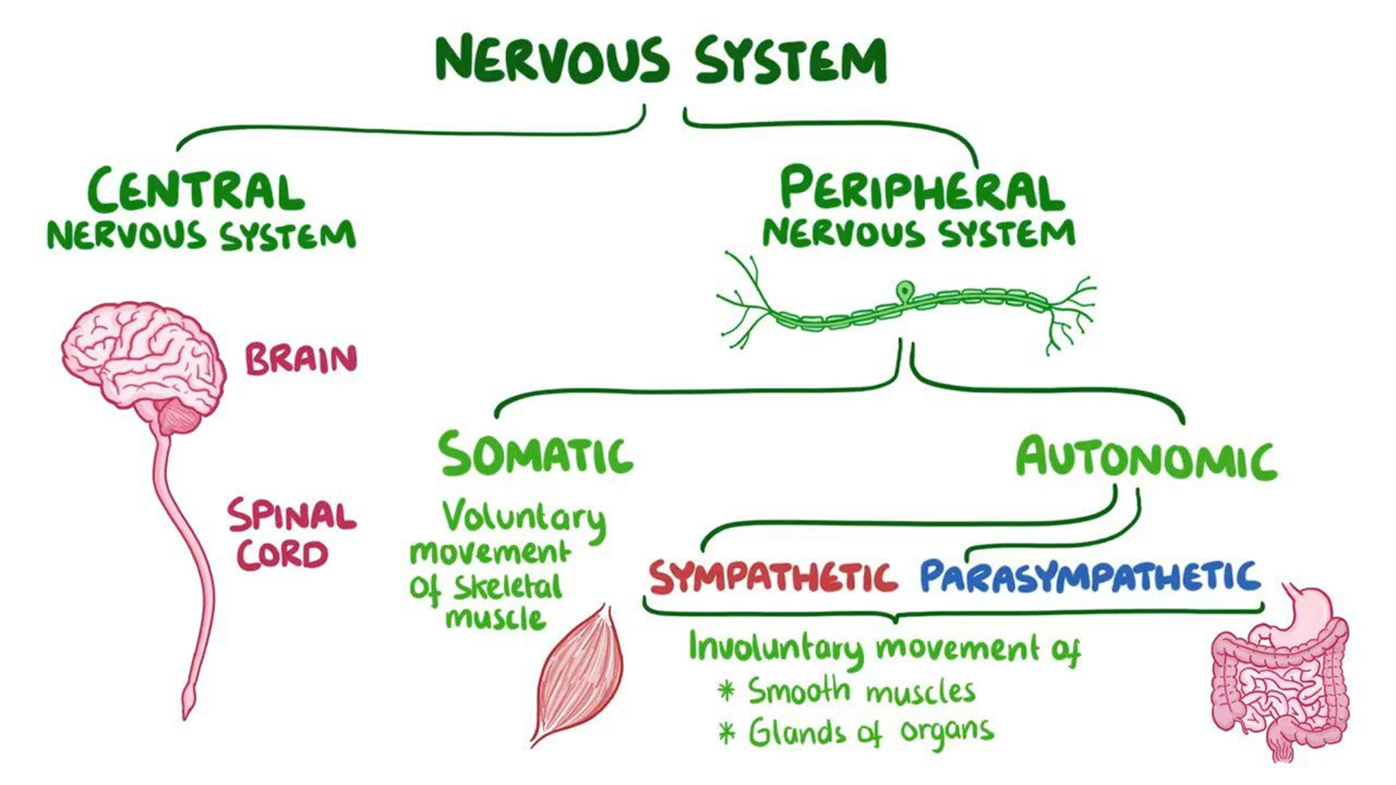
There are two divisions of the nervous system:
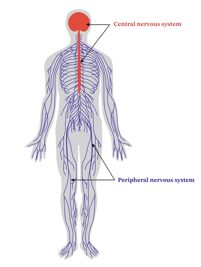
Central nervous system (CNS)
Peripheral nervous system (PNS)
Autonomic nervous system (ANS)
Sympathetic nervous system
Parasympathetic nervous system
Somatic nervous system (SNS)
Central NS
The CNS is made of the brain and spinal cord. It carries sensory information from the PNS, through the spinal cord, to the brain via sensory neurons. The CNS also carries motor messages from the brain to PNS via motor neurons.
The spinal cord:
Delicate cable of nerve fibres from base of brain to lower back. It connects the brain to the rest of the body via its connection to PNS.
Key roles:
Delivering sensory information to spinal cord, and spinal cords sensory neurons transmirt this information up to the brain for processing.
Motor neurons in spinal cord receive motor messages from brain, which it transmorts to the PNS and therefore whole body.
If spinal cord damaged, flow of information between brain and rest of body is interrupted. May result in loss of sensation in specific body part or inability to move specific body part.
Peripheral NS
The PNS includes nerves on the outside of the brain and spinal cord that carry sensory informatio to CNS from body and motor messages from brain to organs and muscles in body.
Its role is to carry sensory information from rest of body to CNS, and motor information from CNS to rest of body.
PNS subdivisions:
Somatic nervous system
Autonomic nervous system
Autonomic NS
A branch of the PNS that carries motor messages from brain to internal glands and orgains via motor neurons, causing involuntary activity. It also carrues sensory messages to the brain about the activity level of glands and organs via sensory neurons.
Contains nerves connected to CNS and smooth (involuntary) muscles that control activity level of internal organs and glands.
By relaying messages between CNS and internal systems, the ANS controls all bodily involuntary internal activities essential to survival. The activities include:
Heart rate
Digestion
Kidney function
Liver function
Glandular activity
Perspiration levels
Somatic NS
A branch of the PNS that carries sensory information received by senosry receptor cells to CNS via sensory neurons. It also carries motor messages from CNS to skeletal muscles via motor neurons.
The SNS hs a sensory role and a motor role:
Sensory role: receiving sensory information from receptor cells located throughout whole body.
Motor role: receives motor messages from CNS and transport to skeletal muscles in specific body regions so our responses to stimuli are appropiate.
The SNS controls voluntary movement through control of skeletal muscles.
Nervous system example
Pain receptors in the skin are simulated by the thumbtack
Sensory messages travel to the spinal cord (part of CNS) via sensory neurons
Sensory messages are sent to the brain via sensory neurons
Motor messages travel via motor neurons from the spinal cord to skeletal muscles in the leg
Skeletal muscles in the leg contract, pulling the foot back up off the thumbtack
The brain registers the sensation and is consciously aware that the reflex response has occurred.
Sympathetic NS
A branch of the ANS that regulates the glands and internal organ function to physically prepare the body for increased activity during heightened physical or emotional arousal. Also known as the “flight or fight” response.
This branch dominates during times of high emotion or intense physical activity. It mobilises thhe body’s internal resources to provide extra energy we need for vigorous action especially in times of stress or threat. It activatesthe body for emergency action by increasing the activity level of some bodily systems while others are slowed down (“flight or fight”).
Parasympathetic NS
A branch of the ANS that reverses bodily functions - calms body and maintains homeostasis.
Once the need for high arousal (created by the sympathetic NS) has passedm the parasympathetic nervous system reverses the effects of the sympathetic nervous system.
This reversal returns our body to its normal level of arousal, or a more relaxed state that is more appropiate when the period of high emotion or need for physical activity has passed. Our heart and breathing rates, the level of sugar and fats in our bloodstream, the size of our pupils, our blood pressure and digestino rate all return to their normal activity levels.
The parasympathetic nervous system plays a vital role in keeping us in a state of wellbeing or homeostasis.
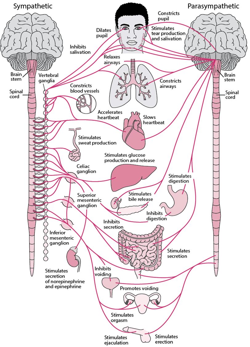
Divisions of the ANS
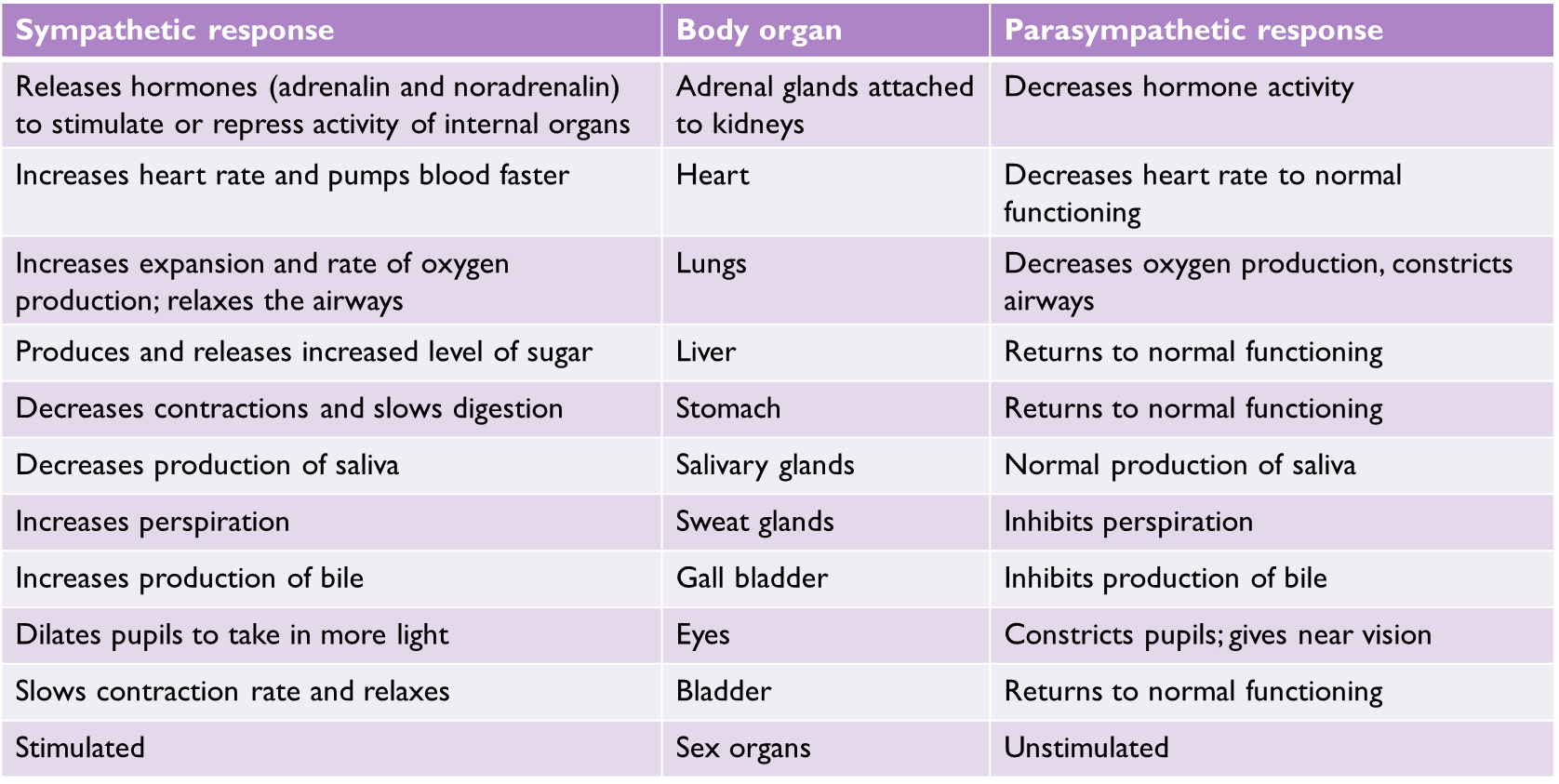
Sympathetic response | Body organ | Parasympathetic response |
Releases hormones (adrenalin and noradrenalin) to stimulate or repress activity of internal organs | Adrenal glands attached to kidneys | Decreases hormone activity |
Increases heart rate and pumps blood faster | Heart | Decreases heart rate to normal functioning |
Increases expansion and rate of oxygen production; relaxes the airways | Lungs | Decreases oxygen production, constricts airways |
Produces and releases increased level of sugar | Liver | Returns to normal functioning |
Decreases contractions and slows digestion | Stomach | Returns to normal functioning |
Decreases production of saliva | Salivary glands | Normal production of saliva |
Increases perspiration | Sweat glands | Inhibits perspiration |
Increases production of bile | Gall bladder | Inhibits production of bile |
Dilates pupils to take in more light | Eyes | Constricts pupils; gives near vision |
Slows contraction rate and relaxes | Bladder | Returns to normal functioning |
Stimulated | Sex organs | Unstimulated |
Neurons
The brain is split into nerve cells called neurons. A neuron is a type of cell that is specialised to receive, transmit and process information.
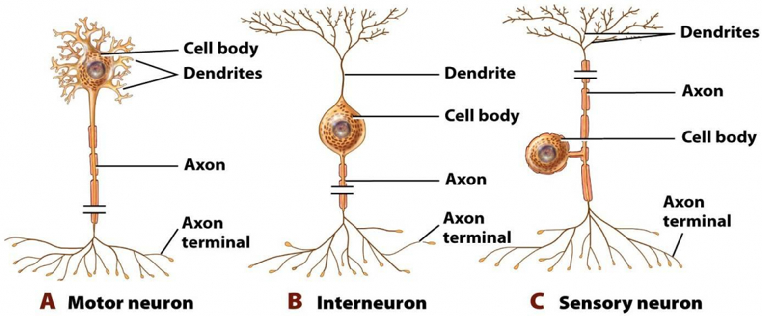
There are three types of neurons:
Motor neurons
Interneurons
Sensory neurons
Features of neurons
Cells of the nervous system that communicate with each other, as well as muscle and gland cells.
Components:
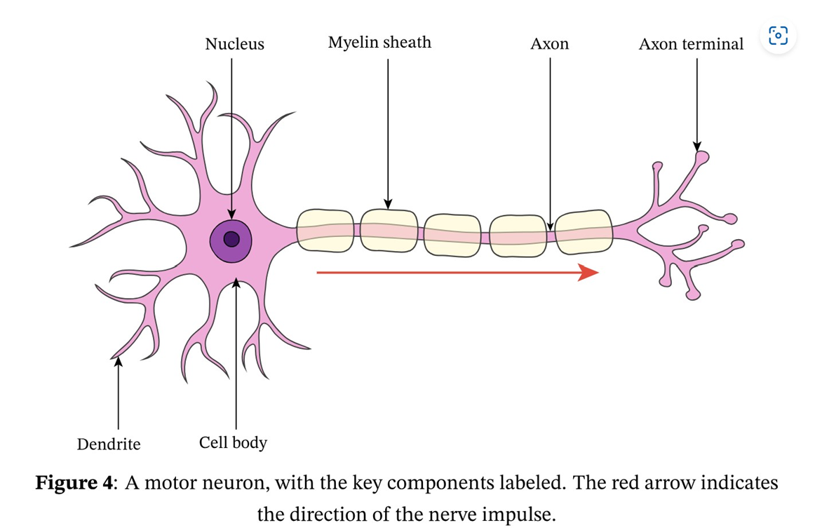
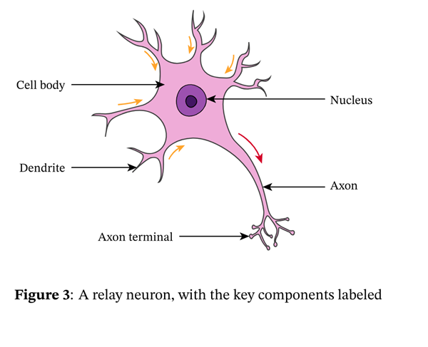
Dendrites - fine branches from cell body/soma that receive information from pre-synaptic neurons and convert them into electrical nerve impulses that are conductied toward the cell body. More dendrites, more information intake capacity.
Cell body/soma - contains nucleus that controls the activities of the neuron. Integrates informated received from many dendrites and passes it to the axon.
Axon - long projection of a neuron that conducts electrical nerve impulses and carries the away from the cell body.
Axon terminals/buttons - a structure found only in neurons, the enlarged end points of an axon branch that store neurotransmitters and release them into the synaptic cleft.
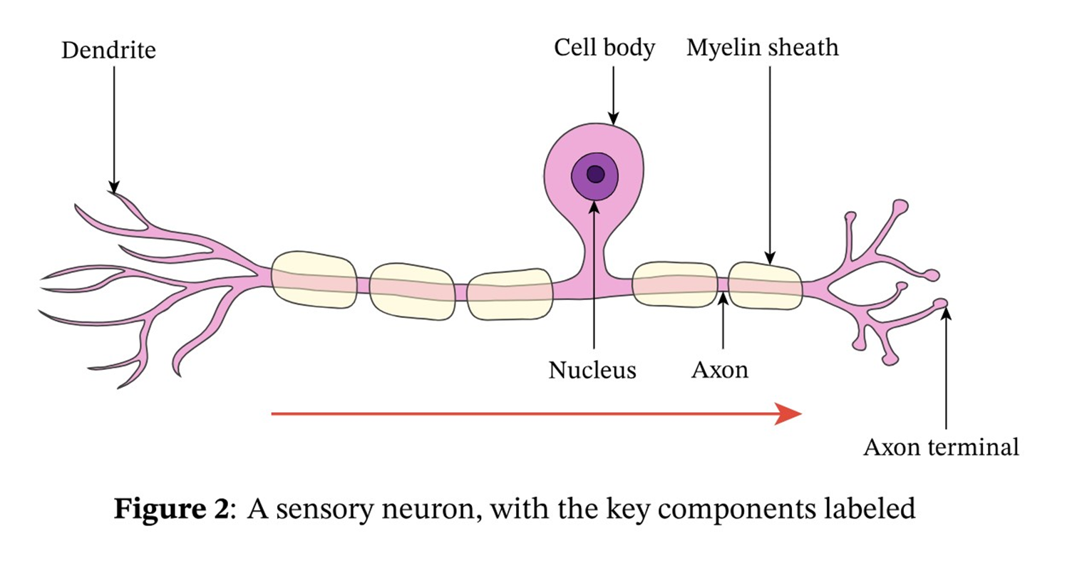
Myelin sheath - in motor and sensory neurons, the axon is covered by a fatty myelin sheath that has gaps periodically where the membrane of the axon is exposed. The fatty layer acts as an insulator, protecting the axon from stimuli that could interfere with electrical nerve impulse transmission. It increases the speed of electrical nerve impulse transmission and helps improve the conduction of the transmission.
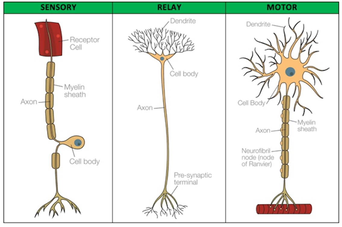
Motor neurons
Motor neurons are part of the CNS and connect to muscles, glands and organs throughout the body.
These neurons transmit impulses from the spinal cord to skeletal and smooth muscles, and so directly control all our muscle movements.
Motor neurons are multipolar, each with one axon and several dendrites.
Sensoy neurons
Seonsory neurons process sensory information from the sense organs and carry the sensory messages to the spinal cord and brain (CNS).
Most sensory neurons are pseudounipolar - they only have one acon which is split into two branches (cell body hangs off the middle of the axon).
Example: when you touch a hot surface with your fingertips, the sensory neurons will be the ones firing and sending off signals to the rest of the nervous system about the infomation they have received.
Interneurons
They act as the connection between sensory + motor neurons and transfers messages from sensory neurons to motor neurons within the CNS.
Determining which neuron
Question | Answer |
1. Is there a myelin sheath on the neuron? | No = Interneuron Yes = Go to Question 2 |
2. Where is the cell body on the neuron? | Within the dendrites on the end of the axon = Motor Neuron Hanging off the middle of the axon = sensory neuron |
Neural Transmission
Direction of transmission
The electrical nerve impulse (action potential) travels in one direction only (starts at dendrites).
The action potential causes the release of neurotransmitters acorss the synaptic cleft (gap).
The myelin sheath can speed up the action potential.
Electro-chemical signal
Action potential is an electro-chemical message that travels through the neuron and enables the diffusion of neurotransmitters between neurons.
Role of the synpase
Synaptic cleft is the space between two neurons.
The purpose of a neural transmission is to convert the electrical message to chemical and then back to electric.
Electrical (pre-cleft) → chemical (cleft) → electrical (post-cleft)
Role of neurotransmitters
Neurotransmitters are molecules that relate information between neurons and to muslces and glands.
Structure of neural transmission
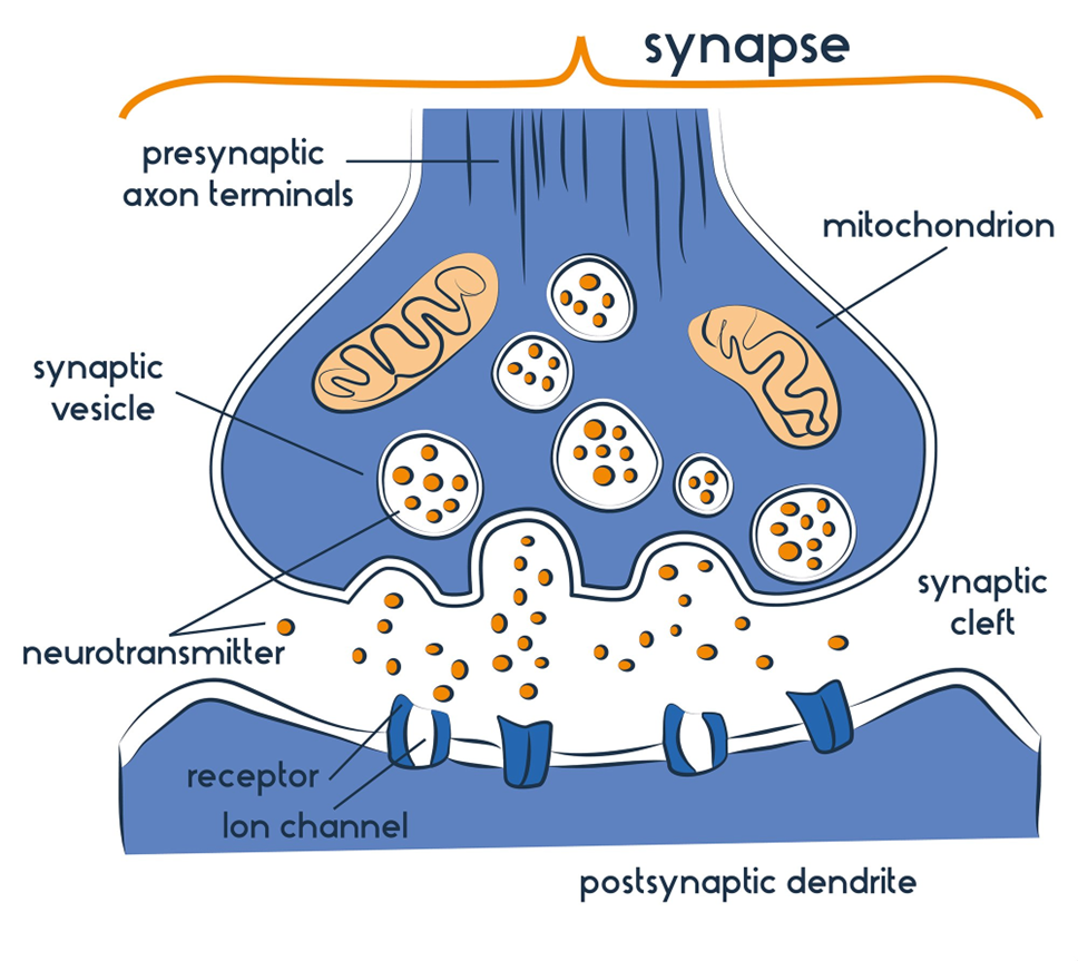
Electrical nerve impulses (action potential) travels to axon terminal.
Action potential causes neurotransmitters to be released from vesicles.
Neurotransmitters diffuse across the synaptic cleft.
Neurotransmitters bind to receptor sites on dendrite of the next neuron.
Dendrite converts chemical message into electrical, then travels to cell body of post synaptic neuron.
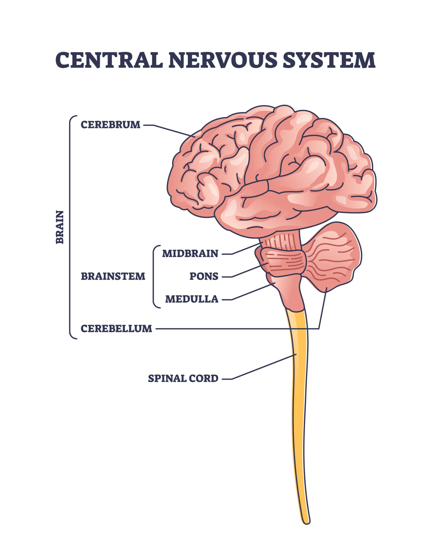
The Brain
Hindbrain
Located at base of brain, near back of skull.
Responsible for unconscious automiatic survival functions and voluntary muscle movements, nalance, coordination and reflexive actions.
Hindbrain - medulla
Located at base of brain, in front of cerebellum.
Relays information between the spine and spinal cord.
The medulla regulateds vital bodily functions by communicating with the ANS.
Damage causes interruptions between neurons which can cause physiological dysfunction and fatalities.
Hindbrain - cerebellum
Located at base of brain and resemles a ‘little brain’.
Controls voluntary movement and balance by relaying motor messages to and from the cerebral cortex.
Examples include judging distance and coordination.
Damage can result in reduced motor control and issues with balance and equillibrium.
Midbrain + reticular formation
Small area of brain located between the forebrain and hindbrain.
Contains reticular information:
Stimulates the brain by bombarding it with sensory information, this keeps the cerebral cortex active and alert.
Also controls our physiological awareness and muscle tone by reglating the ANS = sleep/wake cycle, we can experience a loss of attention/pain.
Damage results in disruption to sleep/wake cycle, loss of attention and pain management and balance.
Forebrain
Largest part of brain.
Key role in cognition, emotion, behaviour and processing of sensory information.
Forebrain - thalamus
Receives sensory information and relays this information from the spinal cord to the brain (vise versa).
Coordinates shifts in attention such as waking up and falling asleep.
Damage includes the loss of senses, problems with movement, sleep and attention.
Forebrain - hypothalamus
Maintains homeostasis by releasing and regulating hormones (connection to endocrine).
This allows for healthy thirst and hunger, sleep/wake cycle, growth, sexual development, motivation and emotion.
Damage can cause disruption to these functions.
Forebrain - cerebrum
Consists of white matter on the inside and grey matter on the inside.
The outer layer is the cerebral cortex which is responsible for higher cognitive function, voluntary movement, emotions adn personality,
There are two cerebral hemispheres.
Cerebral cortex
The thick layer of the cerebrum (wrinkled appearance).
It is the ultimate control and information processing centre in the brain:
Controls voluntary movement.
Integrates sensory information.
Responsible for higher order functions.
Left and right hemispheres
The divisions of the brain are referred to hemispheres.
The hemispheres have contralateral control.
The left hemisphere:
Verbal and analytical functions.
The right hemisphere:
Non-verbal functions.
Corpus callosum
A bundle of nerve fibres, connects two hemispheres.
Allows transmission and processing of information across two hemispheres.
Lobes
The cerebral cortex is divided into four lobes:
Frontal lobe:
Located at the front of the brain.
Responsible for:
Voluntary movement.
Decision making.
Personality.
Parietal lobe:
Located at the top of brain.
Responsible for:
Somative sensations (bodily). Except smell.
Spacial awareness.
Temporal lobe:
Located on either side of the brain.
Responsible for:
Understanding speech.
Interpreting auditory information.
Facial recognition.
Occipital lobes:
Located at the lower back of the brain, responsible for:
Visual perception and processing.
Interpreting visual information.
Localisation of brain functions
Frontal lobe
Primary motor cortex:
Arch of tissue in the frontal lobe, responsible for voluntary motor/muscle movement.
Has contralateral control
Broca’s area:
Left frontal lobe.
To produce clear and articulate speech.
Damage causes Broca’s aphasia, a speech disorder including the inability to produce clear and articulate speech however, they can still comprehend language.
Temporal lobe
Primary auditory cortex:
Registers and process auditory information received by both ears.
Right auditory cortex responsible for processing non-verbal sounds.
Left auditory cortex responsible for processing verbal sounds.
Wernicke’s area:
Left frontal lobe.
Control's comprehension of speech, linked to memory.
Damage causes Wernicke’s aphasia, a speech disorder that involves no comprehension of speech, however the production of speech is clear and fluent.
Parietal lobe
Primary sensory cortex:
Strip of neurons at front of parietal lobe.
Registers and processes sensations detected by the body’s sensory receptors.
Sensory information from left side of body sent to right side of sensory cortex.
Occiptal lobe
Primary visual cortex:
Loacted at base of each occiptal lobe.
Responsible for registering and processing visual information brought in by optic nerve.
Visual cortex assembles many pieces of information into one image we can make sense of.
Historical Research on the Structure and Function of the Brain
Phineas Gage
1848, Phineas gage was a foreman, working at a railroad.
He was compacting powder in a hole when it exploded and a metal rod went through his left frontal lobe.
However, Gage experienced a marked shift in personality.
Before the accident, Gage was described as organised, reasonable and calm. After the accident, he was described as impatient, uncaring, impulsive and he swore oftem.
This case study was notable as it linked brain injury to personality and it linked the frontal lobe to controlling personality.
Roger Sperry
Sperry conducted research involving split brain operations which involved removing th ecorpus callosum.
Sperry’s split brain experiments demonstrated that the corpus callosum is required for full functioning of the brain.
They found that the left hemisphere has a verbal specialisation and the right has a non-verbal for communication between the hemispheres.
Walter Freeman
Freeman and Watts were the first to perform a frontal lobotomy in the USA.
Freeman claimed that he could help people suffering from mental illness by severing the neural pathways between the thalamus and the pre-frontal cortex.
However whilst the procedure reduced agitation in patients, it causes apethy, numbness to emotion and decreased concentration.
Ethics Committee/ethical guidelines
The role of the Ethics Commitee is to review research proposals that plan to use human participants in order to make sure the proposal meets Australian ethical guidelines.
The committee will compare the proposal to existing research and ensures the benefits outweigh the risks.
If approved, the Ethics Committee will continue to monitor.
There are 8 ethical guidelines which governs the code of practise during psychological research:
Protection from harm - researchers must protect the physical and psychological wellbeing of participants.
Informed consent - researchers must obtain written and signed informed consent sheets that inform the nature of the study.
Withdrawal rights - participants can end their participation in a study, or have results removed without pressure or penalty.
Confidentiality - information collected must be stored in a secure manner and then disposed of when no longer required. *The how of information.
Privacy - only information relevant to the study should be collected from participants. *The what of information.
Voluntary participation - participants partake in a study because it is their choice to do so.
Deception - used when participants knowing the true nature/purpose of the study may impact their results.
Debriefing - an explanation given to particpants at conclusion of study.
Selection of participants
Population - the entire group of people that is of interest to the researcher. Example: All college students in the United States.
Sampling - subsection of the population. Example: A researcher selects 200 college students from five different universities across the U.S. to study the effects of stress on academic performance. This group represents a sample of the larger population.
Sampling technique | What |
Convenience sampling | Selecting participants who are easily accessible. |
Snowball sampling | Involves choosing the initial participant, from here, they spread the word about the study and encourage others to join. |
Random sampling | All members of the population have an equal opportunity of being randomly drawn/selected for the sample. |
Stratified sampling | Involves breaking population off into subgroups based on common characteristics, from here, the researcher takes an equal amount of participants from each subgroup. |
Allocation of participants
Random allocation - involves random distribution of participants in the sample into the controlled and experimental group.
Examples include computer generation.
Strength allows for generalisability and prevents selection bias.
Limitation: cannot be used in non-experimental research and equality of characteristics/representation is not guaranteed.
Science inquiry
Aim:
A general statement that addresses the purpose of the study/experiment.
“To investigate” or “to find out”.
The aim must include the population.
It can include the independent/dependent variable if the research is experimental.
Research inquiry question:
A general question based on background research or content.
Must include population.
May include independent/dependent variables.
Variables:
Independent variable (IV) - manipulated variable.
Dependent variable (DV) - measured variable.
Controlled variable (CV) - made to stay consistent.
Extraneous variable (EV) - variable other than IV. Has potential to influence DV.
Hypothesis:
A statement that expresses a possible answer to a research question.
It has four components:
Population
Operationalised IV
Operationalised DV
Directional/non-directional statement
Directional statement - a statement that compares the predicted outcome of each condition.
Create a directional hypothesis when we have context.
Non-directional hypothesis - a statement that declares there is a difference between the conditions but doesn’t state a direction of the difference.
Use this when little to no context is given.
Types of extraneous variables:
Participant variables - types of extraneous variables relating to the individual characteristics of participants.
Environmental variables - type of extraneous variables relating to the environment of the study; where it takes place and how this affects participant responses.
Researcher variables - type of extraneous variables relating to the personality characteristics, appearance and conduct of the researcher that unintentionally impact participant response.
Confounding variable - variables that impact the dependent variable and also have a causal or correlational relationship with the independent variable.
There are three ways to minimise the effects of confounding and extraneous variables:
Random allocation of participants:
Only in experimental research.
Participants in sample have an equal chance of being in the control/experimental group.
Single-blind procedure:
Involves the researcher being aware of which participants are in the control vs experimental group whilst the participants are unaware.
Helps control the effects of demand characteristics because the participants are less likely to deduce/perceive the aim.
The standardisation of procedures and instructions to both the experimental and control group:
This reduces environmental variables.
Types of data | What |
Qualitative data | Data in the form of words. |
Quanitative data | Data in the form of numbers. |
Qualitative data | Quantitative data | |
Data format |
|
|
Data display |
|
|
Question format used to obtain data |
|
|
Method of collection |
|
|
Analysis |
|
|
Contemporary Methods to Study the Brain
/
/
Contemporary methods:
Still pictures:
Single, static, 2D image.
Examples: CT, MRI.
Dynamic pictures:
3D image that can change over real time.
Examples: fMRI.
Structural imaging:
A scan that shows the structure of the brain.
Examples: CT and MRI.
Functional imaging:
A scan that produces information on the function of certain areas in the brain.
Examples: fMRI.
Two types of resolution:
Temporal resolution:
Detects rapid change in brain activity (T for timing).
Examples: EEG.
Spatial resolution:
Detects the area of the brain that is responsible for activity (S for space).
Examples: fMRI.
Developmental Stages + Domains of Development
Lifespan Psychology is the study of continuity and change throughout the lifespan.
Developmental stages
Developmental stages across the lifespan:
Prenatal - conception to birth.
Infancy - birth to 1 year.
Childhood - 1 to 2 years.
Adolescence - 12 to 20 years.
Early adulthood - 20 to 40 years.
Middle age - 40 to 65 years.
Older age - 65+ years.
Domains of development
There are four domains of development that humans experience throughout their lifespan. The domains of development may interrelate and be more dominant in some developmental stages than others.
Domain of development | What |
Physical development (described in fine and gross motor skills) |
|
Cognitive development (ability to think and reason) |
|
Social + developmental (psychological development encompasses changes in a person’s view of themselves, their emotions and relationships with others. |
|
Domain + Stage
Developmental stage | Gross and fine motor skills | Cognitive | Social + emotional |
Prenatal stage |
|
|
|
Infancy |
|
|
|
Childhood |
|
|
|
Adolescence |
|
|
|
Early adulthood |
|
|
|
Middle age |
|
|
|
Older age |
|
|
|
Adaptive and Developmental Plasticity (Infancy)
Brain plasticity
Neuroplasticity is the nervous system’s ability to change its structure and function as a result of experience and a response to injury.
Developmental plasticity
Brain plasticity occurs throughout the lifespan. The most rapid development in the brain occurs during infancy and adolescence. Developmental plasticity is the ability for neural connections to grow and reorganise in response to sensory input from the environment.
Adaptive plasticity
The ability of neural connections to reorganise in response to learning new information, ir to compensate for lost functions and to take advantage of other functions. Infants have a higher chance of full recovery due to brain plasticity being heightened.
Stages of plasticity during infancy
There are 5 stages of plasticity during infancy:
Proliferation
Migration
Circuit formation
Synaptic pruning
Myelination
Proliferation
The overproduction of neurons and synapses in the brain of an infant.
The excess neurons and synapses allow the infant to develop sensory, motor and cognitive skills.
Cell proliferation = increase in the number of cells.
Migration
When newly generated neurons move throughout the brain until they reach their final position.
They do thus by following the chemical trail of similar neurons.
M = migration = movement.
Circuit formation
After neurons have migrated, they form neural circuits whereby the neurons send electro-chemical messages between each other.
In infancy, the most developed neural circuits are in the sensory and visual cortex.
Can be clusters or far apart.
Synaptic pruning
Neurons that do not form connectors with other neurons will die; synaptic pruning allows the brain to develop more in depth, effect and complex functions as fewer connections take place.
“Use it or lose it”.
Myelination
Myelin starts to grow on the axons of sensory and motor neurons.
The myelin sheath insulates the neural connections, speeding up transmission and increase effectiveness.
Brain Plasticity (Adoloscence)
Circuit formation continues through childhood, until age 11 in females and 12 in males.
Synaptic pruning continues through adolescence and into early adulthood, this increases brain efficiency and specialisation.
The neural connections that remain after synaptic pruning undergo myelination.
There are 5 areas of the brain that change in structure during adolescence:
Cerebellum
Reaches its maximum volume (12 for females, 15 for males).
Synaptic pruning affects behaviour and emotion therefore it is common for teenagers to have poor impulse control and regulation of emotions.
Corpus callosum
The thickness of the corpus callosum increases during the process of myelination. Instead of growing uniformly, various regions grow at different rates (hormones influence).
The neural networks strengthen, leading to stronger connection between the hemispheres. We start to see behaviourial and emotional regulation.
Amygdala
The amygdala grows in volume (due to puberty).
It is highly reactive to emotional stimuli (e.g. facial expressions), this leads to teenagers to be more likely to misinterpret emotions of others and social cues.
This leads to increased inappropriate/aggressive behaviour.
Frontal lobe
The frontal lobe s not fully myelinated until the age of 30, therefore teenagers have less white matter than adults and have a reduced ability/efficiency to integrate information between parts of the brain.
This leads to poor impulse control, poor decision making, and the inability to regulate emotions.
Prefrontal cortex
Continues to undergo myelination leading to an increase in white matter.
Synaptic pruning continues, reducing grey matter.
The prefrontal cortex is responsible for problem solving, attention and predicting consequences hence increased risky behaviour in teenage years.
Emotional regulation and prediciton of emotions improve.
Domains of development (Piaget)
Process of schema formation
Schemata refers to the experiences of the world organised as mnetal structures.
Piaget proposed that during cognitive development, the brain builds schemata. Piaget believed infants are born with few schemata based on innate reflexes, such as rooting and sucking. Soon after birth, infants develop schemata based on the interaction between senses and motor skills. Over time, schemata utlises cognitive sc
The two processes involved in altering existing schemata or developing new schemata are called assimilation and accomodation.
Assimilation
When new experiences are combined with existing schemata.
Example: When an infant experiences a new toy for the first time, they may put their mouth and suck on it. - this behaviour demonstrates that the infantn is trying to fit new information about this new toy into their existing schemata of sucking.
Similarly, if a child saw a toy truck for the first time, they would examine it and fit it into their schema of cars. since the truck has similar qualities to a car.
Accomodation
An infatn or child discovers that new information does not fit into the schemata, then accommodation occurs.
Accomodation is wehn new experiences cause schemata to change or modify.
For instance, if an infat discovers that the object they are trying to suck is too big or tastes awful, then these thoughts cause schemata to change and become more complex.
Assimilation | Accomodation |
Integrating of new information into existing schemata without the schemata being altered. | Changing existing schemata to integrate new information, or the creation of new schema when integrating is not possible. |
Most new information an individual is exposed to can be dealth with using the process of assimilation alone. Assimilating new information gathered through interacting with the world, individuals are kept in a state of balance, or equilibrium. When an individual is exposed to new information and is unable to assimilate it into an existing schema, a state of disequilibrium or mental imbalance is experienced. | The mismatch between prior knowledge and new information gathered from the environment is instinctively dissatisfying. To return to the state of equilibirum or mental balance, the process of accomodation either ‘stretches’ existing schemata by incorporating the conflicting informaiton or creates a new schema if the information differs too greatly to be amalgamated. |
No cognitive change occurs due to assimilation because schemata have not been altered. | Cognitive changes are produced. |
!! Equlibrium is the state experienced when existing schemata (based on prior knowledge) can account for new information.
!! Disequilibrium is the state experienced when existing schemata are unable to account for new information.
Example | ||
Assimilation | Mischa is a toddler who had parents that recently adopted two kittens from a cat rescue. Using her senses to see, hear and smell them, along with the fine muscles in her hands to pet them, she developed a mental representation (schema) for cats. Upon visiting a neighbour a few months later, she was able to interact with the neighbour’s cat. Since this cat had very similar characteristics to her pet kittens, the new experience was assimilated into her existing cat schema. | Cognitive change did not occur, and she continued experiencing a state of equilibrium. |
Accomodation | The following week her parents took her to the zoo for the first time. When observing the lions, her parents did not correct her when she called them ‘cats’, but she noticed they were much larger than cats she had seen before. Mischa was unable to assimilate her experience of lions into her chat schema due to their obvious size3 difference, and she felt a state of disequilibrium. | Using the process of accommodation, she modifed the cat schema to account for tiny kittens, through to very large cats (lions) - equilibrium was quickly restored. |
Accomodation | •The next enclosure housed meerkats, and this time her parents corrected her when she called them ‘cats’, pointing out that they are meerkats and stand up on their back legs to look around. She tried to assimilate her understanding of meerkats into her existing ‘cat’ category, but the schema was unable to account for the new information. | The characteristic of meerkats made them too different to cats to be successfully integrated into their existing schema, and this assessment caused a state of disequilibriu m. She used accommodation to create a new schema specifically for meerkats, thus generating cognitive change and intellectual growth, and she returned to the stable state of equilibrium. |
Summary
Piaget’s process of adaption can be thought of as a continual cycle of incorporating, amending and creating schemata as one comes across new information throughout their life.
Stages and developmental changes
Piaget theorised that children move through four stages in order to pass from one stage to the next, they need to be physically mature and be expoesd to appropiate experiences.
The exposure to these experiences allows children to reach their cognitive potential.
Each child, regardless of cultures, passes through the stages in the same order, and there is no going backwards to earlier stages, except as the result of brain damage.
Children move through stages at different ratse due to differences in their genetic make-up and exposure to environmental conditions, therefore the provided ages are only approximate averages.
Piaget described numerous cognitive accomplishments children were required to make within each stage before being able to advance to the nexts stage.
Stage | Description |
Sensorimotor stage |
|
Preoperational stage |
|
Concrete operational stage |
|
Formal operational stage |
|
Summary of the Piagetian tasks:
Invisible displacement - testing object permanence.
Three moutains - testing egocentrism.
Conservation - testing mass and volume conservation.
Pendulum problem - testing abstract logical thinking.