The Circulatory System
Lesson Goals:
Describe the structure of arteries and veins.
Identify important arteries and veins in the circulatory system.
Key Vocabulary:
Arteries: A blood vessel that carries blood from the heart to tissues and organs in the body.
Veins: A blood vessel that carries blood to the heart from tissues and organs in the body.
Capillaries: These tiny blood vessels have thin walls. Oxygen and nutrients from the blood can move through the walls and get into organs and tissues. The capillaries also take waste products away from your tissues. Capillaries are where oxygen and nutrients are exchanged for carbon dioxide and waste.
Introduction:
Blood vessels carry blood from the heart to the lungs and tissues of the body and back to the heart. The system of blood vessels is called the circulatory system and is considered a closed system. Oxygenated blood is carried to tissues where substances are exchanged. Substances needed for cell maintenance and growth move out of the blood while waste products and substances needed for regulation of the body move in.
Arteries and Veins
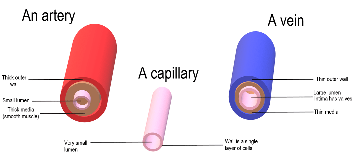
Both arteries and veins have 3 layers which are called the tunica externa or outer layer, tunic media or middle layer and tunic interna or inner layer.
The tunica media is a smooth muscle layer that is thicker in arteries than in veins. This helps with regulating blood pressure and the flow of blood.
Veins have special structures called valves not found in arteries. The valves only allow blood to flow toward the heart. This allows for external forces such as muscle contraction to help push the blood toward the heart.
The smallest vascular structures are the capillaries. These contain only 1 layer of simple squamous epithelium. This thin layer helps to transport substances in and out of the blood. For example, oxygen moves into the tissues and carbon dioxide move out of the tissues and into the blood.
Examine the differences between arteries and veins in the image below:
Arterial Circulation
The circulatory system contains many arteries and veins, and we will cover just a few.
Let’s look at the main circulatory system flow from the heart to the head and upper extremities.
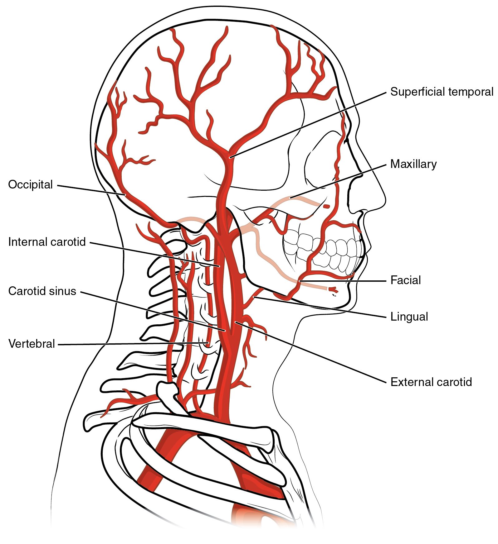
Blood moves from the left ventricle to the aorta. There are 3 primary branches off the aorta which include the right brachiocephalic, left common carotid and left subclavian arteries. The right brachiocephalic divides into the right common carotid and subclavian arteries.
The common carotid artery moves into the neck and head area and divides into internal and externa carotid arteries. The internal carotid provides blood to the brain while the external carotid provides blood to the facial area.
There is a vertebral artery that travels through the transverse foramen of the vertebrae that also supplies the brain with blood.
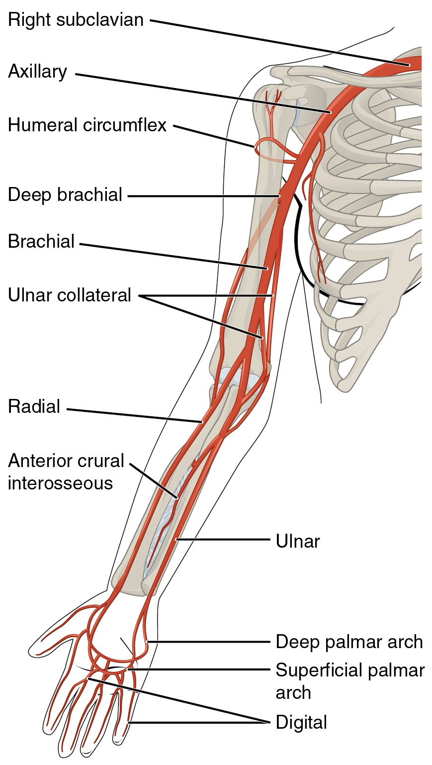
The subclavian artery moves under the clavicle then becomes the axillary artery once it emerges near the upper extremity. The axillary artery then become the brachial artery which divides into the radial and ulnar arteries.
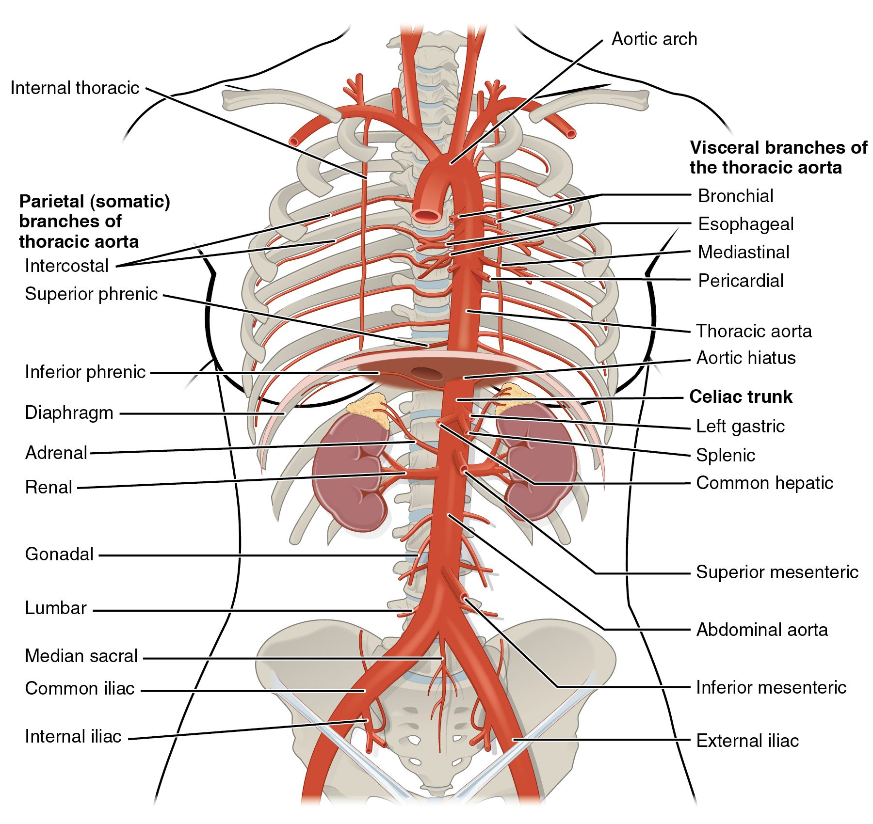
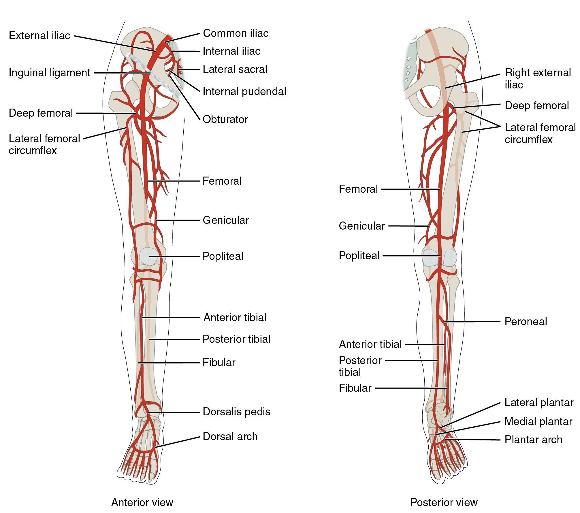
If we follow the arch of the aorta we see it becomes the descending or thoracic aorta. It then moves through the diaphragm to become the abdominal aorta then divides into the right and left common iliac arteries which divide into internal and external iliac arteries.
Some of the branches off the abdominal aorta include:
Inferior phrenic arteries
Celiac trunk
Superior and inferior mesenteric arteries
Renal arteries
The external iliac artery become the femoral artery which travels posterior to become the popliteal artery which then divides into anterior and posterior tibial arteries.
Study the arterial circulation in the images below:
Venous Circulation
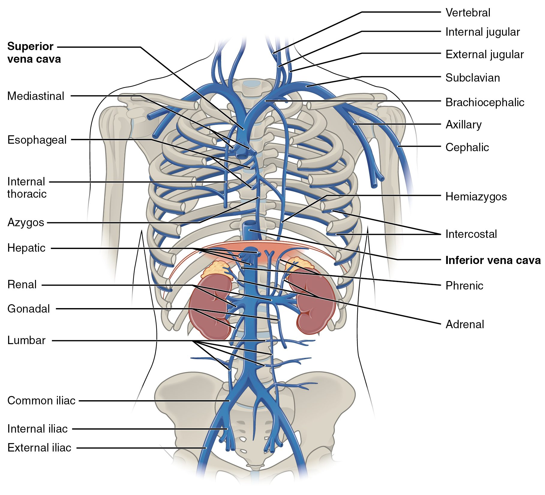
The venous system includes deep and superficial veins. Many of the deep veins have the same names as the arteries.
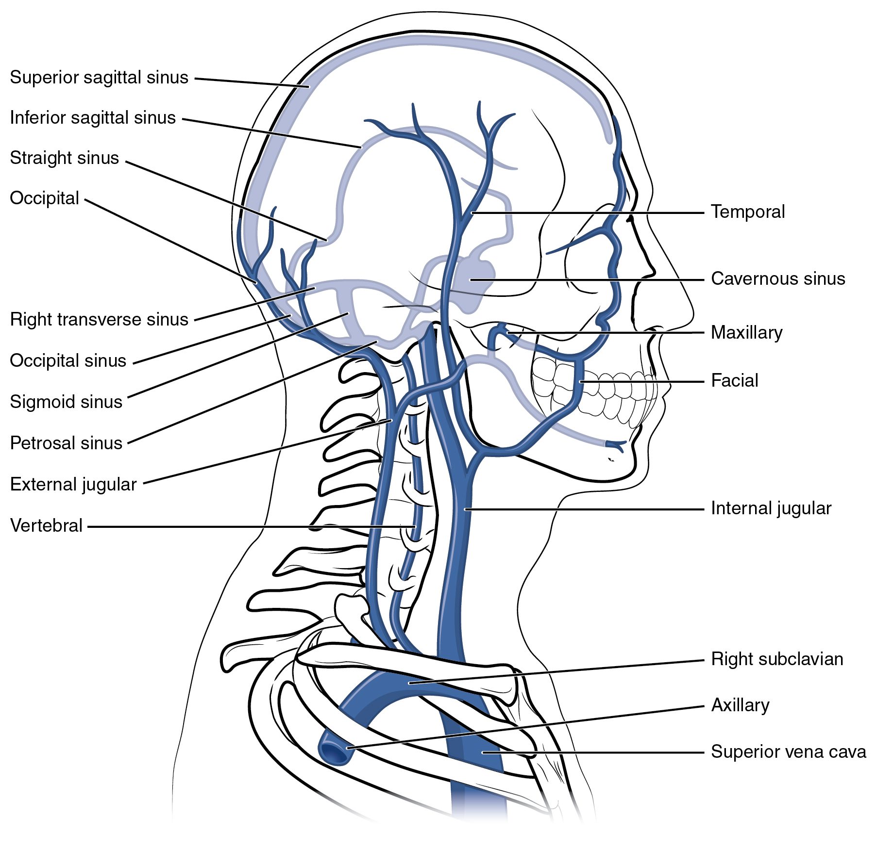
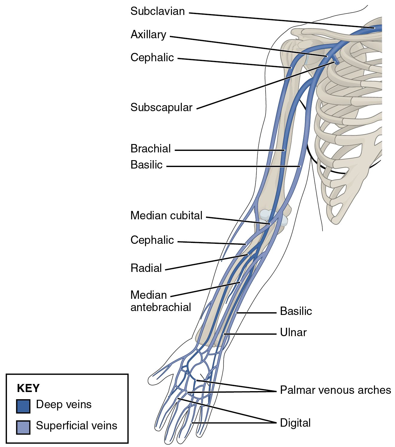
If we follow the superior vena cava we see it branches into right and left brachiocephalic veins. There are internal and external jugular veins. The subclavian vein follows the same course as the artery and becomes the axillary, then brachial, then radial and ulnar veins.
The difference in the venous system is that there are superficial veins. For example, the cephalic and basilic veins are in the arm.
Some of the branches of the inferior vena cava include the renal, gonadal and lumbar veins.
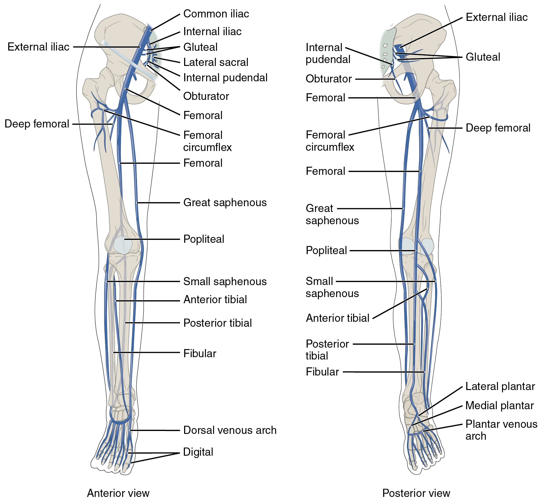
Like the arteries, the inferior vena cava divides into common iliac, internal and external iliac veins. The external iliac becomes the femoral, popliteal and anterior and posterior tibial veins.
One important superficial vein of the lower extremity is the great saphenous vein.
Study the venous circulation in the following images: