Muscle of Soft Palate, Pharynx, and Tongue
Soft Palate
Innervated: Vagus Nerve (cranial nerve X)
EXCEPT tensor veli palatini → trigeminal nerve of mandibular branch 3
5 pairs
Functions
move soft palate up and back to contact throat
seal off nasal cavity from oral cavity (food to such)
Muscle | Description | Origin | Insertion | Action |
|---|---|---|---|---|
Palatoglossal 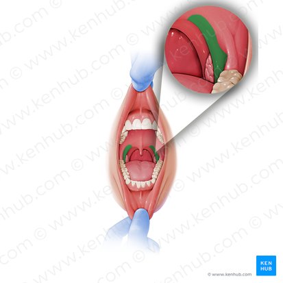 | Forms the anterior faucial pillar; the extrinsic tongue muscle; runs from palate to lateral tongue | Soft palate aponeurosis  | Dorsal and lateral tongue 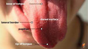 | pulls tongue up and back, soft palate down, and narrow space between pillars |
Palatopharyngeal 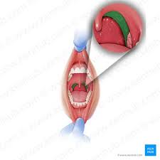 | Forms the posterior faucial pillar; connects palate to pharynx and larynx | Posterolateral soft palate  | Thyroid cartilage of larynx & wall of pharynx 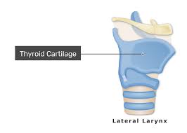 | Elevates and dilates pharynx, narrows fauces, closes nasopharynx during swallowing 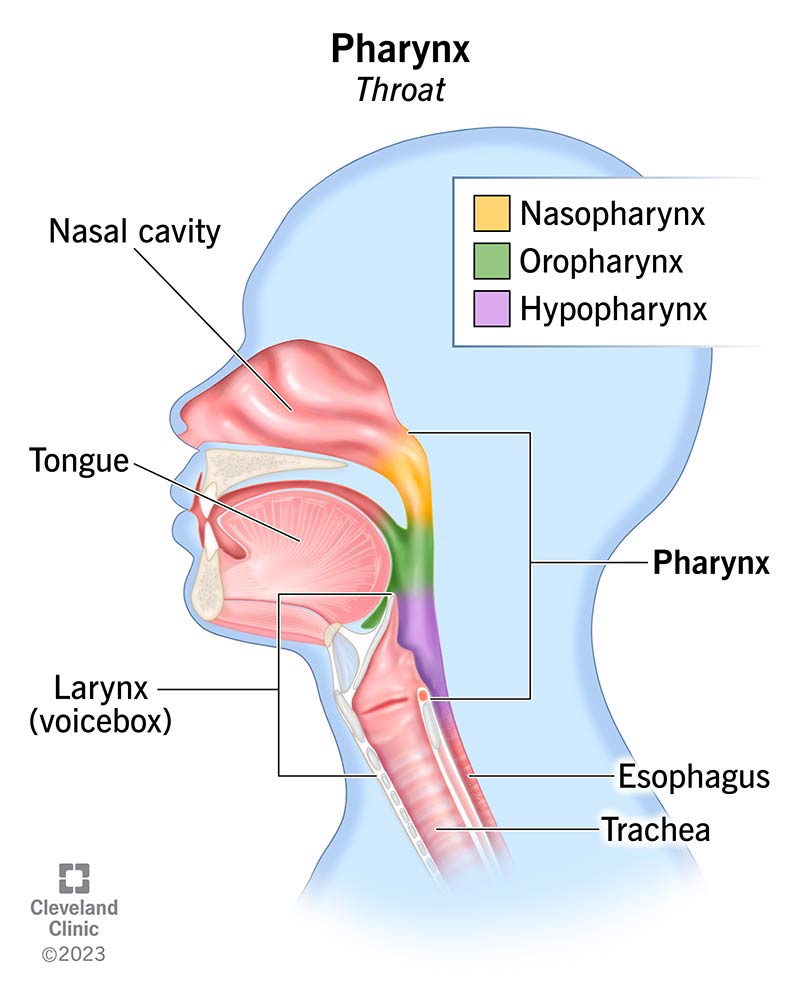 |
Musculus uvulae 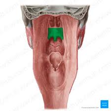 | Small muscle within the uvula of the soft palate | Posterior end of hard palate 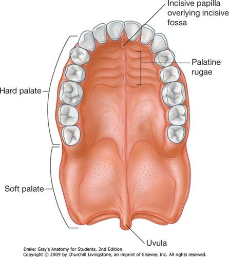 | Tissue of uvula  | Shortens and broadens uvula; helps soft palate close nasopharynx during swallowing  |
Levator veli palatini 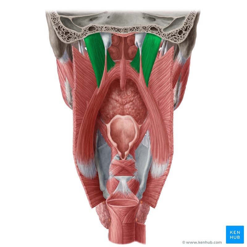 | Main elevator of soft palate; located above soft palate | temporal bone  | Posterior part of soft palate | Elevates and pulls soft palate backward, closes nasal cavity during swallowing, opens auditory tube |
Tensor veli palatini 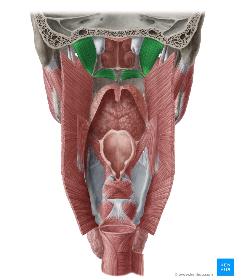 | Ribbon-like muscle that tenses soft palate | Medial pterygoid plate and auditory tube | Palatine aponeurosis at junction of hard and soft palate | Tenses and lowers soft palate, opens auditory tube, equalizes middle ear pressure 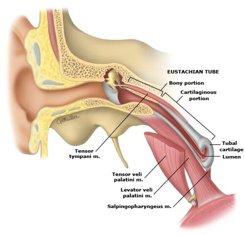 |
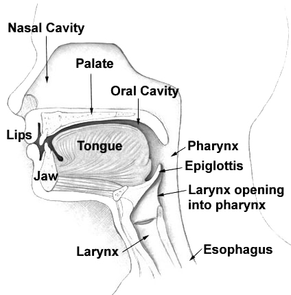
Pharynx
a part of the respiratory and digestive tract
connected to both the nasal and oral cavities
involved in speaking, swallowing, and middle ear function
3 parts:
nasopharynx
oropharynx
laryngeal pharynx
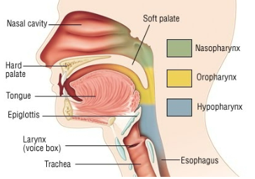
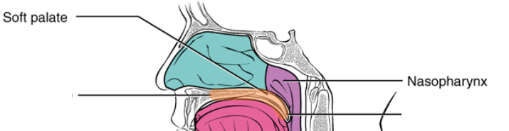
Nasopharynx
above soft palate; continuous with nasal cavity
auditory tube opens to lateral wall
pharyngeal tonsils located on the nasopharynx (posterior wall)
mass of lymphoid tissue
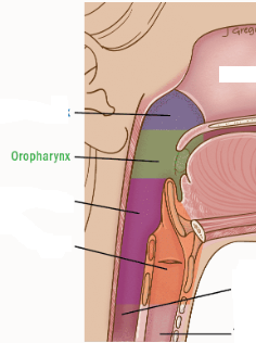
Oropharynx
region from soft palate to epiglottis
continuous through the fauces
fauces bound laterally by the palatoglossus and the palatopharyngeal fold
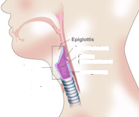
Epiglottis
lower border of the tongue
extend upward during breathing
covers the opening during swallowing so food goes to the esophagus
Laryngeal Pharynx
below tongue where digestive and respiratory system branch into esophagus and larynx
Pharyngeal Constrictor Muscles
forms the lateral and posterior wall
3 paired muscles
inferior is most superficial when overlapped
ALL insert into median pharyngeal raphe
tendinous band of the posterior wall of the pharynx
ALL innervated by pharyngeal plexus; cranial nerves IX, X, and XI
Muscle | Description | Origin | Insertion | Action |
|---|---|---|---|---|
Superior Pharyngeal Constrictor 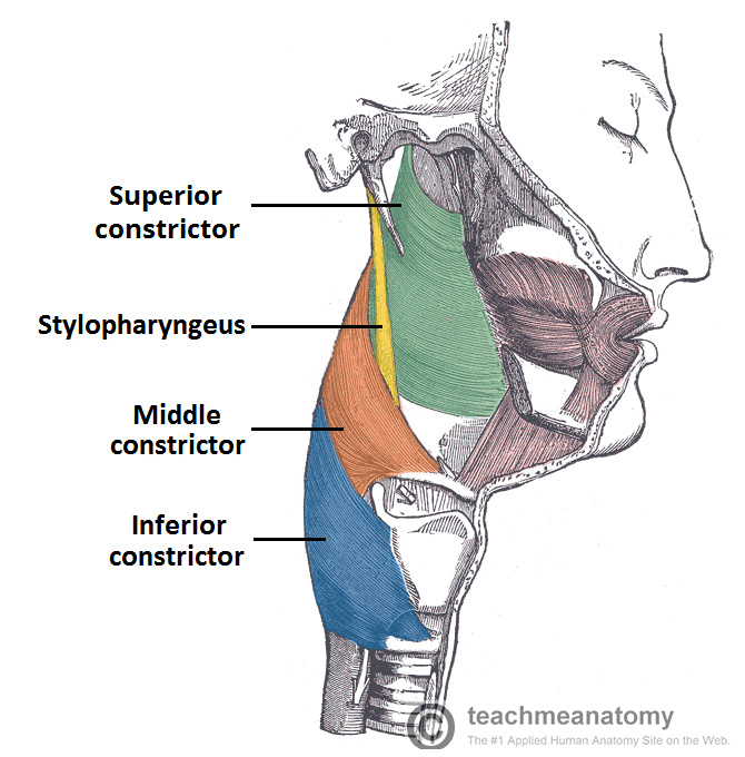 | lower part of the Medial pterygoid plate, mandible, pterygomandibular raphe 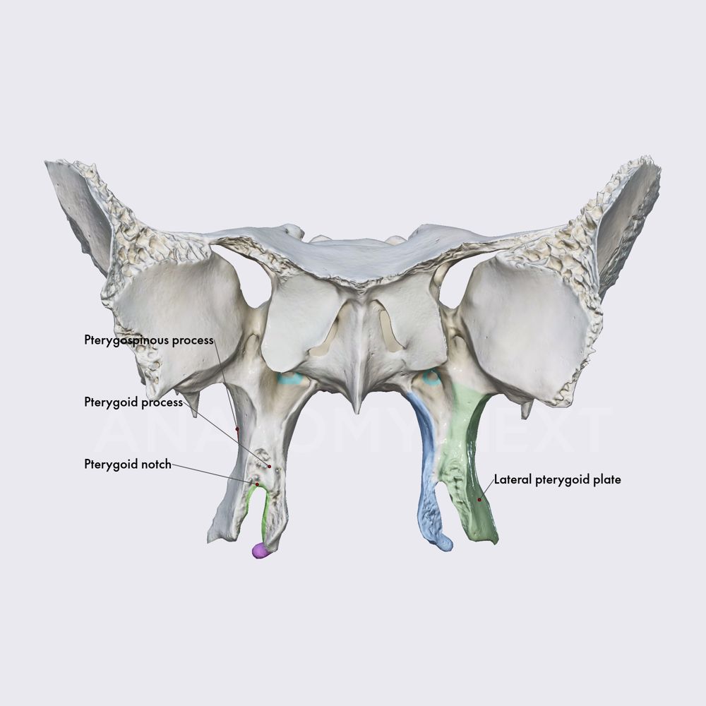 | Median pharyngeal raphe  | Constricts upper pharynx during swallowing | |
Middle Pharyngeal Constrictor 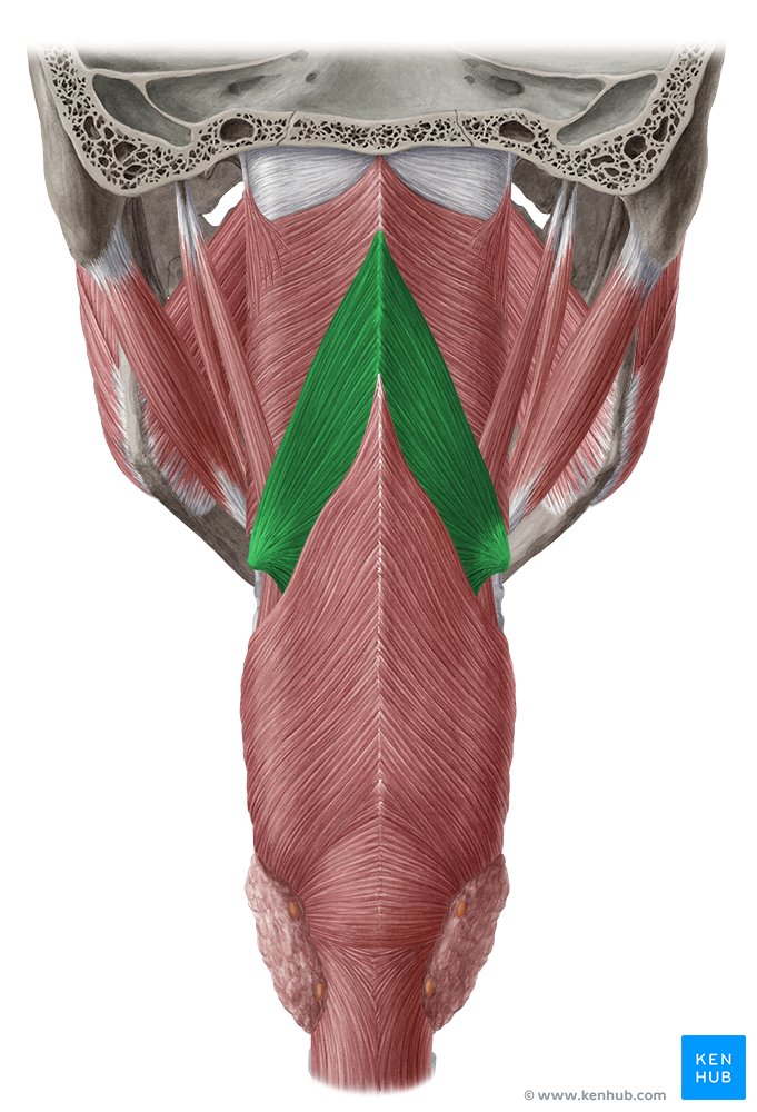 | Greater and lesser horns of hyoid bone, stylohyoid ligament 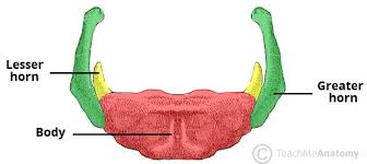 | Median pharyngeal raphe  | Constricts pharynx and pushes food into esophagus | |
Inferior Pharyngeal Constrictor | Lowest and most superficial constrictor | Posterior part of larynx 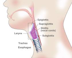 | Median pharyngeal raphe  | Constricts lower pharynx and forces food ALL the way esophagus |
Pharyngeal Elevators and dilatators
palatopharyngeal muscle → soft palate
Muscle | Description | Origin | Insertion | Action |
|---|---|---|---|---|
Stylopharyngeal (CN IX) | 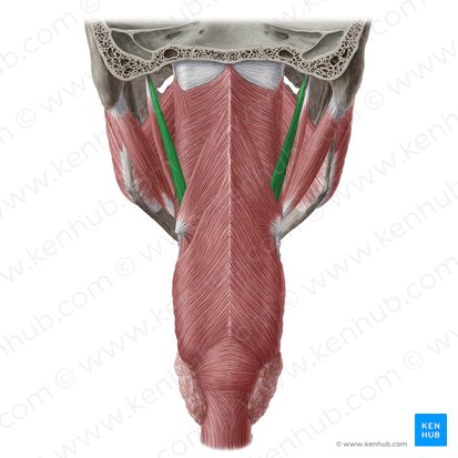 | Styloid process 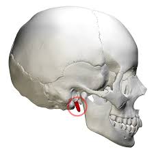 | Lateral pharyngeal wall & thyroid cartilage | Elevates and dilates pharynx |
Salpingopharyngeal (CN IX, X, and XI) | 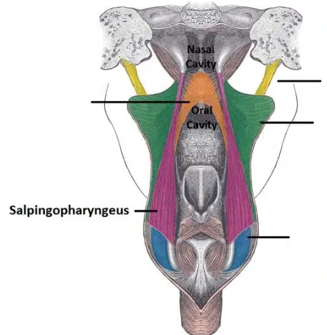 | Near opening of auditory tube | Blends with palatopharyngeal muscle and lateral pharyngeal | Elevates pharyngeal wall |
Muscle of the tongue
intrinsic muscle
located inside the tongue
changes the shape of the tongue
extrinsic muscles
origin outside the tongue
insertions are INSIDE the tongue
moves the tongue while suspending and anchoring the tongue to body structures
Innervation: cranial nerve XII (hypoglossal nerve)
Intrinsic Tongue
4 pairs
grouped by orientation
Muscle | Description | Origin | Insertion | Action |
|---|---|---|---|---|
Superior Longitudinal | Superficial muscle running from base to tip (oblique and longitudinal direction) | 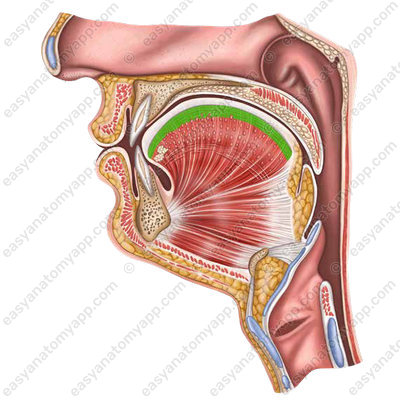 | 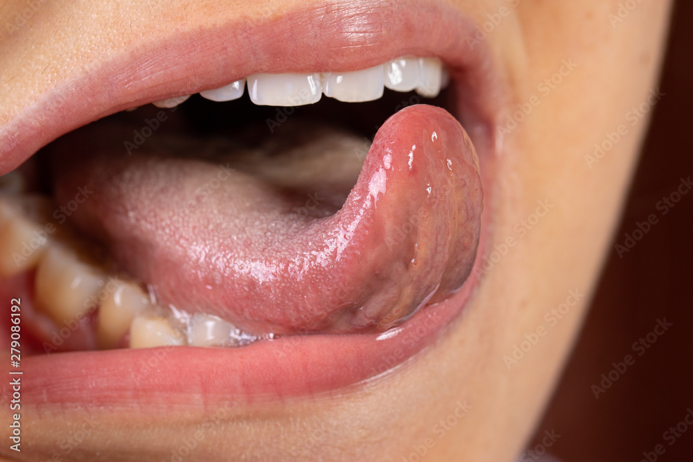 | Shortens tongue and curls tip upward |
Inferior Longitudinal | runs longitudinal direction from base to apex |  | 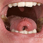 | Shortens tongue and pulls tip downward |
Transverse Fibers | medial septum to lateral border; Runs side to side |  | 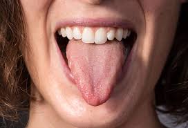 | Narrows tongue |
Vertical | Runs from top to bottom of tongue | 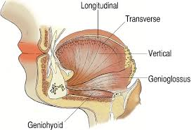 |  | Flattens and widens tongue |
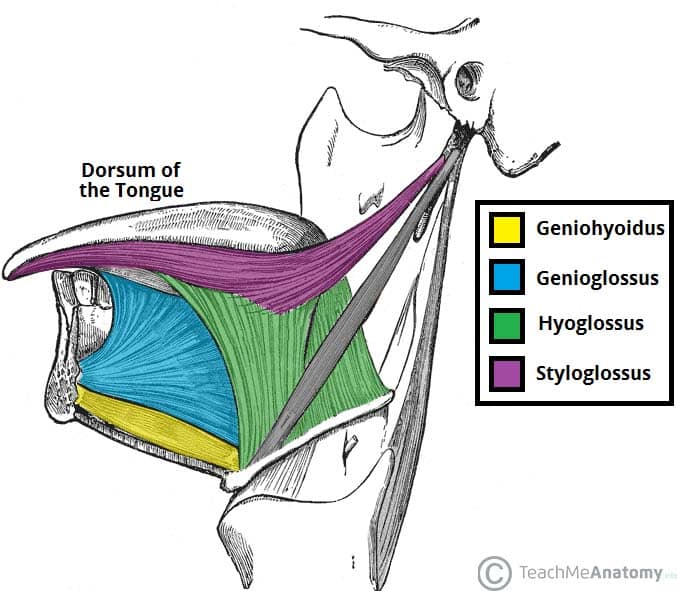
Extrinsic Tongue Muscles
4 pairs
named by location and insertion (glossus)
Note: Genioglossus is used to test the 12 cranial or hypoglossal nerve by sticking out the tongue; it deviates on the paralyzed side
Muscle | Description | Origin | Insertion | Action |
|---|---|---|---|---|
Genioglossus 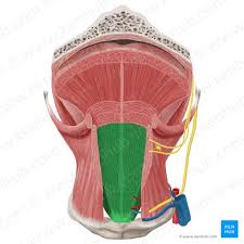 | Large fan-shaped tongue muscle; separated by median septum | Genial tubercles of mandible 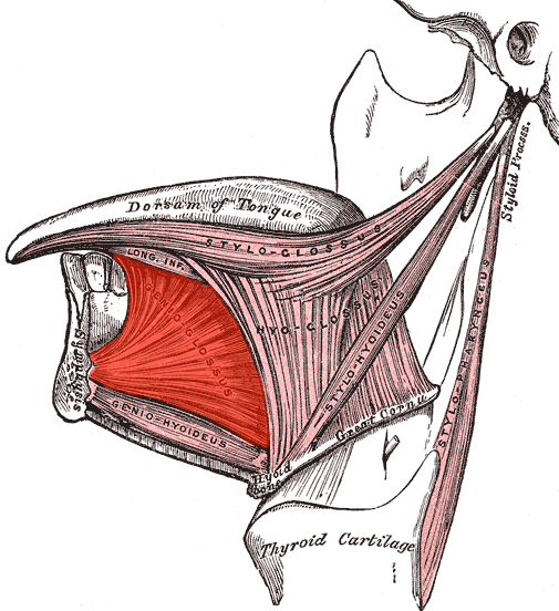 | Body of tongue | Protrudes and depresses tongue; prevents tongue from falling back |
Hyoglossus 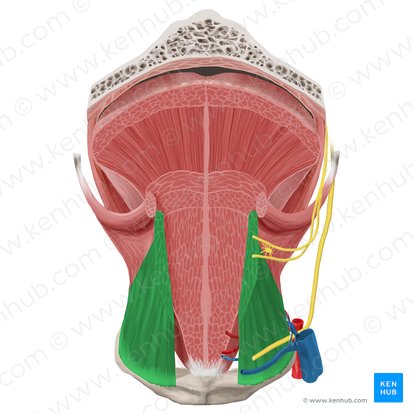 | Flat muscle connecting hyoid to tongue | Hyoid bone 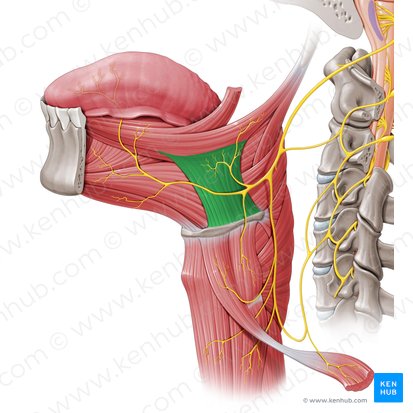 | Body of tongue | Depresses tongue and pulls sides downward |
Styloglossus 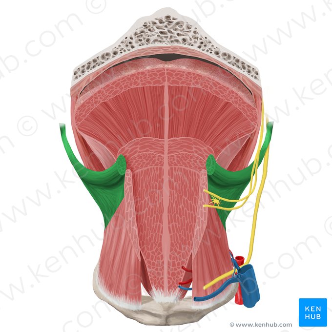 | Long muscle from skull to tongue | Styloid process of temporal bone 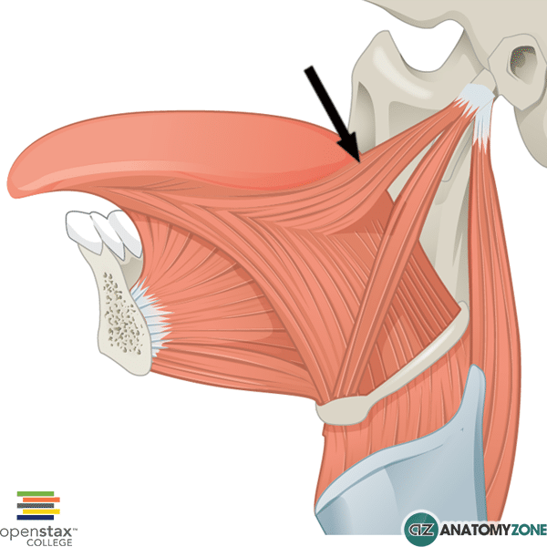 | Lateral tongue | Retracts tongue upward and backward |
Palatoglossus 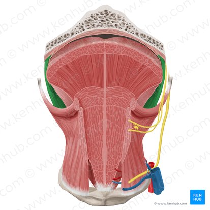 | Connects palate and tongue | Soft palate 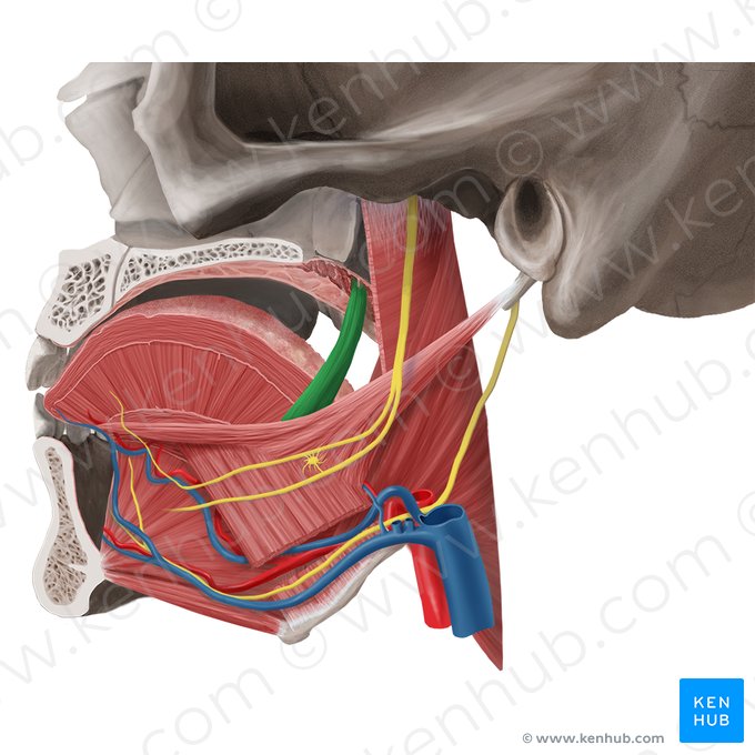 | Lateral tongue | Elevates tongue |
Blood Supply to the tongue
lingual artery
branch of the external carotid artery
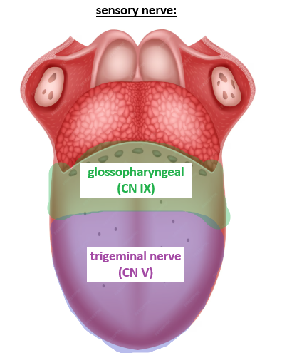
sensory: afferent
anterior 2/3
Lingual branch of the mandibular division of CN V
posterior 1/3
glossopharyngeal CN IX
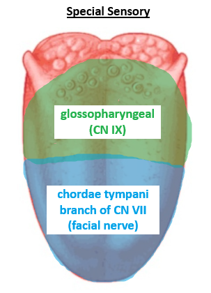
special sensory: taste
anterior 2/3: chordae tympani branch of CN VII
posterior 1/3: glossopharyngeal CN IX
Sensory and special sensory come from the vagus nerve
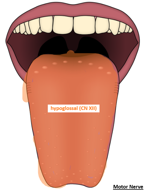
motor
extrinsic and intrinsic muscles
hypoglossal CN XII
Structure | Main Nerve | Exception |
|---|---|---|
Tongue | CN XII (hypoglossal) | Palatoglossus |
Soft palate | CN X (vagus) | Tensor veli palatini (trigeminal) |
Pharynx | CN X (vagus) | Stylopharyngeus |