DEVELOPMENT OF THE VEINS
three embryonic
venous systems comprising:
1. The vitelline system, draining the
yolk sac and gut tube
2. The umbilical system, bringing
oxygen-rich blood from the
placenta
3. The cardinal system, draining the
body wall, neck, and head
1. The remodeling makes
the right-side dominant
for the vitelline and
cardinal systems,
2. The umbilical vein on the
right-side degenerates
3. Remodeling results in
the formation of the
superior and inferior
venae cavae (SVC & IVC)
4.Makes the left side dominant for
the umbilical system, although it
still drains into the right side of the
heart
5. A shunt (vessel) develops in the liver
as the ductus venosus which allows
blood in the left umbilical vein to
drain into the inferior vena cava
6. Remodeling of venous system is more
complex than in the arterial system
RVV and LVV come from yolk sac
Little capillaries(sinusoids) form after V V passes through the GI tract and enters the liver
NB: S—Trasnversum becomes the diaphragm
Sinusoids drain into hepatic vein( L and R hepatic from iVC)
Splenic + SMV +IMV= hepatic portal vein
SMV and IMV are capillary networks formed in GI tract
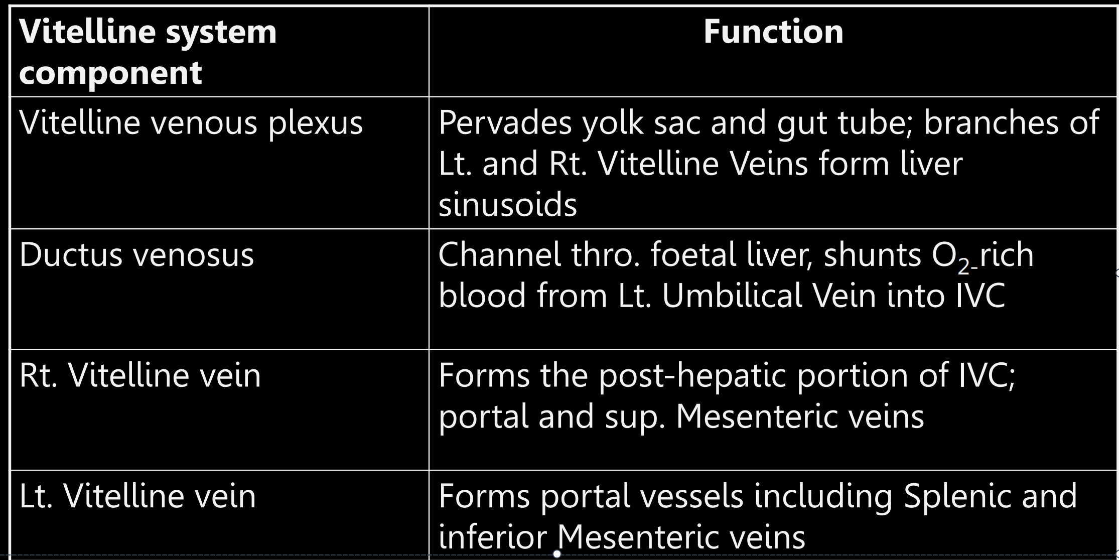
Vitelline produces 4 main; i. liver sinusoids,
ii. ductus venosus
iii. the portal venous system and
iv. the cranial (post-hepatic) part
of the Inferior Vena Cava(IVC)
Umbilical vein derivatives
• The right umbilical vein
degenerates
• The left umbilical vein loses
its connection to the left
sinus horn
• It drains into the IVC via the
ductus venosus
NB: reminant of L umbilical vein is Ligamentum Teres
Dev of viteline and umbilical occurs second to 3rd week
THE CARDINAL VEIN SYSTEM
Starts around 5th week
The cardinal system initially consists
of symmetric, paired anterior and
posterior cardinal veins
• They drain into the sinus horns via
short common cardinal veins
• The anterior cardinals drain the head
and neck
• The posterior cardinals drain the body
wall
Posterior cardinal veins degenerate and become subcardinal(medial dorsal body wall especially kidney and gonads) and supracardinal(drains thoracic wall) veins
RIGHT SUBCARDINAL VEIN
The right subcardinal
vein forms the next most
caudal part of the IVC
after the right vitelline
vein
• The left cardinal vein will
then drain into this part of
the inferior vena cava
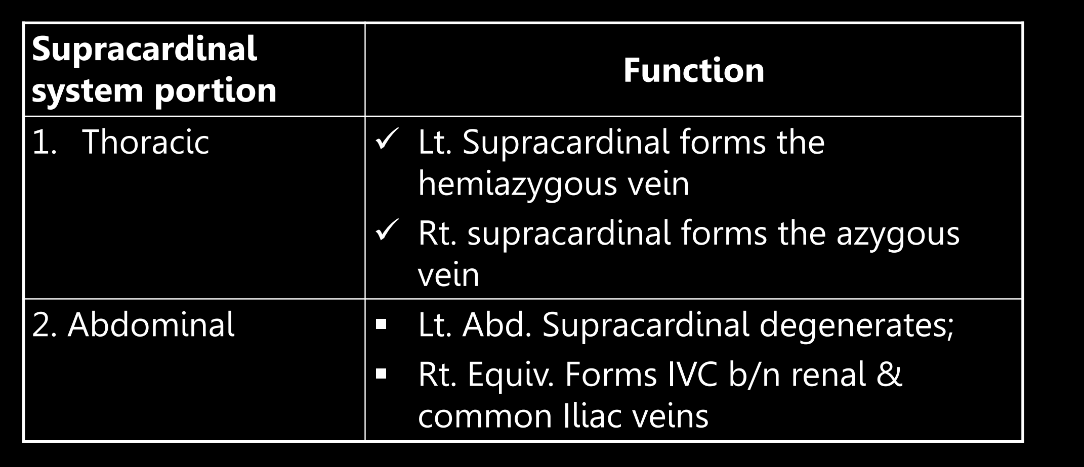
DERIVATIVE OF THE RT. ABDOMINAL SUPRACARDINAL
VEIN
It forms the inferior vena
cava (IVC) between renal
tributary and bifurcation of
iliac veins
FORMATION OF THE IVC
The inferior vena cava
(IVC), from cranial to
caudal, is derived from:
i. The right vitelline vein
ii. The right subcardinal vein
iii. The right supracardinal
vein
iv. The posterior cardinal
ANTERIOR (OR PRE-) CARDINAL VEIN 1
The anterior cardinal
system
drains the head, neck
and upper limbs
• The distal Ant. Cardinal
vein forms internal
jugular veins (on either
side)
ANTERIOR CARDINAL VEIN (2)
The Right common cardinal vein
[yellow circle] with contribution
from the proximal right
anterior cardinal becomes
Superior vena cava (SVC) [Red
circle]
ii. The proximal Left anterior
cardinal vein degenerates, so the
left brachiocephalic
(innominate) vein forms to
drain the left internal jugular
vein into the SVC
NB; supracardinal vein become azygous, hemiazygous and accessory hemiazygous vein and the subcardianl major iVC