Session 5: PNS & ANS Notes
Peripheral Nervous System (PNS) & Autonomic Nervous System (ANS)
Learning Objectives: The Peripheral Nervous System
Name the 12 pairs of cranial nerves; indicate the body region and structures innervated by each.
Describe the general structure of a spinal nerve.
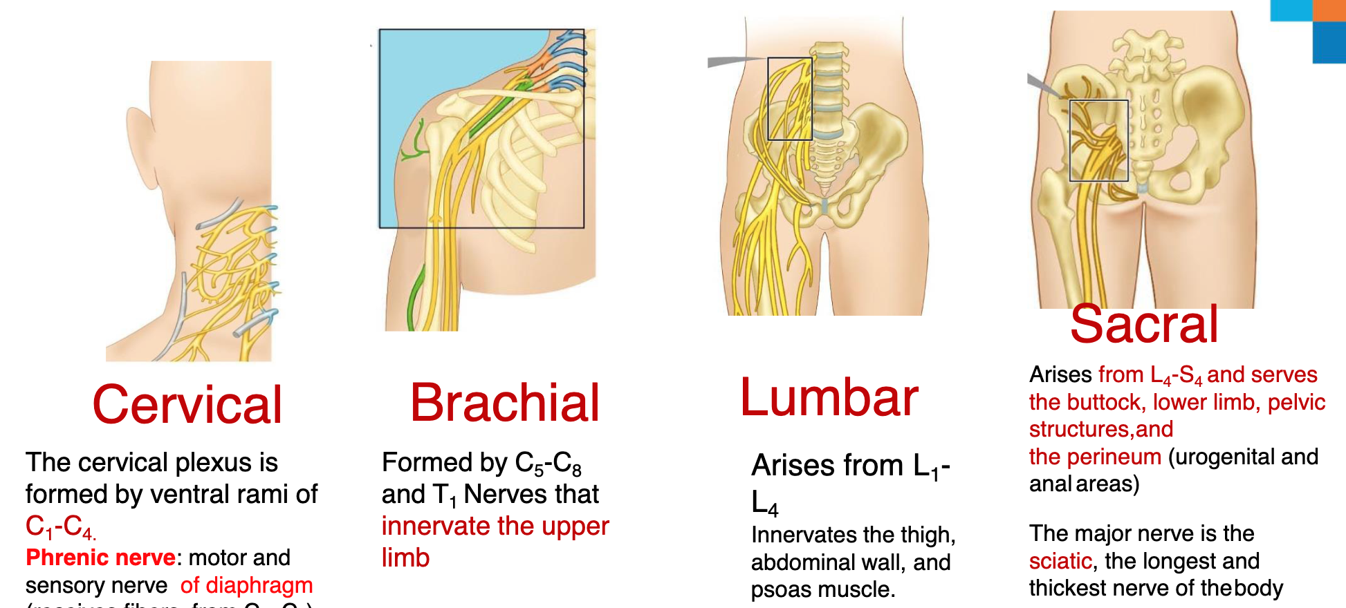
Define spinal plexus. Name the four major spinal plexuses and describe the general distribution and function of the peripheral nerves arising from each plexus.
Name the components of a reflex arc and distinguish between somatic and visceral.
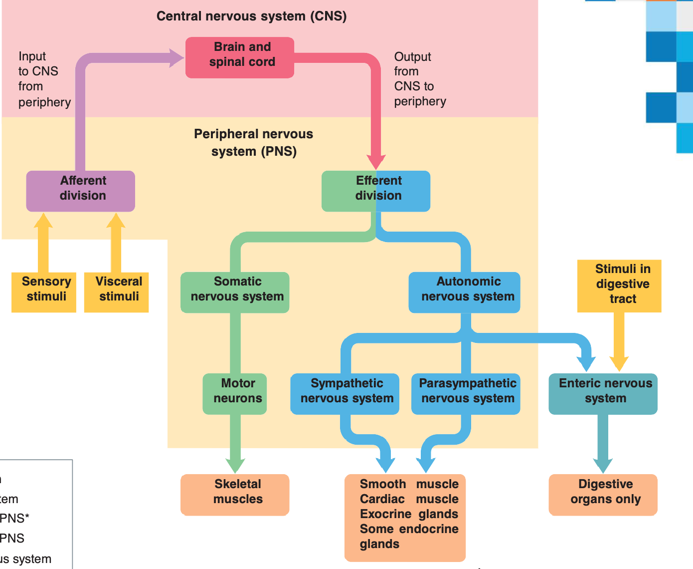
Somatic vs. Autonomic Nervous Systems
Autonomic nervous system
Involuntary branch of the peripheral efferent division
Somatic nervous system
Branch of the efferent division subject to voluntary control
The AFFERENT division is included in the sensory pathway for both systems.
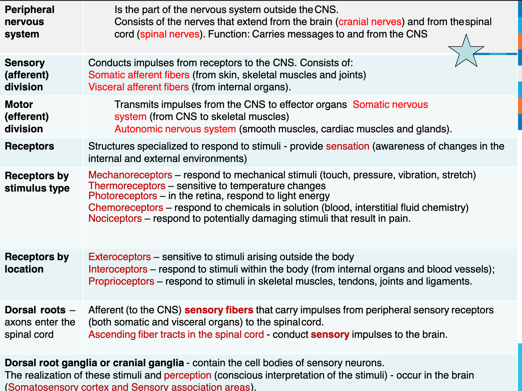
Peripheral Nervous System (PNS)
Consists of nerve fibres and cell bodies outside of the central nervous system (CNS)
Organised into nerves which connect the CNS with the body
12 pairs of cranial nerves
31 pairs of spinal nerves
Note the use of the term pairs: they occur on the left and right
Nerves and Associated Ganglia
Classification according to the nerve origin:
Spinal nerve from spinal cord
Cranial nerve - from brain (brainstem)
Classification according to the direction nerves transmit impulses
Sensory (afferent) nerves: impulses only toward CNS
Motor (efferent) nerves: impulses only away from CNS
Mixed nerves: contain both sensory and motor fibers
Impulses travel both to and from CNS
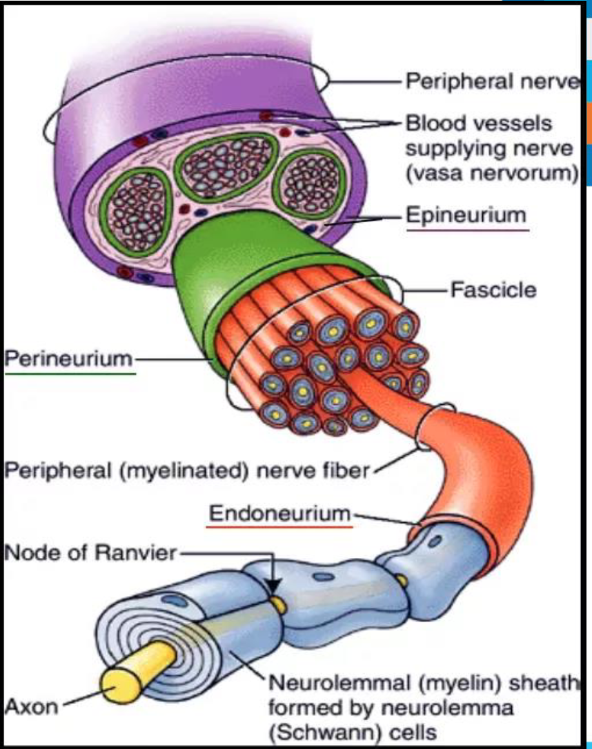
Nerve:
Cordlike organ of the PNS consisting of axons/dendrites enclosed by connective tissue
Anatomy of a Nerve
What are the differences between: a neuron, a nerve fiber, a nerve fascicle and a whole peripheral nerve?
Name the connective tissue membranes that cover the:
Axon (Nerve Fiber)
Nerve fascicle
Entire peripheral nerve
Cranial Nerves
12 pairs
Cranial nerves are in the PNS
12 pairs of cranial nerves
11 pairs arise from the brain, 1 pair arise from the spinal cord
Pass through the foramina of the skull
Named in two ways
A descriptive name eg. optic nerve, abducens nerve, accessory nerve
Via roman numerals CN II, CN VI, CN XI
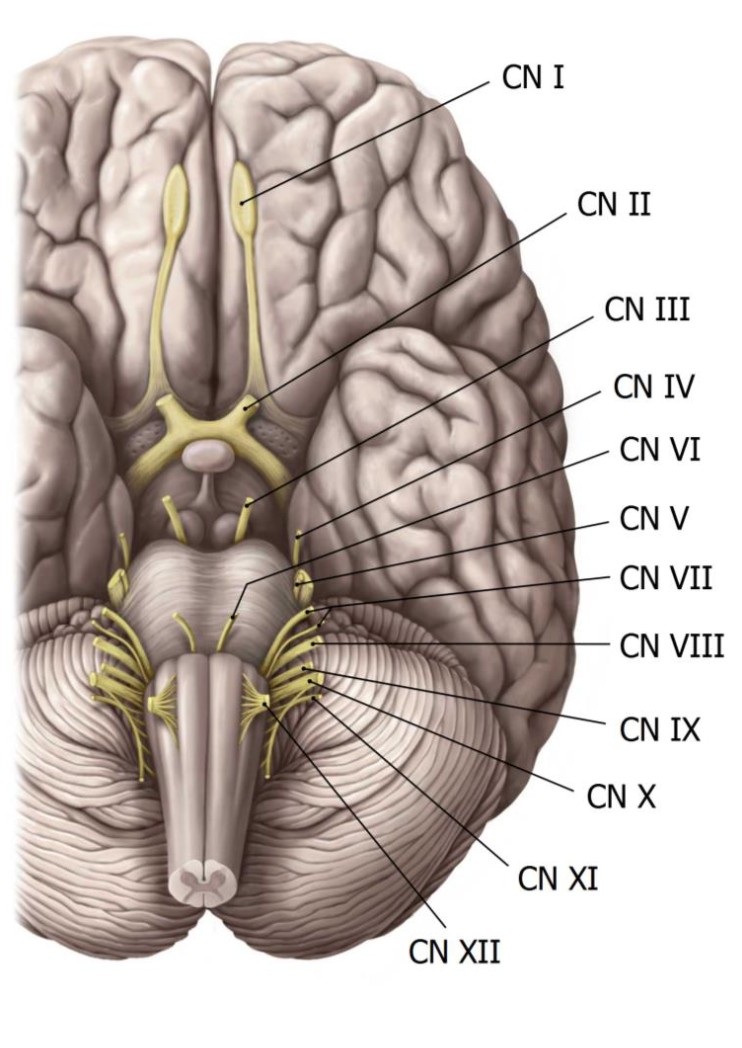
Cranial Nerves List
CN I Olfactory n.
CN II Optic n.
CN III Oculomotor n.
CN IV Trochlear n.
CN V Trigeminal n.
CN VI Abducens n.
CN VII Facial n.
CN VIII Vestibulocholear n.
CN IX Glossopharyngeal n.
CN X Vagus n.
CN XI Accessory n.
CN XII Hypoglossal n.
Distribution of Cranial Nerves
Mnemonic devices to remember the names of the cranial nerves:
"On occasion, our trusty truck acts funny—very good vehicle anyhow"
"Oh once one takes the anatomy final, very good vacations are heavenly"
Functions of Cranial Nerves
Number | Name | Fiber Types | Functions |
|---|---|---|---|
I | Olfactory | Sensory | Smell |
II | Optic | Sensory | Vision |
III | Oculomotor | Mixed (mainly motor) | Eyeball and eyelid movement, pupil constriction, change of lens shape for near vision. Proprioception (awareness of position of body parts) |
IV | Trochlear | Mixed (mainly motor) | Eyeball movement Proprioception |
V | Trigeminal | Mixed | Chewing Somatic sensations (touch, pressure, pain, and temperature) of face and mouth |
VI | Abducens | Mixed (mainly motor) | Eyeball movement Proprioception |
VII | Facial | Mixed | Facial expression, secretion of saliva and tears Taste from front of tongue |
VIII | Vestibulocochlear | Sensory | Hearing, sense of equilibrium |
IX | Glossopharyngeal | Mixed | Swallowing, secretion of saliva Taste from back of tongue, somatic sensation of oral cavity, blood-pressure monitoring Efferent output for skeletal muscles of pharynx and larynx |
X | Vagus | Mixed | Efferent output for skeletal muscles of pharynx, larynx and for smooth muscle and glands of thoracic and abdominal organs and for cardiac muscle of heart. Afferent input from thoracic and abdominal organs, blood-pressure monitoring |
XI | Accessory | Motor | Efferent output for skeletal muscles of pharynx, larynx, neck, and shoulder |
XII | Hypoglossal | Motor | Tongue movement |
Carried by afferent fibers.
Carried by efferent fibers.
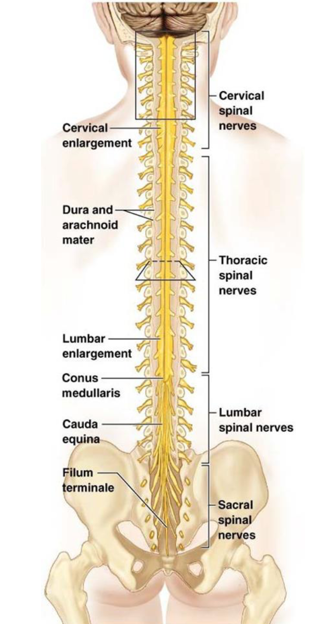
Spinal Nerves Numbering
31 pairs of spinal nerves exiting the spinal cord
8 cervical (8 pairs)
12 thoracic (12 pairs)
5 lumbar (5 pairs)
5 sacral (5 pairs)
1 coccygeal (1 pair)
Somatic Plexus (refers to a network of intersecting nerves in the somatic nervous system, which controls voluntary movements and sensory perception in the body wall and limbs)
Ventral rami (larger branches and carry both motor and sensory fibers) from adjacent spinal nerves merge to form networks called a plexus
Multisegmental peripheral nerves (nerves that receive input from more than one spinal cord segment) arise from a plexus
Primarily occurs to supply limbs
Innervation of Specific Body Regions
Spinal Nerves

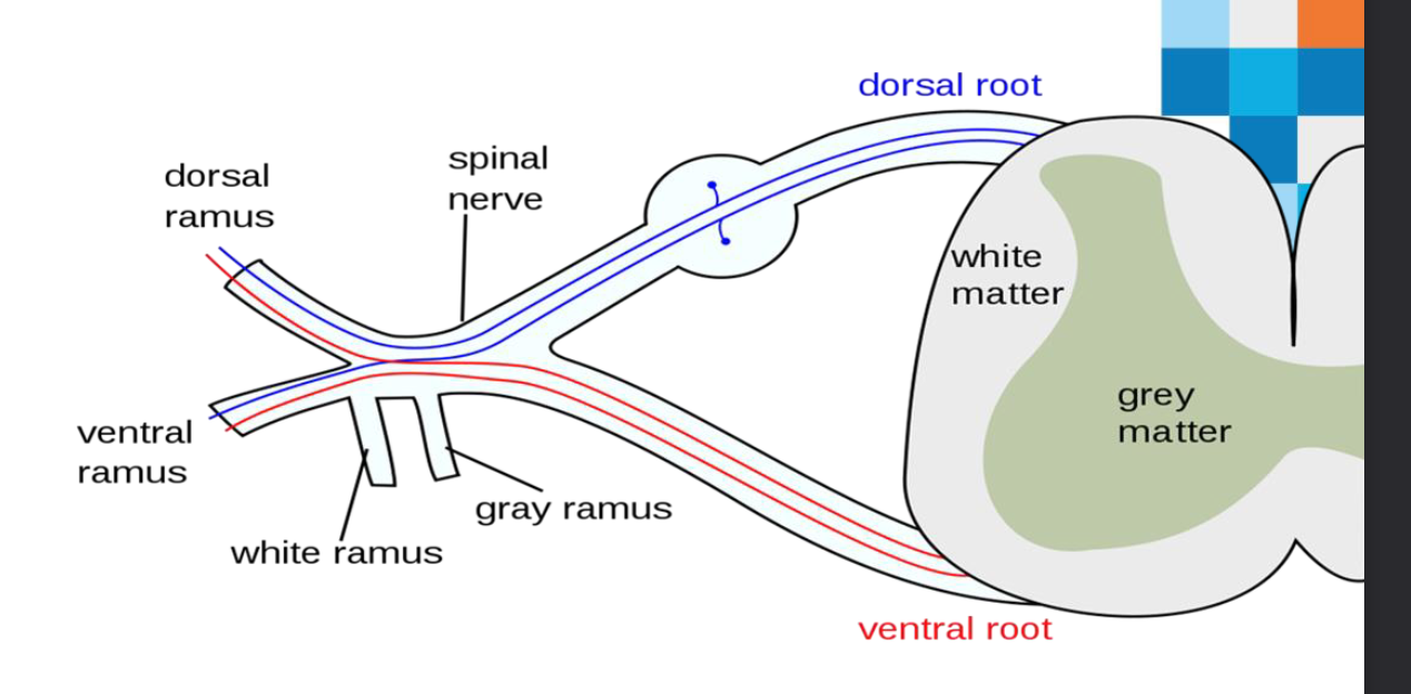
Gray Matter and Spinal Roots
Gray commissure: bridge of gray matter that connects masses of gray matter on either side
Encloses central canal
Spinal nerves: formed by fusion of dorsal and ventral roots
Dorsal roots: sensory input to cord
Dorsal root (spinal) ganglia: cell bodies of sensory neurons
Ventral roots: bundle of motor neuron axons that exit the spinal cord
Afferent Vs Efferent
Memory trick to remember the difference between afferent and efferent neurons.
S – Sensory
A – Afferent
M – Motor
E – Efferent
D – Dorsal (enter the dorsal aspect of the spinal cord)
A – Afferent
V – Ventral (exits the ventral aspect of the spinal cord)
E - Efferent
Spinal Nerve Formation
Dorsal Root Ganglion
Dorsal Horn
Dorsal Ramus
Mixed
Dorsal Roots
Sensory
Ventral Spinal Nerve
Ventral Root
Ventral Horn
Ventral Ramus
Mixed Motor
Mixed
Spinal Nerves - Neurons
Blue neurons – sensory
Red neurons – motor
Note that the spinal nerve and rami contain both motor and sensory neurons
Structure and Classification (cont.)
Ganglia: contain neuron cell bodies associated with nerves in PNS
Ganglia associated with afferent nerve fibers contain cell bodies of sensory neurons
Dorsal root ganglia (sensory, somatic)
Ganglia associated with efferent nerve fibers contain autonomic motor neurons
Autonomic ganglia (motor, visceral)
Spinal Cord and Innate Reflexes
A reflex is an involuntary, stereotypical response of the effector tissue from the stimulation of receptors.
These reflexes are executed by the successive activation (chain reaction) of a certain number of neurons that are mutually connected at the level of spinal cord.
If there are two neurons with one synapse involved= monosynaptic
If there are more than 2 neurons and more than one synapse= polysynaptic
The last neuron generally innervates the effector tissue, which is usually a muscle.
These reflexes do NOT include brain
The spinal cord is responsible for the integration of many innate reflexes
Components of a Reflex Arc
Components of a reflex arc (neural path)
Receptor: site of stimulus action
Sensory neuron: transmits afferent impulses to CNS
Integration center: either monosynaptic or polysynaptic region within CNS
Motor neuron: conducts efferent impulses from integration center to effector organ
Effector: muscle fiber or gland cell that responds to efferent impulses by contracting or secreting
Components of a Reflex Arc (cont.)
Reflexes are classified functionally as:
Somatic reflexes (Spinal reflexes)
Activate skeletal muscle
Autonomic (visceral) reflexes (homeostatic regulation & feedback loop)
Activate visceral effectors (smooth or cardiac muscle or glands)
Spinal Reflexes
Spinal reflexes occur without direct involvement of higher brain centers
Brain is still advised of spinal reflex activity and may have an effect on the reflex
Testing of somatic reflexes important clinically to assess condition of nervous system
If exaggerated, distorted, or absent, may indicate degeneration or pathology of specific nervous system regions
Most commonly assessed reflexes are stretch, flexor, and superficial reflexes
Sensory Processing
Survival depends upon:
Sensation: the awareness of changes in the internal and external environment
Perception: the conscious interpretation of those stimuli
Somatosensory system: part of sensory system serving body wall and limbs
Receives inputs from: Exteroceptors, proprioceptors, and interoceptors
Input is relayed toward head, but processed along the way
Sensory Processing (cont.)
Processing at the receptor level
Generating a signal: For sensation to occur, the stimulus must excite a receptor, and the AP must reach CNS
Stimulus energy must match receptor specificity (touch receptors do not respond to light)
Stimulus must be applied within receptive field (the nerve that innervates that area)
Transduction must occur—energy of stimulus is converted into graded potential called generator potential (in general receptors) or receptor potential (in special sense receptors)
Graded potentials must reach threshold → AP
Key Concept: Sensory Receptors
Sensory receptors: specialized to respond to changes in environment (stimuli)
Converts mechanical, physical, chemical change into Action potential (opens sodium channels)
Classification by Stimulus Type:
Mechanoreceptors—respond to touch, pressure, vibration and stretch
Thermoreceptors—sensitive to changes in temperature
Photoreceptors—respond to light energy (example: retina)
Chemoreceptors—respond to chemicals (examples: smell, taste, changes in blood chemistry)
Nociceptors—sensitive to pain-causing stimuli (examples: extreme heat or cold, excessive pressure, inflammatory chemicals)
Classification by Location
Exteroceptors
Respond to stimuli arising outside body
Receptors in skin for touch, pressure, pain, and temperature
Interoceptors (visceroceptors)
Respond to stimuli arising in internal viscera and blood vessels
Sensitive to chemical changes, tissue stretch, and temperature changes
Sometimes cause discomfort but usually person is unaware of their workings
Proprioceptors
Respond to stretch in skeletal muscles, tendons, joints, ligaments, and connective tissue coverings of bones and muscles
Inform brain of one's movements
Peripheral Nervous System Summary
The ANS and CNS
Many regions of the CNS are involved in the control of autonomic activities
Some autonomic reflexes are integrated at the spinal- cord level
Medulla oblongata within the brain stem is the region most directly responsible for autonomic output
Hypothalamus plays a role in integrating autonomic, somatic, and endocrine responses
Autonomic & Somatic Nervous Systems
Autonomic nervous system
Involuntary branch of the peripheral efferent division
Somatic nervous system
Branch of the efferent division subject to voluntary control
Somatic Nervous System
Motor neurons supply skeletal muscle
Bring about movement
Axons of motor neurons originate in the CNS and end on skeletal muscle. Consist of a one-neuron chain.
Motor-neuron axon terminals release ACh to stimulate muscle contraction
Motor neurons are the final common pathway
Pathways and Divisions of the ANS
An autonomic nerve pathway consists of a two-neuron chain
Preganglionic neuron: synapses with the cell body of the postganglionic fiber in a ganglion outside the CNS
Postganglionic neuron: sends axons that end on the effector organ
Autonomic nervous system has two subdivisions
Sympathetic and parasympathetic
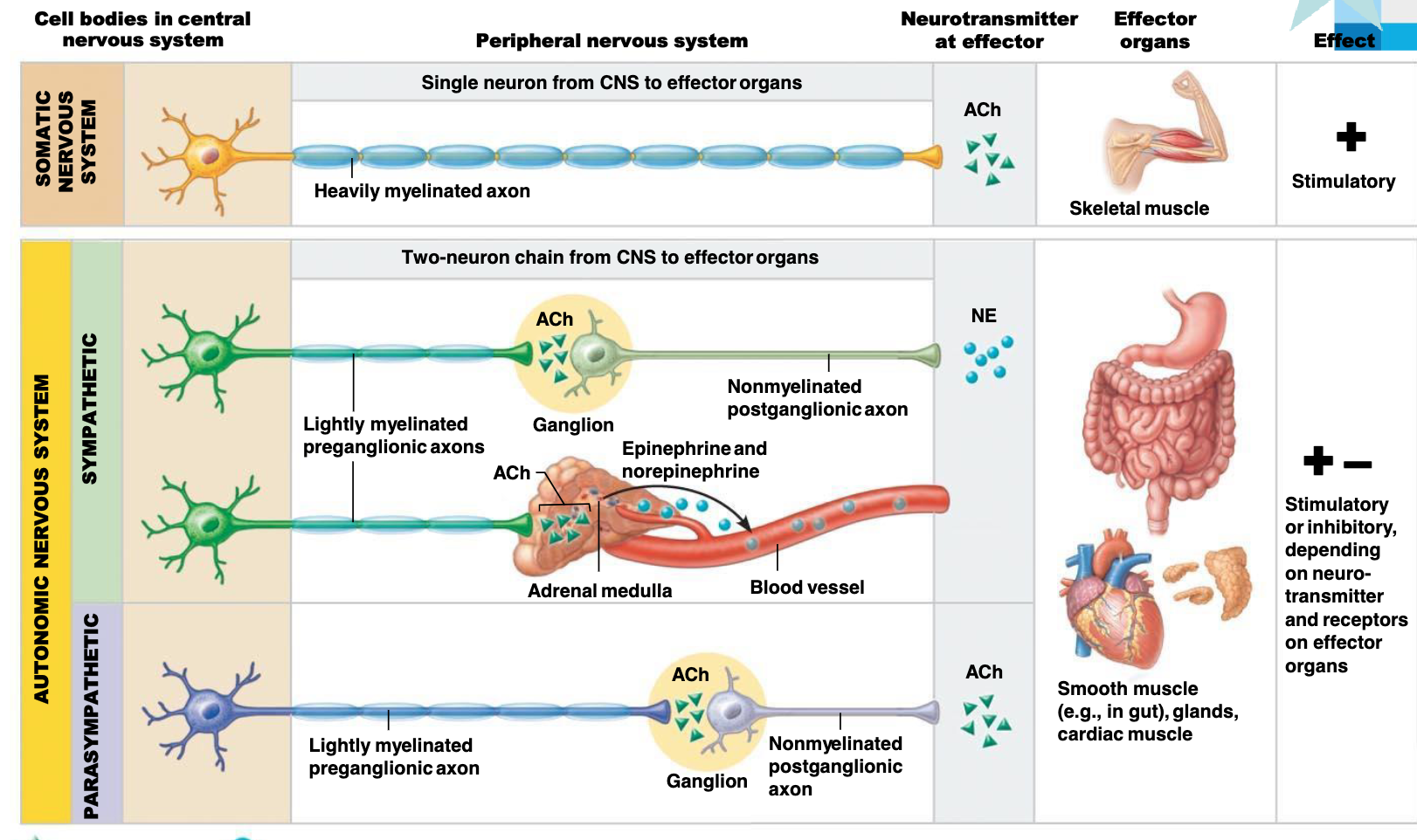
ANS vs. Somatic Nervous System
Feature | Somatic | Autonomic |
|---|---|---|
Effectors | Skeletal muscles | Cardiac muscle, smooth muscle, and glands |
Efferent Pathways | Heavily myelinated axons | Axons of the ANS are a two-neuron chain 1) preganglionic neuron has a lightly myelinated axon 2) ganglionic neuron extends to effector |
Neurotransmitter | All release Acetylcholine | Preganglionic fibers release ACh. Postganglionic fibers release norepinephrine (NE) or Ach and the effect is either stimulatory or inhibitory. |
Effects | Always have an excitatory effect | ANS effect on the target organ depends on neurotransmitter released and the receptor type of the effector |
Anatomy of ANS
Feature | Parasympathetic | Sympathetic |
|---|---|---|
Origin of Fibers | Brain and sacral spinal cord | Thoracolumbar region of the spinal cord |
Length of Fibers | Long preganglionic, short postganglionic | Short preganglionic, long postganglionic |
Location of Ganglia | In the visceral effector organs | In the visceral effector organs, close to the spinal cord |
Key Anatomical Differences Between ANS Divisions
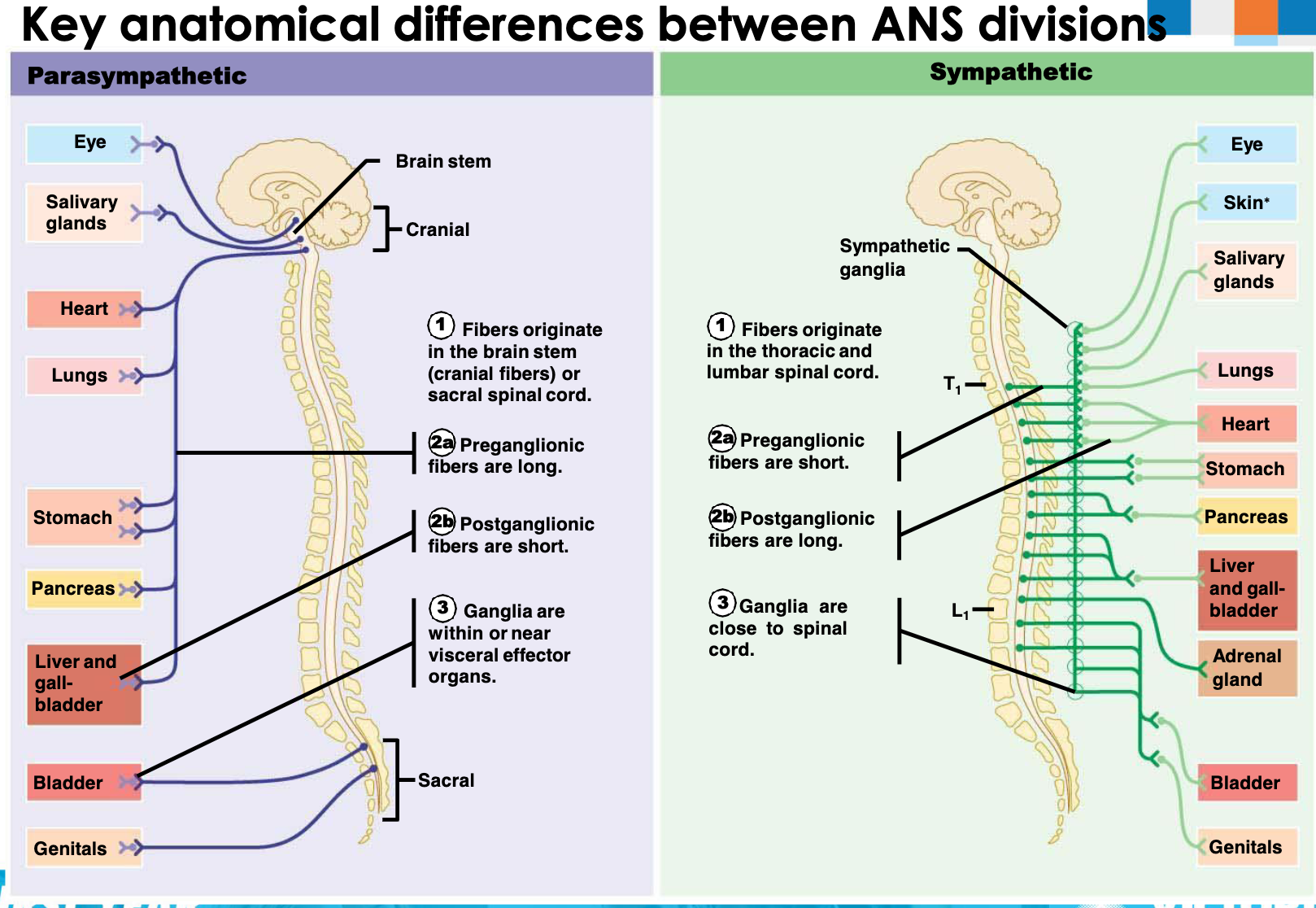
Divisions of the ANS
Feature | Parasympathetic | Sympathetic |
|---|---|---|
Origin of Fibers | Brain (brainstem nuclei of cranial nerves III, VII, IX, X) and sacral spinal cord (S2-S4) | Thoracolumbar region (T1-L2) of the spinal cord |
Length of Fibers | Long preganglionic and short postganglionic fibers | Short preganglionic and long postganglionic fibers |
Location of Ganglia | Very close to or within the visceral effector organs | Close to the spinal cord |
Neurotransmitters | All release Acetylcholine (=cholinergic fibres) | All preganglionic axons release Acetylcholine; most postganglionic =adrenergic |
Functional Role | Performs maintenance activities and conserves body energy | Mobilizes the body during activity and extreme situations |
Sympathetic and Parasympathetic Systems
Parasympathetic postganglionic fibers release acetylcholine
Cholinergic fibers
Sympathetic postganglionic fibers release noradrenaline
Adrenergic fibers
The Adrenal Medulla
The adrenal medulla is a modified part of the sympathetic nervous system
Adrenal medulla secretes catecholamine hormones on stimulation – epinephrine (80%) & norepinephrine (20%)
Dual Innervation
Sympathetic and parasympathetic nervous systems dually innervate most visceral organs
Dual innervation: innervation of a single organ by both branches of the autonomic nervous system
Times of sympathetic dominance: “fight-or-flight” response
Times of parasympathetic dominance: “rest-and-digest” response
Illustration of Autonomic Innervation
Parasympathetic and sympathetic innervation of various organs
Cranial nerves involved: III, VII, IX, X
Spinal nerves involved: S2, S3, S4, L1, L2, T1-T12
Organs include: Nasal mucosa, lacrimal gland, parotid gland, sublingual and submandibular glands, eye, trachea, lung, heart, liver, stomach, spleen, gall bladder, adrenal gland, spinal nerves, kidney, pancreas, colon, small intestine, rectum, urinary bladder, genitalia
Effects of Autonomic Nervous System on Organs
Organ | Sympathetic Stimulation (Adrenergic Receptors) | Parasympathetic Stimulation |
|---|---|---|
Heart | Increases rate/force (\beta_1) | Decreases rate/force (atria only) |
Blood Vessels | Constricts (\alpha_1) | Dilates vessels of penis/clitoris only |
Digestive Tract | Decreases motility (\alpha2, \beta2), contracts sphincters (\alpha1), inhibits secretions (\alpha2) | Increases motility, relaxes sphincters, stimulates secretions |
Urinary Bladder | Relaxes (\beta_2) | Contracts (emptying) |
Eye | Dilates pupil (\alpha1), far vision (\beta2) | Constricts pupil, near vision |
Liver (glycogen) | Glycogenolysis (\beta_2) | None |
Adipose Cells (fat) | Lipolysis (\beta_2) | None |
Exocrine Pancreas | Inhibits secretion (\alpha_2) | Stimulates secretion |
Sweat Glands | Stimulates (cholinergic, \,alpha_1) | None |
Salivary Glands | Small volume, thick saliva (\alpha_1) | Large volume, watery saliva |
Adrenal Medulla | Epinephrine/norepinephrine (cholinergic) | None |
Endocrine Pancreas | Inhibits insulin, stimulates glucagon (\alpha_2) | Stimulates insulin and glucagon |
Genitals | Ejaculation/orgasm (\alpha_1) | Erection |
Brain Activity | Increases alertness | None |
Receptors of the ANS
Several receptor types are available for each autonomic neurotransmitter:
Cholinergic receptors: nicotinic and muscarinic receptors
Adrenergic receptors: alpha and beta receptors
Autonomic agonists and antagonists: agonist binds to the neurotransmitter’s receptor and an antagonist binds with the receptor