Week 6 Scrotum and Testes Part 1
Learning Objectives
Identify and describe anatomical structures of the scrotum and testes, including the spermatic cord.
Describe the normal sonographic appearance of the scrotum, testes, and spermatic cord.
Anatomy Overview
Structures Involved:
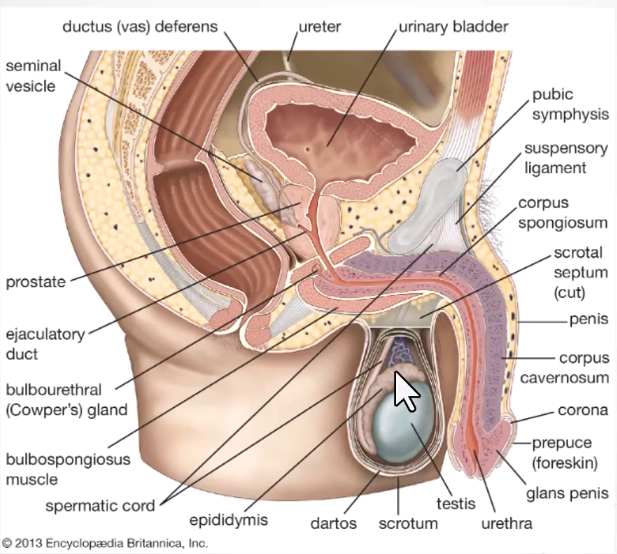
Scrotum
Description:
Dual chambered protrusion of skin and muscle containing the testes, epididymis, and spermatic cord.
Located between the penis and anus, extending from the perineum.
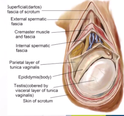
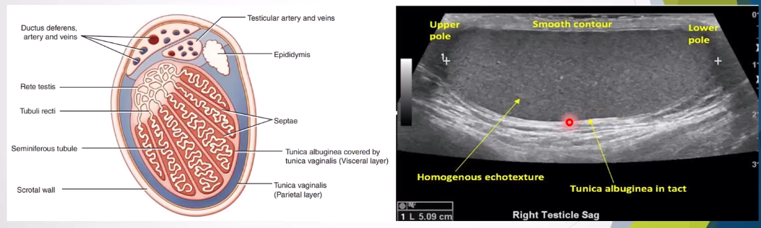
Wall Structure:
Composed of several layers (2-8 mm thick):
Superficial to deep:
Pigmented skin with rugal folds
Superficial fascia and dartos muscle
External spermatic fascia
Cremasteric fascia
Internal spermatic fascia
Tunica vaginalis
Tunica albuginea (visceral layer covers testes)
Scrotal Contents:
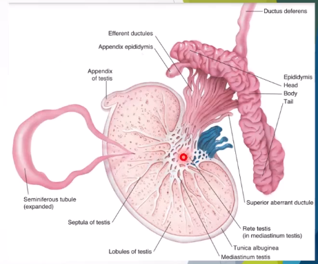
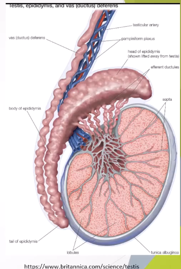
Testes
Epididymis
Testicular and epididymal appendages
Distal part of spermatic cord
Arterial Supply:
Posterior scrotal branches of the perineal artery
Anterior scrotal branches of the deep external pudendal artery
Cremasteric artery
Venous Drainage:
Accompanied by scrotal veins
Testes
Function: Production of sperm and testosterone.
Anatomical Details:
Paired ovoid shape measuring approximately:
Length:
Anteroposterior (AP):
Transverse (TR):
Volume:
Size decreases with age.
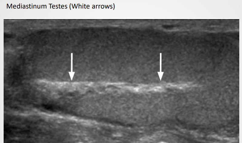
Spermatozoa produced by seminiferous tubules, transported to rete testes in mediastinum.
Efferent tubules convey sperm to epididymis for storage.
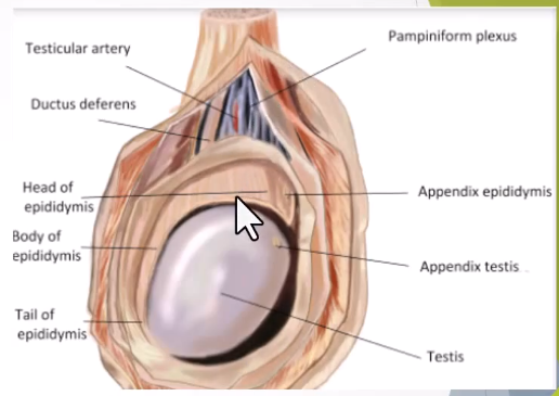
Appendix testis represents developmental remnants of the mullerian duct.
Layers Covering Testes:
Tunica Vaginalis:
Covers anterior surface and sides, derived from peritoneum with lubricating fluid.
Tunica Albuginea:
The fibrous capsule that protects the testes.
Arterial Supply:
Supplied through testicular arteries and branches of the cremasteric artery.
Venous Drainage:
Via pampiniform plexus; left testicular vein drains into left renal vein, while right drains directly to inferior vena cava (IVC)
Embryology
Development:
Testes arise from the gonadal ridge near mesonephric ridge of intermediate cell mass.
Week 8: Testis attaches to lower abdominal wall; descent starts with the processus vaginalis forming.
Fetal Development Stages:
4th month: Testes near deep inguinal ring.
7th month: Testes within the deep inguinal ring.
Following weeks: Descent into scrotum.
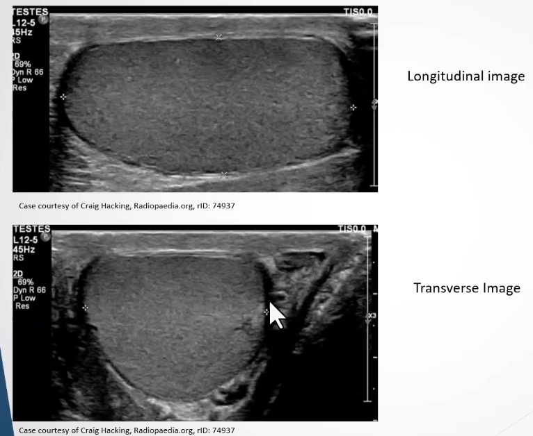
Ultrasound Appearance
Testes:
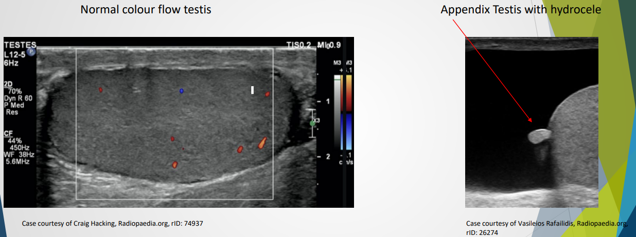
Medium homogeneous appearance.
Tunica albuginea visible as a thin echogenic line surroudning the testes
Tunica invaginates to form a linear ehcogenic mediastinum testis
Rete testis may appear as a hypoechoic region near mediastinum and easily visible if dilated
Appnedix testis can be seen if combined with a hydrocele
Epididymis:
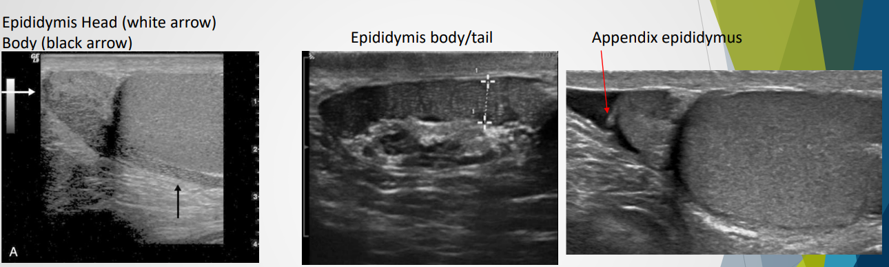
Size: 6-7 cm, structure divided into head, body, and tail.
Begins superiorly and runs posterolateral to the testes
Head: superior to upper of testes, contains 10 - 15 efferent ductucles from the rete testis which forms a single duct in body and tail. This duct becomes the vas deferens and moves into the spermatic cord
Body: smaller than the head and follows the posterorlateral aspect of the testes from upper to lower pole
Tail: slightly larger than the body and sits posterior to the lower portion of testes
Appendix of the epididymis: small protuberance from the head
Ultrasound appearance: similiar or hypoechoic to testes; coarse echotexture
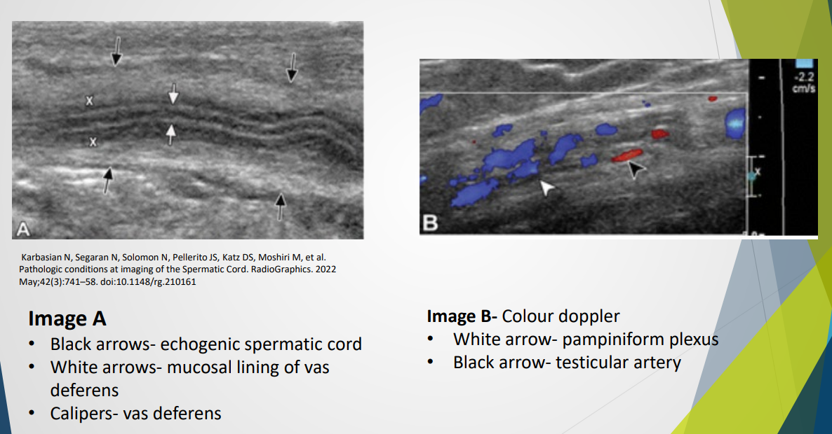
Spermatic Cord:
Contains various structures:
Ductus deferens, testicular artery, pampiniform plexus, lymphatics, nerves.
Appears echogenic on ultrasound; color Doppler shows testicular artery and plexus.