Homeostasis - An Introduction to the Kidney
Homeostasis
“A constant internal environment is a necessary condition for life under varying external conditions”
Importance of maintaining fluid around cells within narrow limits to ensure proper body function.
Small fluctuations can disrupt biochemical activities; larger fluctuations can result in cell death.
Maintenance of a constant internal environment - Homeostasis
Greek for 'staying the same.'
Key Features of Internal Environment
Chemical composition: Ions, glucose.
Blood pH: 7.35 to 7.45
CO2 + H2O ⇌ H2CO3 ⇌ H+ + HCO3-
Osmotic pressure: Osmoregulation.
Temperature regulation:
Endotherms (birds, mammals) maintain temperature.
Ectotherms (most fish, amphibians) not regulated tempreture
Temperature Regulation Mechanism
regional heterothermy - tuna
some parts for swimming are warmer than other parts
preflight warm up - moths/bees
start shivering to get energy and warm, them up
Behavioural regulation in an ectotherm - lizard
radiates heat on rocks
regulation and conforming
river otter - regulator
largemouth bath - conformer
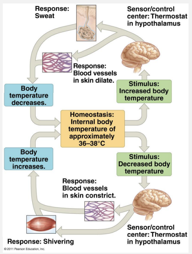
Control System
Stimulus: Temperature change.
Receptors: Skin and hypothalamus.
Control Centre - set point: Hypothalamus.
Effectors: Skin blood vessels, sweat glands, hair (fur), muscles.
Responses: Constriction/dilation of blood vessels, sweat production, piloerection, shivering thermogenesis.
Osmosis and Solutions
Hyperosmotic Solution
More solutes than other solution - less water.
Hypo-osmotic Solution
Less solutes than other solution - more water.
Water moves by osmosis from low to high concentration
into a hyperosmotic solution, out of a hypo-osmotic solution
A solution with few solutes in it will have lower osmoticpressure than one withmany solutes
Osmolarity : Number of osmoles of solute per liter of solution.
analogy - squash in high concentrations has more solute, less solution and less water = hyper. squash in low concentrations has less solute, more solution, more water = hypo.
Water will move from a low to a high osmotic pressure solution (opposite)
Functions of the Kidney
Nitrogenous waste removal: Metabolism of proteins/nucleic acids.
Water content regulation: Osmoregulation.
Salt balance regulation: Concentrations of Na+, K+, Cl-, Ca2+, Mg2+ (ionic regulation).
Nitrogenous Waste Products
Ammonia (NH3): Highly toxic waste product.
Urea: Less toxic, excreted mostly by mammals.
Uric Acid: Very low toxicity, excreted by birds and reptiles.
Comparison of Nitrogenous Wastes
Waste Type | Toxicity | Solubility (ml/g N) | Metabolic Cost | Examples |
Ammonia | High | 500 | None | Fish, Aquatic Amphibians |
Urea | Low | 50 | Some | Mammals |
Uric Acid | Very Low | 1 | High | Birds, Terrestrial Reptiles |
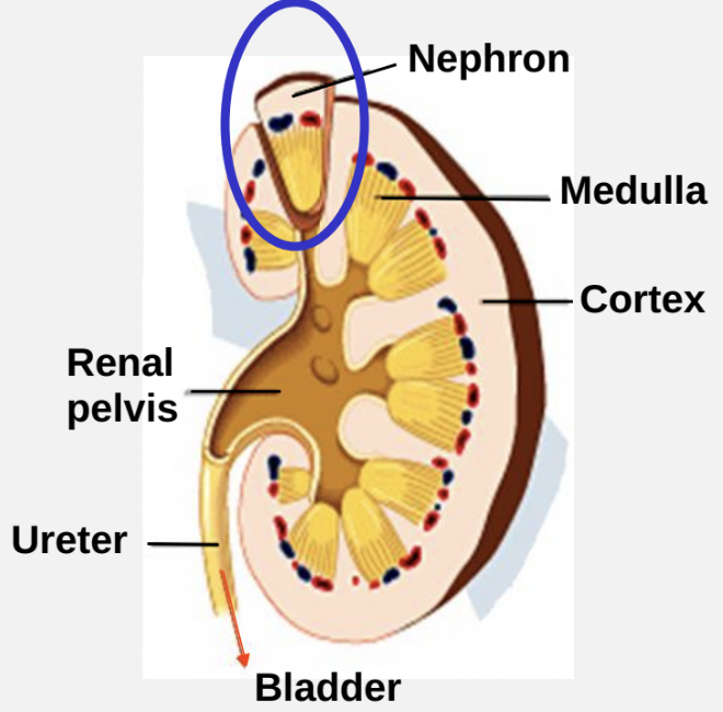
Structure of the Kidneys
Two bean-shaped organs, each the size of a clenched fist, located against the back wall of the abdomen.
Kidneys comprise only 1% of body weight but receive 25% of cardiac output (1.25 liters/min).
Control chemical composition of blood.
connects to bladder with ureters which prevent urine from reentering the kidney
Kidney Anatomy
Parts: Renal pelvis, ureter, cortex, medulla.
Nephron: Over 2 million nephrons per kidney; approximately 60 km in total length.
Nephron Structure
Two types of nephron
Juxta-medullary (concentrated urine)
cortical (less concentrated urine).
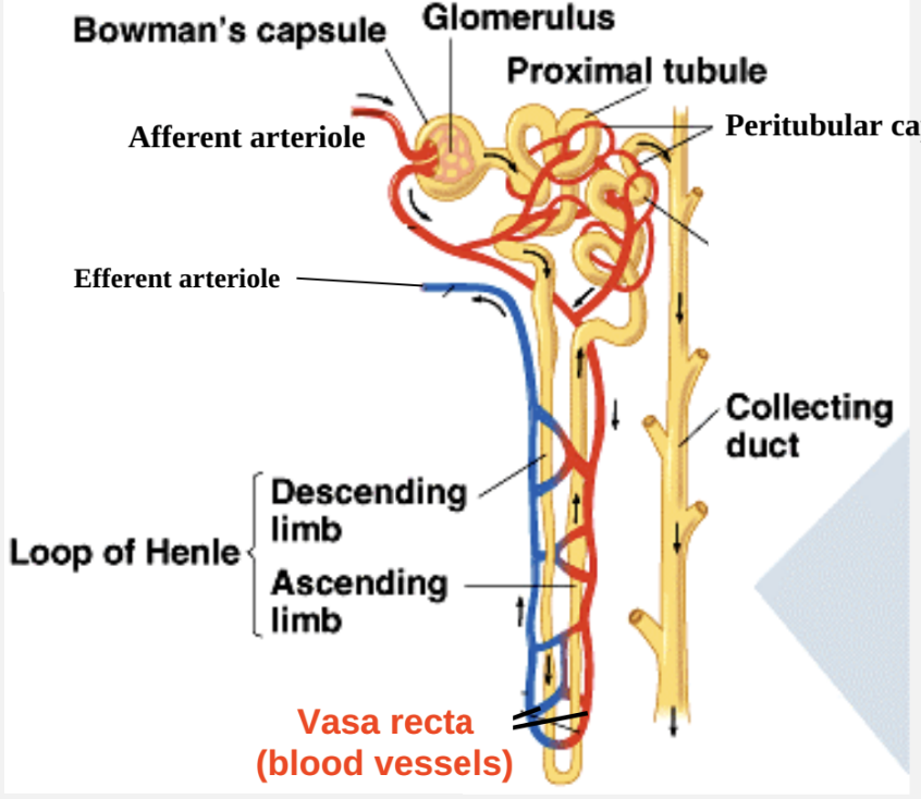
Key Components:
Vasa recta (blood vessels)
Afferent arteriole
Efferent arteriole
Peritubular capillaries.
85% of nephrons are cortical; 15% are juxta-medullary (only found in birds and mammals for concentrated urine).
Bowman’s Capsule
Fluid moves via ultrafiltration.
Hydrostatic pressure of blood plasma forces fluid and solutes through glomerular capillaries into Bowman’s capsule.
The process is passive (no energy needed).
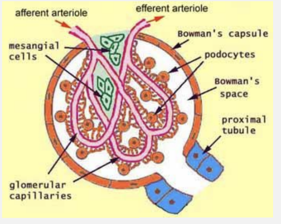
Substances Passing Through Bowman’s Capsule
Molecules <1.8 nm filter freely (water, glucose, urea, amino acids).
Molecules between 1.8 – 4.2 nm filter but less freely.
Molecules >4.2 nm do not pass (e.g., proteins).
Filtration Mechanism and Forces
Hydrostatic Force: Blood pressure in glomerular capillaries is high due to:
Low resistance input pathway (large diameter arteries).
Constriction of arteriole increases pressure.
High resistance due to numerous thin capillaries.
Promoting Filtration:
Glomerular hydrostatic pressure = 55 mm Hg (promotes fluid movement out of plasma and into Bowman’s capsule).
Opposing Filtration:
Capsular hydrostatic pressure = 15 mm Hg (resists filtration).
Glomerular colloid osmotic pressure = 30 mm Hg (osmotic pressure in capillaries).
Net Filtration Pressure: 10 mm Hg (calculated as 55 - 15 = 40, 40-30 = 10).