thorax vi
The outermost layer of the serous pericardium is termed the ________.
A) parietal pericardium
B) fibrous pericardium
C) visceral pericardium
D) endocardium
E) epicardium
Prominent muscular ridges, which extend along the inner surface of the right auricle, across the adjacent anterior wall of the right atrium are called ________.
A) moderator bands
B) pectinate muscles
C) trabeculae carneae
D) papillary muscles
E) chordae tendineae
The fibrous skeleton reinforces the valves and helps prevent overexpansion of the heart.
TRUE
Since the heart is rotated slightly to the left, the anterior surface or sternocostal surface, consists primarily of both atria.
FALSE
The right lymphatic duct ________.
A) carries lymph to the right brachiocephalic vein
B) carries lymph originating in tissues superior to the diaphragm on the right side of the body
C) carries lymph from tissues inferior to the diaphragm
D) varies very little from individual to individual
E) empties into the axillary vein
The thymus produces thymosin, which is the name originally given to a thymic extract that promoted the development and maturation of lymphocytes and thus increased the effectiveness of the immune system.
TRUE
On the right side, the roots of the lungs are located anterior to which vertebra?
A) T5
B) T7
C) C5
D) T3
E) C7
The ________ are a series of irregular muscular folds formed on the internal surface of the ventricles.
A) trabeculae carneae
B) papillary muscles
C) chordae tendineae
D) moderator bands
E) pectinate muscles
The myocardium consists of multiple, interlocking layers of cardiac muscle tissue, with associated connective tissue, blood vessels, and nerves.
TRUE
The major branches of the right coronary artery are the atrial branches, which supply the right atrium and a portion of the left atrium; the ventricular branches, which supply the interventricular septum and adjacent portions of the ventricles; and the branches to the conducting system, which supply the SA node and the AV node.
FALSE
The medulla of the thymus contains ________.
A) thymic corpuscles
B) B cells
C) macrophages
D) granulocytes
E) lymphoid stem cells
Preganglionic neurons in the thoracolumbar division of the ANS are located in the ________ of spinal segments T1-L2.
A) dorsal white columns
B) lateral gray horns
C) dorsal root ganglia
D) ventral roots
E) anterior gray horns
The most medial posterior structure(s) observed in a cross section at the level of vertebra T12 is/are the ________.
A) transverse colon
B) latissimus dorsi muscles
C) spinalis thoracis muscle
D) T12- L1 intervertebral discs
E) longissimus thoracis muscle
Which of the following is/are the largest and most easily recognized structure(s) on the lateral portion(s) of a cross-section at the level of the middle thoracic vertebra (T8)?
A) heart
B) lungs
C) esophagus
D) trachea
E) latissimus dorsi muscles
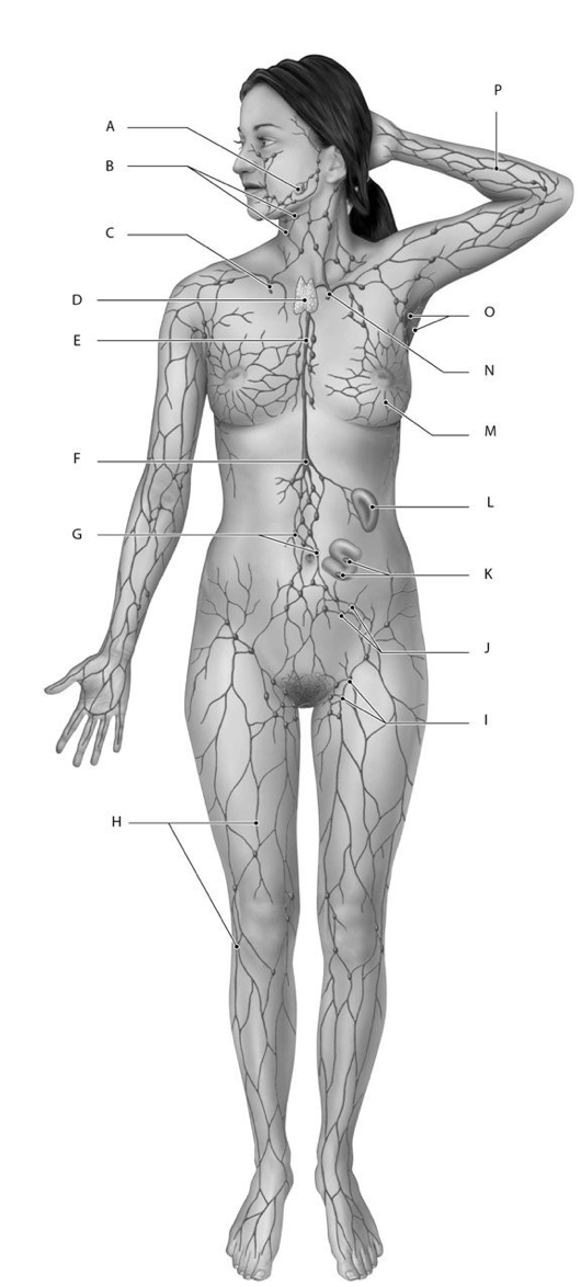
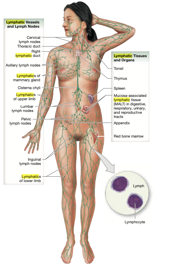
Identify the structure(s) indicated by Label C.
A) Spleen
B) Right lymphatic duct
C) Tonsil
D) Cisterna chyli
E) Thymus
Identify the structure(s) indicated by Label D.
A) Right lymphatic duct
B) Cisterna chyli
C) Thymus
D) Tonsil
E) Spleen
Identify the structure(s) indicated by Label E.
A) Lumbar lymph nodes
B) Cervical lymph nodes
C) Right lymphatic duct
D) Cisterna chyli
E) Thoracic duct
Identify the structure(s) indicated by Label N.
A) Cervical lymph nodes
B) Cisterna chyli
C) Thoracic (left lymphatic) duct
D) Lumbar lymph nodes
E) Right lymphatic duct
Identify the structure(s) indicated by Label O.
A) Right lymphatic duct
B) Lumbar lymph node
C) Cisterna chyli
D) Cervical lymph nodes
E) Axillary lymph nodes
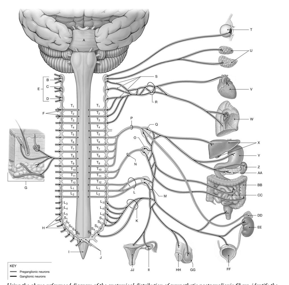
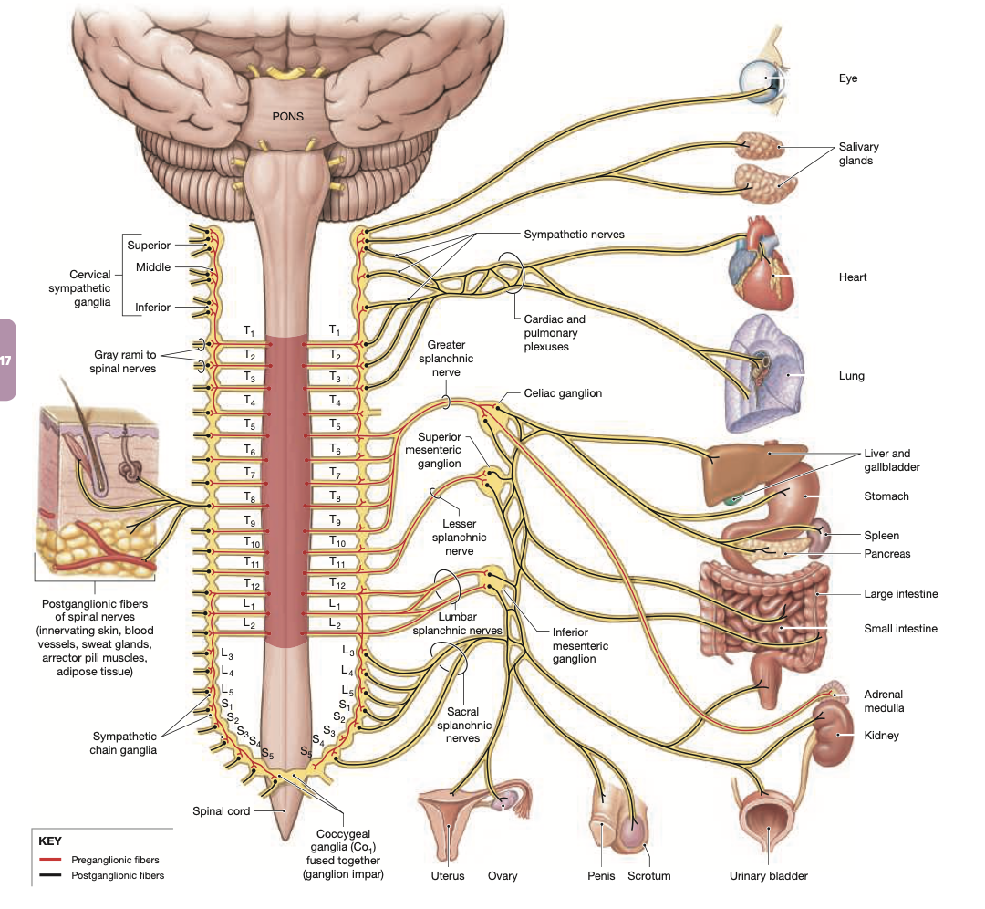
Identify the structure(s) indicated by Label R.
A) Cardiac and pulmonary plexuses
B) Lumbar and cardiac plexuses
C) Esophageal plexus
D) Pulmonary and sacral plexuses
E) Lumbar and sacral plexuses
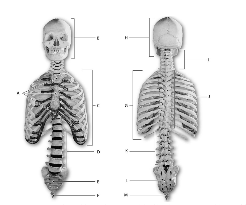
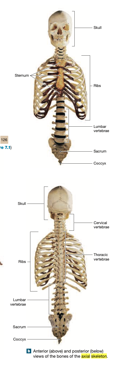
Identify the structure indicated by Label A.
A) Cranium
B) Ribs
C) Clavicle
D) Sternum
E) Skull
Identify the structure indicated by Label C.
A) Thoracic vertebrae
B) Scapula
C) Clavicle
D) Cranium
E) Ribs