Muscular System
Myosin and Actin
Myosin comprise a family of ATP-dependent motor proteins. They’re known for their role in muscle contraction and their involvement in a wide range of other motility processes
- Myosin II (has two heads) is an ATPase involved in actin-based motility (muscle contraction)
Contraction of muscle
- Myosin (thick filaments) is bound to the actin filament. ATP then binds to myosin “head” and myosin releases actin (thin filaments)
- ATP hydrolyzes (→ ATP+Pi+energy). This ‘cocks’ the myosin protein to high energy conformation (loads the spring)
- Phosphate group is released from myosin which releases the energy of the cocked position and causes it to push on the actin filament, it releases the spring as a power stroke that creates mechanical energy
- ADP is released, and myosin is still bound to the actin, but one stroke further along the actin filament. Chemical energy has turn into mechanical energy
- This action contracts the muscle cell, and through the synchronous process in many muscle cells, contracts the entire muscle
Tropomyosin and Troponin and Muscle Contraction Regulation
How do we stop movement of myosin along the actin when we don’t want to contract our muscle?
- Tropomyosin and troponin
Tropomyosin protein coils around the actin, and its attached by a protein complex called troponin
- when a muscle is contracting, tropomyosin keeps the myosin from crawling up the actin
- it blocks the myosin from being able to attach to the acting in its usual place OR if myosin is already bound, tropomyosin keeps it from moving and walking up the actin
- The only way to make the troponin unlock myosin is for the troponin to change its shape which only happens when there is a high concentration of calcium ions in the cell
- calcium binds to troponin and changes it conformation enough that tropomyosin is moved out of the way and myosin can bind and walk up the actin (contraction)
- if calcium concentrations get low, the troponin will go back to the standard conformation and makes the tropomyosin block the myosin again so contraction doesn’t happen (relaxation)
Sarcoplasmic Reticulum
The sarcoplasmic reticulum allows the nervous system to tell the cells to change calcium concentrations
The membrane of the muscle cell is the sarcolemma, and in it is a fold called the T-tubule
- inside of the muscle cell is an organelle called the sarcoplasmic reticulum, whose function is purely storage. ATP-fueled channels on the SR pump in calcium, so in a resting muscle, you have a very high concentration of calcium within the SR
- a protein complex connects the T-tubule to the sarcoplasmic reticulum
- When a muscle is contracting, the SR will release the calcium into the cell (where actin, myosin, etc. are)
- Motor neuron synapses allow SR to know when to release the calcium
- Motor neuron synapses on a muscle cell
- neuron is synapsed, sending an action potential down its axon, which eventually leads to an axon terminal where acetylcholine (in this case for the muscle cell) which causes membrane depolarization and action potential across the cell due to the opening of gated ion channels
- eventually the action potential travels along the membrane and eventually leads to the t-tubule
- when it reaches the t-tubule, the protein complex triggers all the calcium ions to be dumped from the SR into the cell, which causes calcium to bind to troponin and change the complex conformation to allow for contraction
- once the signal is over, the door releases the calcium closes and the SR gains back all its calcium
Anatomy of the Muscle Cell
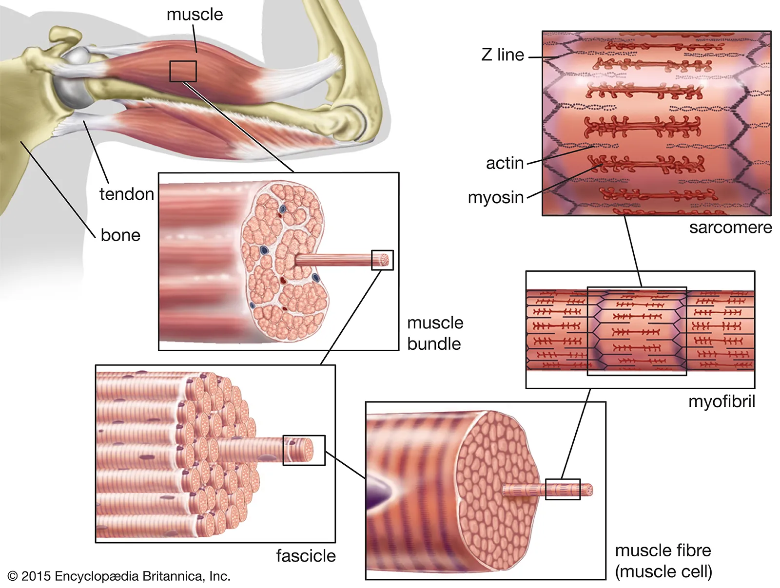
Tendons on either side of the muscle anchors it to bone. Tendons are just a type of connective tissue, and its somewhat continuous with the connective tissue that forms the outer layer of the muscle, the epimysium (which protects the muscles)
A second layer of connective tissue, called the perimysium, is right under the protective layer which covers the subunits of muscle, including the fascicles (fasciculus)
- within each fascicle there’s another layer of connective tissue called the endomysium, which covers individual muscle cells (myofibers)
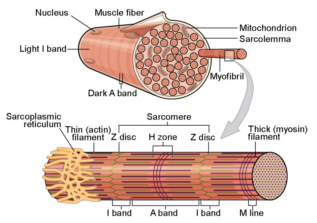
The myofiber has bumps on the outside, which are where the nuclei sit, on the periphery of the cell.
- the cytoplasm of the myofiber is called the sarcoplasm
Within the myofiber is a further division called a myofibril- where the contraction actually occurs
- microscopically on the myofiber there are striations (another name for skeletal muscle is ‘striated’ skeletal muscle).
- These striations are the z-lines or z-disks and the space between the two z-lines is called the sarcomere, the most basic unit of muscle contraction
Actin filaments are anchored to the z-line, and myosin is attached to the actin (myosin is anchored in the sarcomere by the protein titin)
The sarcomere has two different parts: A-bands and I-bands
- A-bands, myosin and actin
- I-bands, SOLELY actin
In contraction, it’s the actin filaments that move, so the z-lines move closer together and towards the center of the sarcomere and the I band is shortened. The A-band remains the same length
3 Types of Muscle
There are three types of muscle- smooth, skeletal, and cardiac- which are involved in basically all movement of the body
- skeletal muscle: most are attached to tendon and bone, but not all are attached to a tendon, or the ‘tendon’ is just a sheet of fibrous tissue called aponeurosis
- voluntary and are the fastest type of muscle
- they’re also straight and have many nuclei that show up as bumps (on the periphery) of the cells
- striated
- cardiac muscle: in the heart and this is where they can only be found.
- involuntary
- branched (which makes them easy to spot) and have 1-2 nuclei in the middle of the cell
- striated
- smooth muscle: largely found in the walls of hollow organs (stomach, bowels, etc.) and blood vessels
- involuntary, slowest muscle
- often described as ‘spindle shaped’ smooth muscle cells just have one nucleus
- not striated
Type 1 and Type 2 Muscle Fibers
Golden rule: mitochondria are more prevalent in type 1 muscle than type 2
Type 1: what allow marathon runners and long distance cyclists to go for hours at a time
- AKA: red slow twitch, red oxidative
- Speed of contraction: slow
- Force generated: low
- Mitochondria: many
- Capillaries: very dense
- Fatigue resistance: high, hours of use
Type 2 A:
- AKA: intermediate, fast twitch oxidative
- Speed of contraction: intermediate
- Force generated: medium
- Mitochondria: some (aerobic but can switch to anaerobic)
- Capillaries: Medium
- Fatigue resistance: Medium, 30 minutes of use
Type 2 B: what jump shots/pole vaults need- explosive force
- AKA: white fast twitch
- Speed of contraction: very fast
- Force generated: high
- Mitochondria: few (anaerobic)
- Capillaries: very few
- Fatigue resistance: low, 1 minute of use
Muscle Innervation
Voluntary contractions are those of skeletal muscle
- controlled by me so I use cerebral cortex or the spinal cord
Involuntary contractions include those of cardiac and smooth muscles
- these are beyond me, so I use the brainstem, or ganglia beside the spinal cord
- the brainstem is responsible for involuntary contractions through the sympathetic or parasympathetic mechanism. The sympathetic ganglia (cell body/soma of neurons that sit outside the brain and spinal cord) are involved in involuntary contractions
Thermoregulation
When the skin perceives that its hot outside, it will send a neuronal signal to the hypothalamus in the brain
- the anterior part of the hypothalamus responds to hot temperatures
- the posterior part of the hypothalamus responds to cold temperatures
the brain then sends a signal back to the smooth muscles and skeletal muscles as needed to help us maintain core body temperatures
- in hot environments, smooth muscle relaxes and vasodilates the arterioles. This allows the blood flow to the skin to increase which allows us to dissipate heat
- in cold environments, smooth muscle will contract and vasoconstrict the arterioles. This shrinks the heat filled blood vessels away from the skin so less heat is lost
- our skeletal muscles (ones in the core) shiver when we’re cold.
- this is because when skeletal muscles contract, an exothermic reaction is created to releases energy in the form of heat