OLD Corneal Dystrophies and Degenerations
Overview
Dystrophies: Inherited conditions present at birth or arising later in life.
Degenerations: Changes resulting from external factors (noxious influences) like age, nutrition, trauma, or post-inflammatory processes.
Genetic Inheritance Patterns
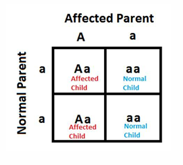
Autosomal Dominance:
Typically, at least one parent is affected.
Each child of an affected person has a 50% chance of inheriting the condition, regardless of gender.
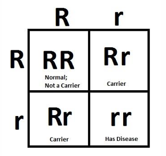
Autosomal Recessive:
Both parents are usually carriers but clinically normal.
Each child has a 25% chance of being affected, a 50% chance of being a carrier, and a 25% chance of being completely normal, irrespective of gender.
Consanguineous marriages (e.g., between first cousins) increase the likelihood of this inheritance pattern.
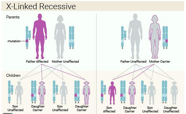
X-linked Recessive:
Typically, the female carries the gene and has a 50% chance of passing it to her sons (who will be affected) on average.
Daughters have a 50% chance of becoming carriers on average.
Father-to-son inheritance does not occur because the Y chromosome is passed from father to son, and the disease gene is located on the X chromosome.
All daughters of an affected male will be carriers.
Importance of Slit Lamp Examination
Some corneal conditions can be very obvious under slit lamp examination, while others are subtle.
Proficiency in slit lamp examination is crucial for accurate diagnosis.
Corneal Degeneration
first three conditions don’t really have any symptoms and no treatment is required.
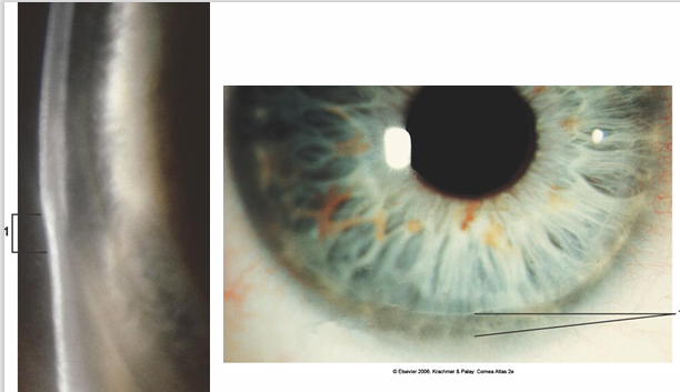
Corneal Arcus
Description: Common age-related lipid degeneration of the peripheral cornea. Occurs in stromal layer
Signs:
Bilateral condition.
Lipid deposits initially in the inferior cornea, then superior, and eventually circumferential.
Appears as a roughly 1mm wide whitish band in the peripheral cornea.
A clear interval exists between the arcus and the limbus (lucid zone or lucid interval of Vogt).
Possible mild non-progressive thinning of the clear marginal zone.
Sharp outer outline with a diffuse central boundary.
Symptoms: Usually asymptomatic, requiring no treatment.
Other Considerations:
Association with raised serum lipids and cholesterol, especially in younger patients (under 50 years old).
Unilateral arcus may suggest carotid disease on the unaffected side.
both situations require referral.
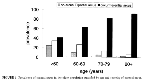
Blue Mountains Eye Study:
Study of 3654 non-institutionalized residents aged 49 years or older.
Included fasting blood samples for measuring total cholesterol, high-density lipoprotein cholesterol, and triglycerides.
Arcus was defined as absent, partial, or circumferential.
Association with Lipid Levels: do not remember numbers.
assessed associated between arcus and hyperlipidaemia, with adjustments for age, sex, hypertension, diabetes and smoking
Arcus (either partial or circumferential) is associated with 60% increased odds for total cholesterol levels > 6.0 mmol/l (adjusted OR 1.6, 95% CI 1.1 – 2.3).
Absence of arcus is associated with reduced odds for total cholesterol > 6.0 mmol/l (OR 0.6, 95% CI 0.5 - 0.9) and for triglycerides > 3.0 mmol/l (OR 0.5, 95% CI 0.3 – 0.9).
If you have a partial arcus, then you have up to 2.3 times greater risk of having cholesterol above or equal to 8.
If you have a circumferential arcus, then up to 4.6 times greater risk of having cholesterol above or equal to 8.
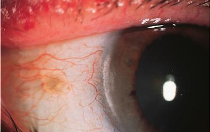
Vogt’s Limbal Girdle
Description: Very common, innocuous age-related condition.
Signs:
Bilateral.
Chalky-white crescentic deposits running in the interpalpebral fissure.
May or may not be separate from the limbus.
Symptoms: Asymptomatic, requiring no treatment.
Other Considerations: Age-related and benign
no systemic association
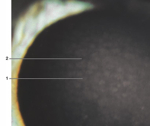
Shagreen (Crocodile Shagreen)
Signs:
Gray-white polygonal stromal opacities separated by clear spaces.
Located in the anterior (generally) or posterior stroma.
Bilateral and symmetrical.
Symptoms: Asymptomatic, requiring no treatment.
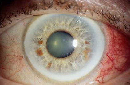
Band Keratopathy
Description: Interpalpebral subepithelial precipitation of calcium salts causing opacities at Bowman's layer, with characteristic black holes.
Signs:
Calcium deposits in the sub-epithelium, Bowman's layer, and anterior stroma.
Interpalpebral plaque (