random
Chapter 12
Introduction:
Shock → not enough oxygen getting to the cells (oxygen supply & oxygen demand are not matching well [imbalanced])
Shock can result from:
Inadequate blood volume → Hypovolemic shock (hemorrhage, dehydration, burns)
Inadequate vascular tone → Distributive shock (septic, anaphylactic, neurogenic — massive vasodilation causes relative hypovolemia)
Ineffective cardiac function → Cardiogenic shock (heart can't pump effectively)
In shock, ALL body systems are affected (not just one organ system)
shock can progress to MODS (multiple organ dysfunction syndrome)
MODS can progress to death
Review of Anatomy & Physiology:
Cardiovascular system: heart, blood, vascular bed
vascular bed: arteries, arterioles, capillaries, venules, veins'
arterioles & venules → deliver O2 & nutrients to cells & removes waste products of cellular metabolism & regulates blood volumes
metarterioles can adjust their diameter & regulate blood flow into capillaries
The cardiovascular system's job is to deliver oxygen to cells. The microcirculation — the tiny vessels between arterioles and venules — is where that delivery actually happens
Here's the path blood takes:
Arteriole → lined with smooth muscle, can change diameter to control how much blood flows forward
Metarteriole → smaller branch off the arteriole, also has some smooth muscle to adjust flow
Precapillary sphincter → a muscular "gate" that controls whether blood enters a capillary bed
Capillary → thin-walled network where oxygen and nutrients leave the blood and enter cells, and waste products come back in
Venule → carries blood away from capillaries; can also dilate or constrict to regulate flow
Key control point: The precapillary sphincter is the only structure that decides whether blood flows into a capillary bed. When it constricts, blood is diverted away from that capillary bed and sent somewhere else that needs oxygen more urgently.
Why capillaries are special:
They're narrow and branched → this increases surface area for exchange and slows blood flow so there's more time for oxygen to diffuse into cells
They have no smooth muscle and can't contract on their own
The precapillary sphincter is the gatekeeper
Why this matters in shock: When the body is in shock, it tries to protect vital organs (brain, heart) by constricting precapillary sphincters in less vital areas (like skin, gut, kidneys). Blood gets shunted away from those tissues. That's why shock patients have cool, pale skin and why prolonged shock leads to organ failure.
After the capillaries: Venules and veinsBlood leaves the capillaries through venules, which can also constrict or dilate to control blood flow — called postcapillary resistance. Then it flows into larger veins and back to the heart.
Arteriovenous anastomoses (shortcuts)
These are direct connections between arterioles and venules that bypass capillaries entirely. When open, blood skips the capillary bed and goes straight through — useful when a tissue doesn't need oxygen right now, or when the body needs to shunt blood quickly to another area.
Oxygen delivery (DO₂) — the formula
For cells to make energy (ATP), they need oxygen delivered. DO₂ depends on three things:
Hemoglobin — carries oxygen
Lung function (SaO₂) — oxygenates the blood
Cardiac output (CO) — pumps the oxygenated blood to tissues
If any of these fail, oxygen delivery drops → cells can't make ATP → lactic acid builds up → organs fail → death.
In shock: DO₂ is impaired, oxygen reserve (SvO₂ → venous oxygen saturation) drops, and tissues become hypoxic.

Pathophysiology of Shock:
Shock occurs when there is an alteration in at least one of these four circulatory components:
blood volume
myocardial contractility
blood flow
vascular resistance
when healthy, these four work together to maintain circulatory homeostasis
when one of these components fail, the others compensate
if more than one fail, shock occurs
Adequate oxygen delivery fails → nutrients decrease → impaired cellular metabolism occurs → cells convert from aerobic to anaerobic metabolism → tissue acidosis → further impair cellular metabolism
Stages of Shock:
poor perfusion → cells can’t get oxygen or glucose → cells can’t make ATP (energy) → worse perfusion
Oxygen pathway:
Cells switch to anaerobic metabolism
ATP drops → sodium-potassium pump fails
Sodium and water flood into cells (edema)
Fluid leaks out of blood vessels → less circulating volume
Lactate builds up → acidosis
Inflammation and cell damage worsen
Glucose pathway:
Body releases stress hormones
Breaks down fat and protein for fuel
More lactate → more acidosis
Muscle and albumin depleted
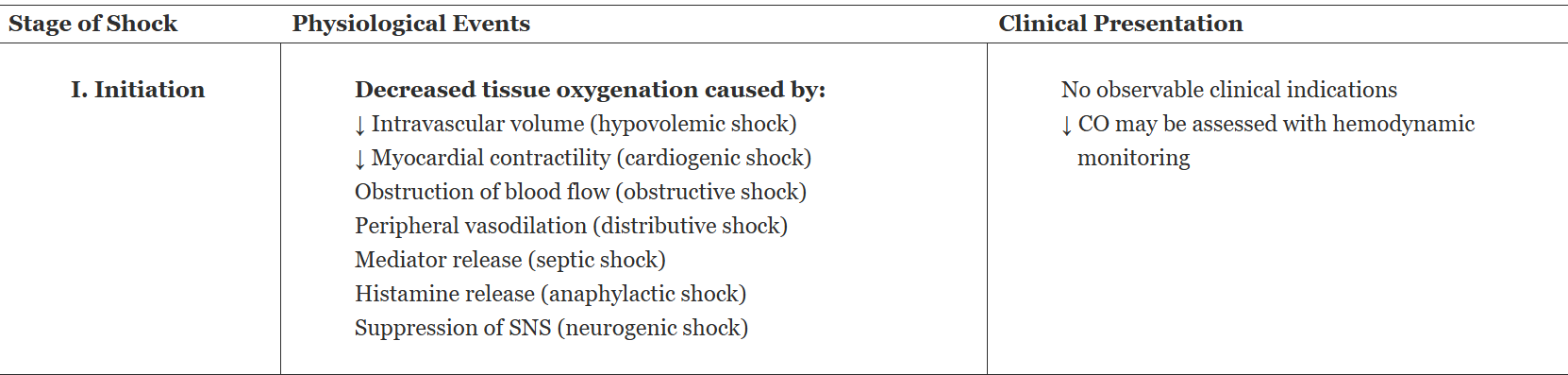
Stage I: Initiation
tissue perfusion begins to drop HOWEVER no obvious signs of this are seen
i.e. decreased cardiac output (can be a slight decline or a steady decline)
Stage II: Compensatory
body senses tissue perfusion dropping & activates 3 systems:
neural compensation
baroreceptors and chemoreceptors sense low BP → sympathetic nervous system fires → catecholamines (epinephrine, norepinephrine) flood the system.
Result: HR and contractility go up, vessels constrict to shunt blood to heart and brain, bronchi dilate, glucose mobilizes
endocrine compensation
hypothalamus → pituitary → releases ACTH (stimulates cortisol and aldosterone) and ADH.
Kidneys sense low pressure → release renin → RAAS cascade kicks in.
Result: sodium and water retention, blood volume increases, vasoconstriction
chemical compensation
chemoreceptors sense low oxygen → respiratory rate and depth increase → hyperventilation blows off CO₂ → respiratory alkalosis → cerebral vasoconstriction 4.
This can worsen brain perfusion even while trying to fix oxygenation
Body is working hard to maintain BP & tissue perfusion to VITAL organs
Clinical symptoms begin to show up: tachycardia, cool skin, decreased urine output
Patient can still recover WITHOUT major organ damage

Stage III: Progressive
the stage where compensation stops helping & starts spiraling towards organ failure
arterioles stay clamped to protect brain & heart = prolonged vasoconstriction causes:
Ischemia in the periphery — extremities cold, pulses weak or gone, skin mottled
Cellular hypoxia → cells switch to anaerobic metabolism → lactate accumulates → metabolic acidosis
LACTIC ACID PRODUCED
ATP drops → sodium-potassium pump fails → sodium and water flood into cells → cellular swelling and dysfunction
The microcirculation does the OPPOSITE — it dilates
This is the key paradox. While the big arterioles stay constricted, the tiny precapillary sphincters relax (because local tissues are screaming for oxygen). But the postcapillary sphincters stay constricted.
Result: Blood flows into the capillary bed easily but can't flow out. It pools there.
Now:
Capillary hydrostatic pressure skyrockets
Fluid gets pushed out into the interstitial space ("third-spacing")
Blood volume in the circulation drops even more
3. Inflammation makes it worse
Hypoxia and acidosis trigger the release of histamine and inflammatory mediators. These:
Increase capillary permeability → proteins leak out through enlarged pores
Capillary oncotic pressure drops (because albumin is gone)
Even more fluid shifts out of vessels
Now the blood gets thick and sludgy (more viscous) because there's less plasma. RBCs, platelets, and proteins clump together — this is capillary sludging. Blood flow slows to a crawl.
4. The heart starts to fail
Less blood returns to the heart (reduced venous return )→ Cardiac output drops
Coronary artery perfusion drops → the heart itself becomes ischemic
The ischemic pancreas releases myocardial depressant factor (MDF)
MDF directly impairs myocardial contractility
Cardiac output, BP, and tissue perfusion spiral downward
Prolonged vasoconstriction → cold, mottled extremities, weak/absent pulses
(You assess this — it tells you shock is progressing)Anaerobic metabolism → lactic acidosis
(Rising lactate = worsening shock. You'll see this on labs and need to interpret it)Third-spacing: fluid shifts out of vessels into interstitial space
(Why fluid boluses may not work as expected — the fluid leaks out)Decreased venous return → decreased cardiac output → BP drops
(The vicious cycle — explains why shock spirals despite treatment)
Why this stage is different:
In Stage II, the body was buying time. In Stage III, those same mechanisms are now destroying the patient. Vasoconstriction that once protected vital organs now causes ischemia everywhere. Capillary pooling that was supposed to be temporary is now permanent. The heart that was compensating is now failing.

Stage IV: Refractory
prolonged poor perfusion → MODS (multiple organ dysfunction syndrome) → death
Blood remains pooled in capillaries → BP too low to perfuse vital organs → therapy doesn't work anymore
What you'll see system by system:
Heart:
Dysrhythmias (pump failure, no ATP, severe acidosis) → cardiac failureLungs:
Capillary + surfactant damage → ARDS (Acute Respiratory Distress Syndrome) → pulmonary edema, V/Q mismatch (ventilation & perfusion mismatch), pulmonary hypertensionBrain:
Ischemia → loss of autoregulation → possible infarction
SNS (sympathetic nervous system) collapses → bradycardia, bradypnea (the flip from tachy to brady = bad sign), can't regulate tempKidneys:
Ischemia → acute tubular necrosis → oliguric/anuric, worsening acidosisLiver:
Can't detoxify, make clotting factors, or mobilize glucose → hypoglycemia, coagulopathyImmune system:
Can't clear bacteria → sepsis riskGlucose problem:
Stress hormones → high blood sugar → you pee out fluid and electrolytes → dehydration
Body uses up all clotting factors → DIC (bleeding and clotting at the same time)
The timeline:
Lungs fail first (12–24 hrs) → clotting issues → kidneys → liver → GI.
Cardiovascular or neuro failure = usually fatal.Bottom line for nursing:
Refractory shock = MODS. You're supporting failing organs (vent, dialysis, pressors, transfusions) while the team decides goals of care.
You just walked through the downward spiral of refractory shock: hypotension → SNS kicks in → vasoconstriction makes things worse → acidosis weakens the heart → brain stops getting oxygen → SNS collapses → vasodilation and bradycardia → death
SNS tries to save you early on (vasoconstriction, tachycardia), but that same response starves your skin, kidneys, and gut. Then when your brain runs out of oxygen, the SNS shuts down completely — and now you get the opposite problem: vasodilation, bradycardia, total cardiovascular collapse

Quiz:
1. The patient arrives at the critical care unit with a diagnosis of pneumonia. He has an increased heart rate, oliguria, and cool, moist skin. A glucose level is obtained, and the patient has hyperglycemia. An accurate history is challenging to obtain, as the patient is restless with intermittent confusion. The patient’s clinical presentation indicates which of the following stages of shock?
A. Initiation
B. Compensatory
C. Progressive
D. Refractory
Answer: B. Compensatory
The patient shows classic signs of compensatory mechanisms at work: increased heart rate, oliguria from renal compensation, cool/moist skin from vasoconstriction, and hyperglycemia from endocrine response — all attempting to maintain perfusion before decompensation occurs.
Systemic Inflammatory Response Syndrome (SIRS):
can result from or lead to MODS
SIRS = body-wide inflammation (not just in one spot)
Lots of different injuries or illnesses can flip the switch: infection, trauma, shock, pancreatitis, ARDS, ischemia
body releases inflammatory mediators (chemical messengers like cytokines) that cause three big problems:
Leaky blood vessels
The walls of your capillaries become permeable (full of holes)
Fluid leaks out of your blood vessels into the tissues
Result: relative hypovolemia — not enough fluid left in the bloodstream, even though total body fluid hasn't changed
Tiny clots everywhere
Mediators trigger clotting in the microcirculation (capillaries)
The body also can't break down clots properly (impaired fibrinolysis)
Result: blood flow gets blocked in tiny vessels → organs don't get oxygen
Widespread vasodilation
Blood vessels relax and get wider
Blood pressure drops
Combined with fluid leaking out, this makes the hypovolemia even worse
Assessment:
Assessment focuses on three areas:
history
clinical presentation
patient’s age or stage of life may alter the clinical presentation
Assessment findings in the initial stages of shock include increased heart rate and respiratory rate; decreased urine output; cool, moist skin; decreased capillary refill; and restlessness or confusion
Later signs of shock include hypotension, dysrhythmia(s), respiratory distress, decreased responsiveness, anuria, and skin mottling
Initial responses to shock include restlessness, agitation, and anxiety. As the shock state progresses, the patient becomes confused and lethargic, then unresponsive if left untreated.
know patient baseline BP
assess the jugular vein
assess capillary refill
assess intake & output
A pulmonary artery (PA) catheter can help diagnose & treat patients in shock
early shock → respirations are rapid & deep
Evaluate skin color, temperature, texture, turgor, and moisture level
skin mottling is a common sign of hypoperfusion
laboratory studies
elevated levels of lactate indicate shock
higher the lactate level the more significant the hypoperfusion
Management:
treat the CAUSE of shock
Care is directed toward correcting the altered circulatory component (e.g., blood volume, myocardial contractility, obstruction, vascular resistance) and reversing tissue hypoxia
Interventions include increasing the cardiac output, hemoglobin level, and arterial oxygen saturation while minimizing oxygen consumption
Resuscitation:
goal = maintain circulating blood volume and an adequate hemoglobin level
The choice of blood or fluid, volume, and rate of infusion depends on the type of fluid lost, the patient’s hemodynamic status, and coexisting conditions
Benefits of IV fluid administration include increased intravascular volume, increased venous return to the right side of the heart, optimal stretching of the ventricles, and increased cardiac output
Fluid administration is adjusted based on changes in blood pressure, urine output, hemodynamic values, diagnostic test results, and the clinical picture of the patient’s response to treatment
If blood pressure remains inadequate after optimal fluid resuscitation, then vasopressors or inotropes are used to achieve blood pressure and cardiac output goals
Chapter 10
Chapter 15