Muscle Physiology
The CNS regulates both voluntary and involuntary efferent branches
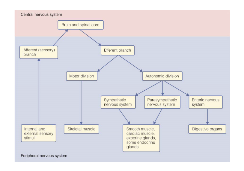
Somatic motor neuron innervate skeletal muscle for voluntary movements
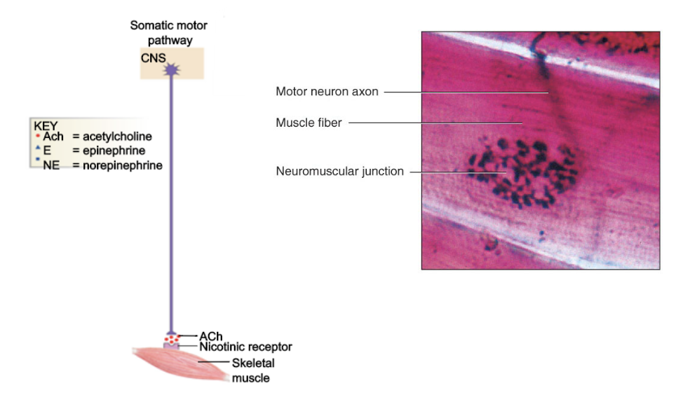
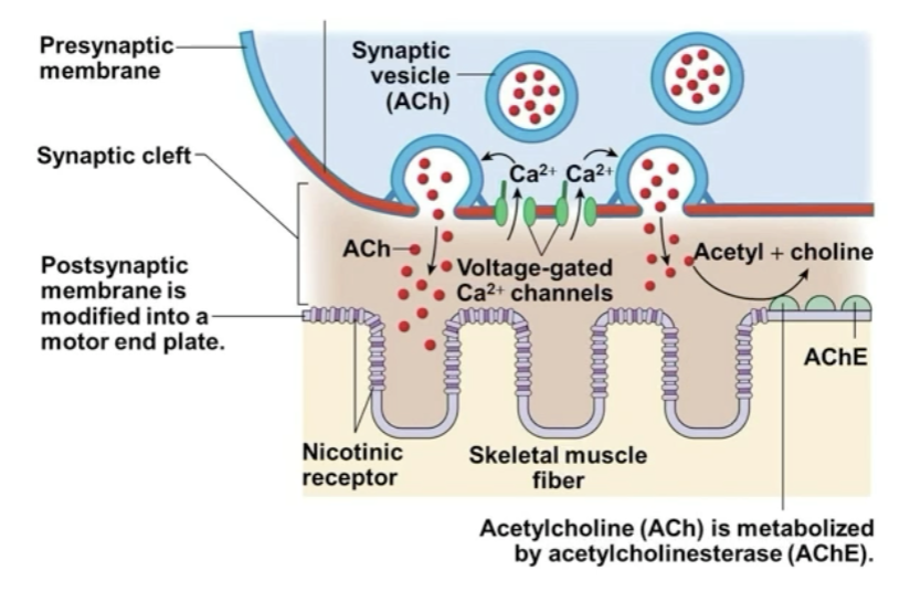
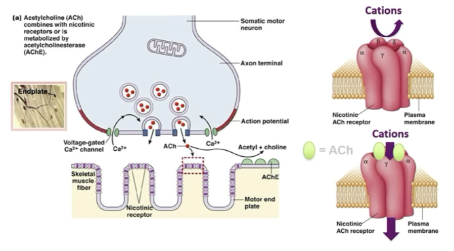
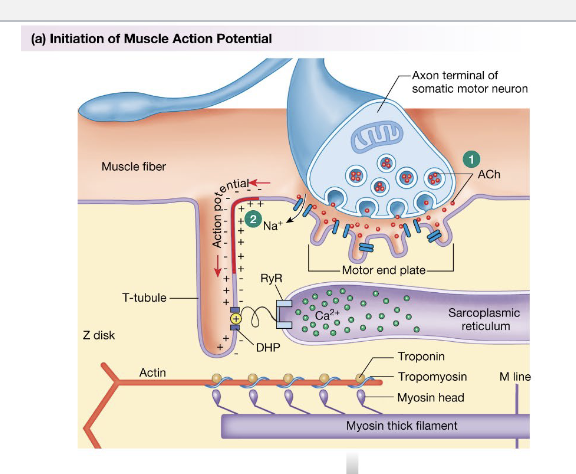
Neurogenic muscle is excited at the neuromuscular junction (NMJ)
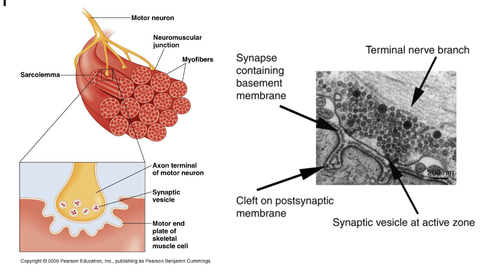
Vertebrate motor neuron release ACh at the NMJ
Structure and characteristics of muscle
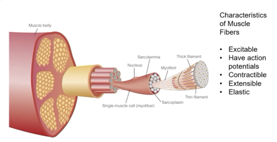
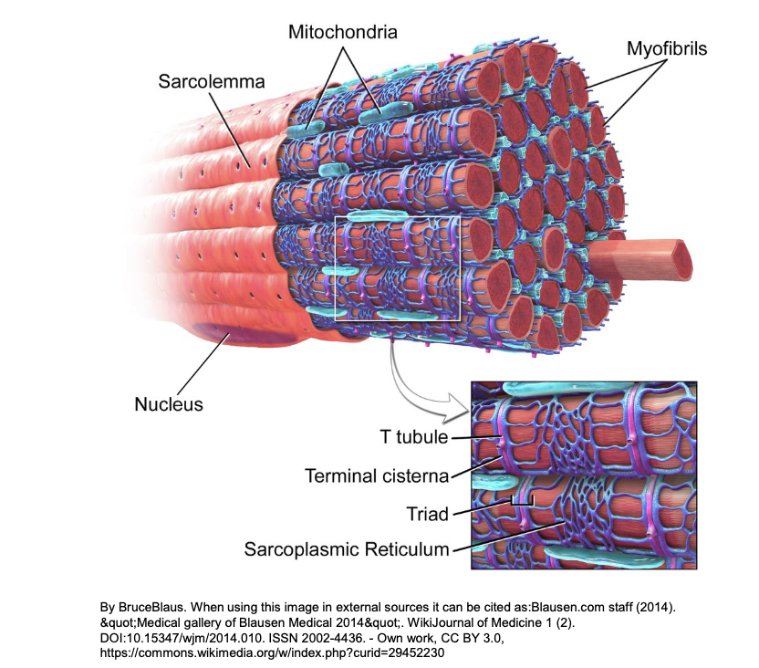
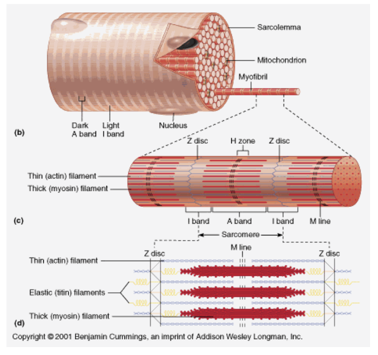
Sarcolemma is the cell membrane of a muscle fiber, essential for muscle contraction, transmitting nerve signals, and regulating the movement of substances in and out of the cell.
Sarcoplasm is the cytoplasm of a muscle cell containing organelles and myofibrils. It plays a crucial role in storing calcium ions for muscle contraction and providing energy for muscle activity.
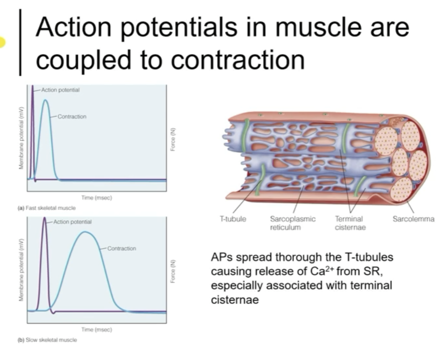
T-tubules are invaginations of the cell membrane in muscle cells, aiding in the conduction of action potentials deep into the cell. They play a crucial role in coordinating muscle contractions by allowing for simultaneous calcium release throughout the cell.
Extension of the sarcolemma that allows APs to spread deeply within the muscle fiber
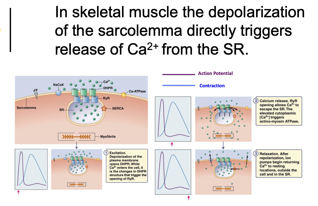
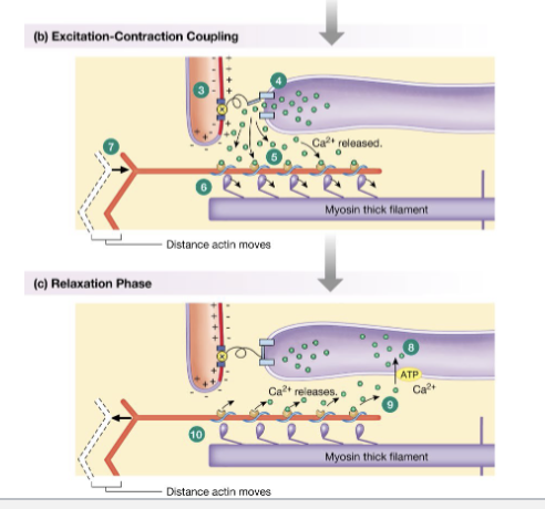
AP spread thorough the T-tubules causing release of Ca2+ from SR, especially associated with terminal cisternae
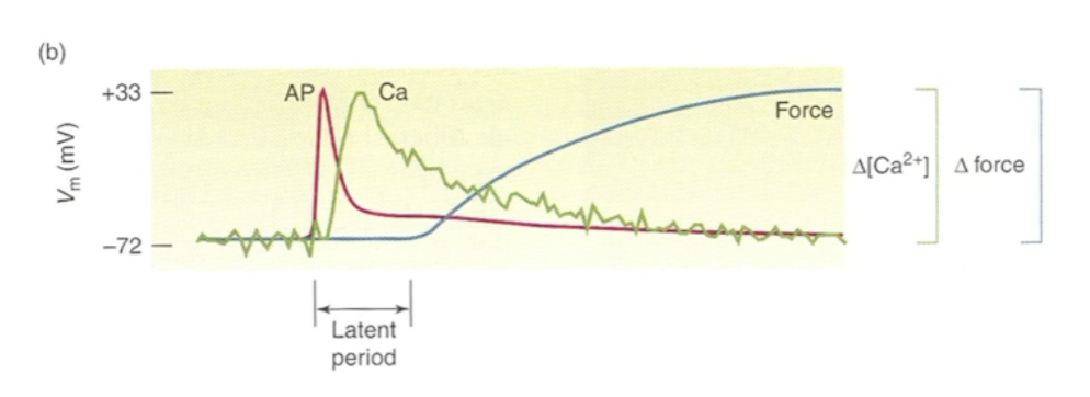
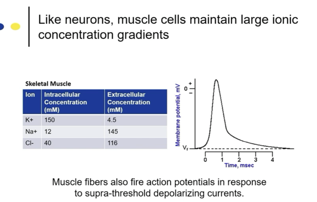
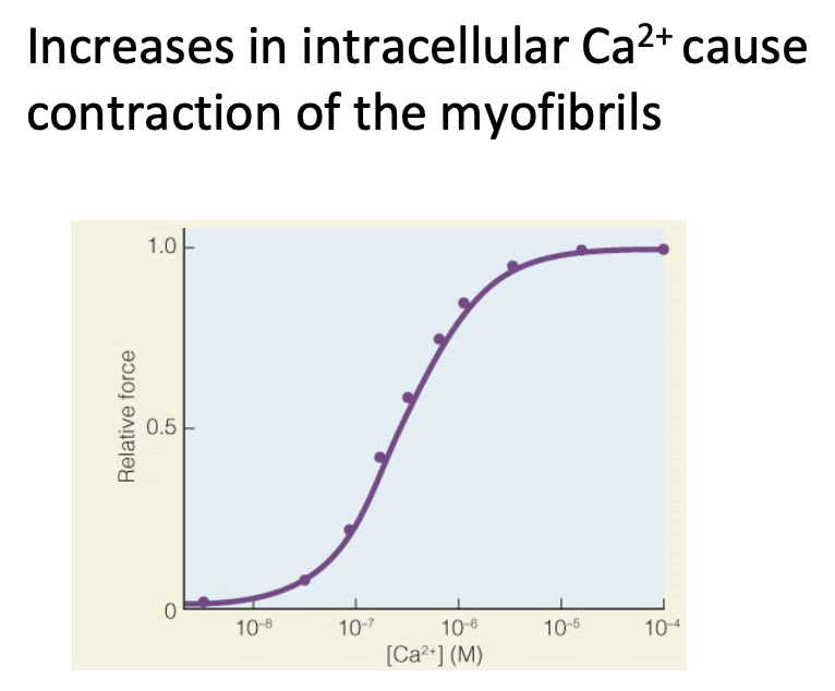
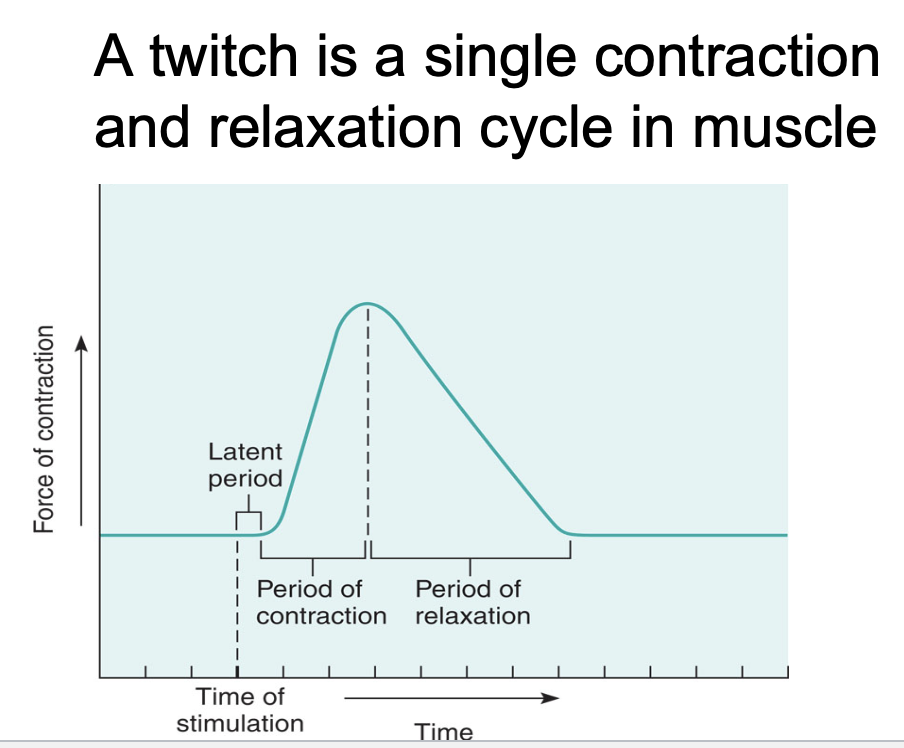
muscle fiber AP is followed by Ca2+ increase and then contraction
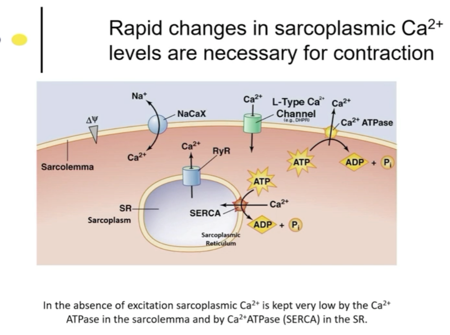
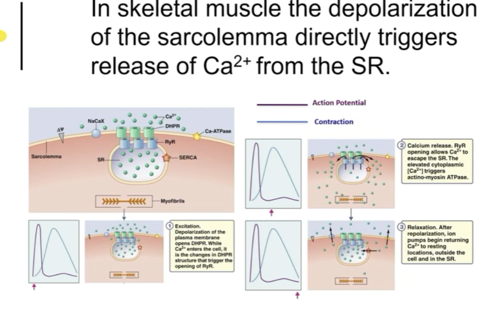
The sarcoplasmic reticulum is an organelle in muscle cells that stores and releases calcium ions necessary for muscle contractions. It plays a crucial role in regulating muscle function by controlling the levels of calcium ions in the cell, which are essential for muscle contraction and relaxation.
Terminal Cisternae: Terminal cisternae are enlarged areas of the sarcoplasmic reticulum in muscle cells where calcium ions are stored. They play a crucial role in muscle contraction by releasing calcium ions when stimulated, allowing for the interaction of actin and myosin filaments.
Sarcomeres are the basic contractile units of muscle fibers composed of actin and myosin filaments. They are crucial for muscle contraction and movement in the body.
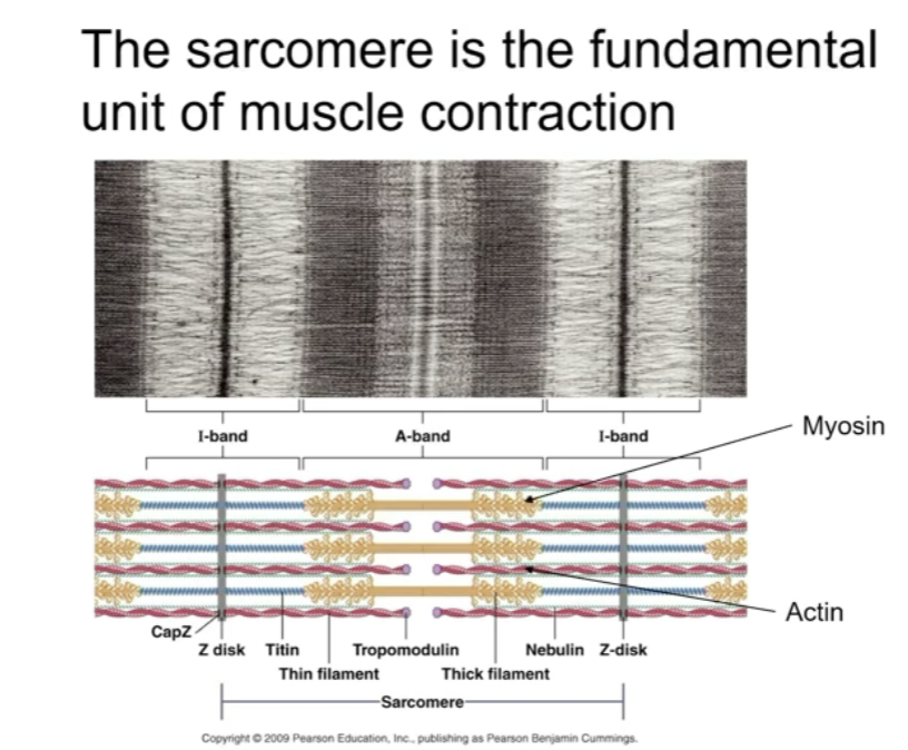
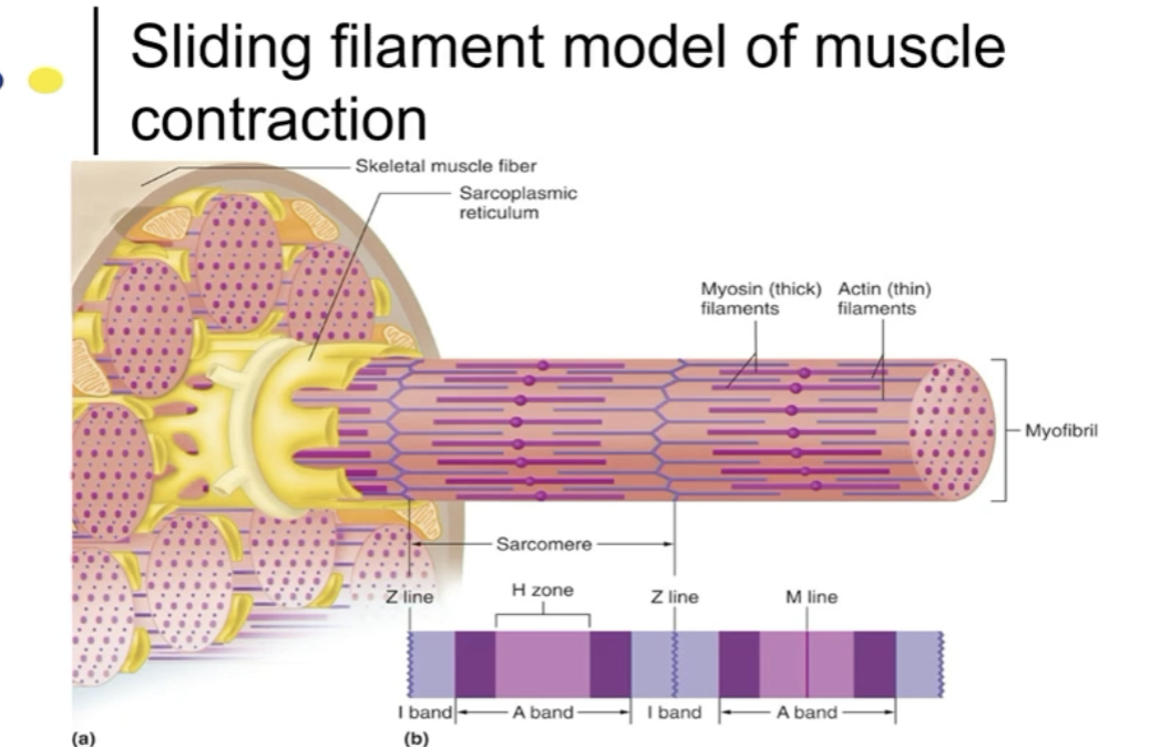
A band : myosin filaments
I-band: actin filaments and Z line
Myofibrils have thick (myosin) and thin (actin) filaments
Structure of the myofiber is specialized for the rapid spread of action potentials and for rapid changes in Ca2+
Each muscle fiber is controlled by efferent neuron
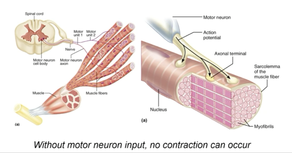
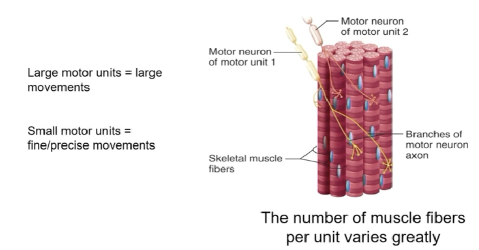
a motor unit is one motor neuron and all the muscle fibers it innervates
motor neuron release acetylcholine into specialized synapses
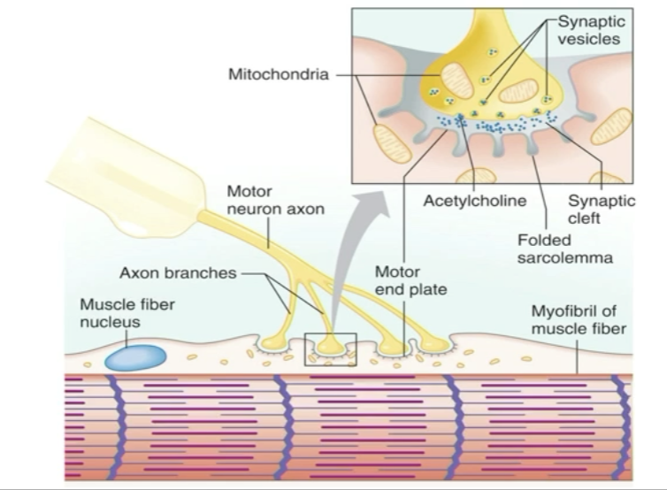
ACh binds nicotinic ACh receptors at motor endplates
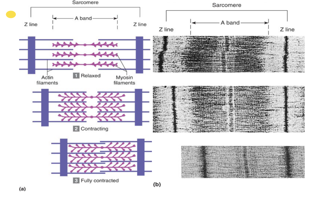
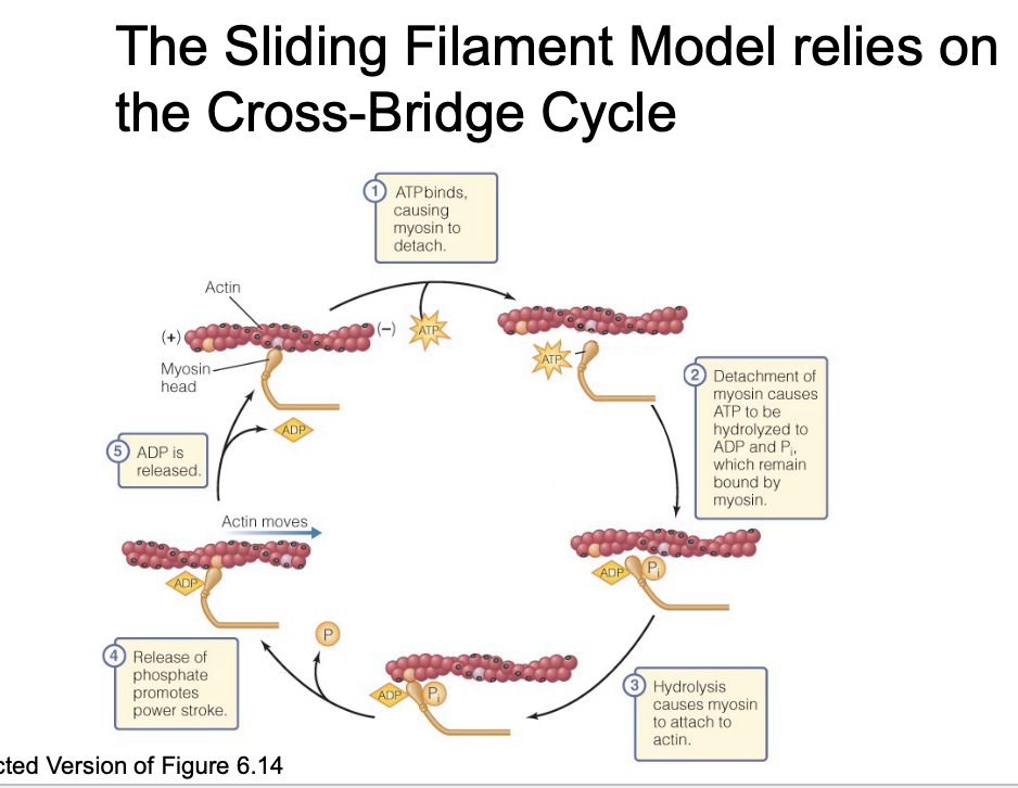
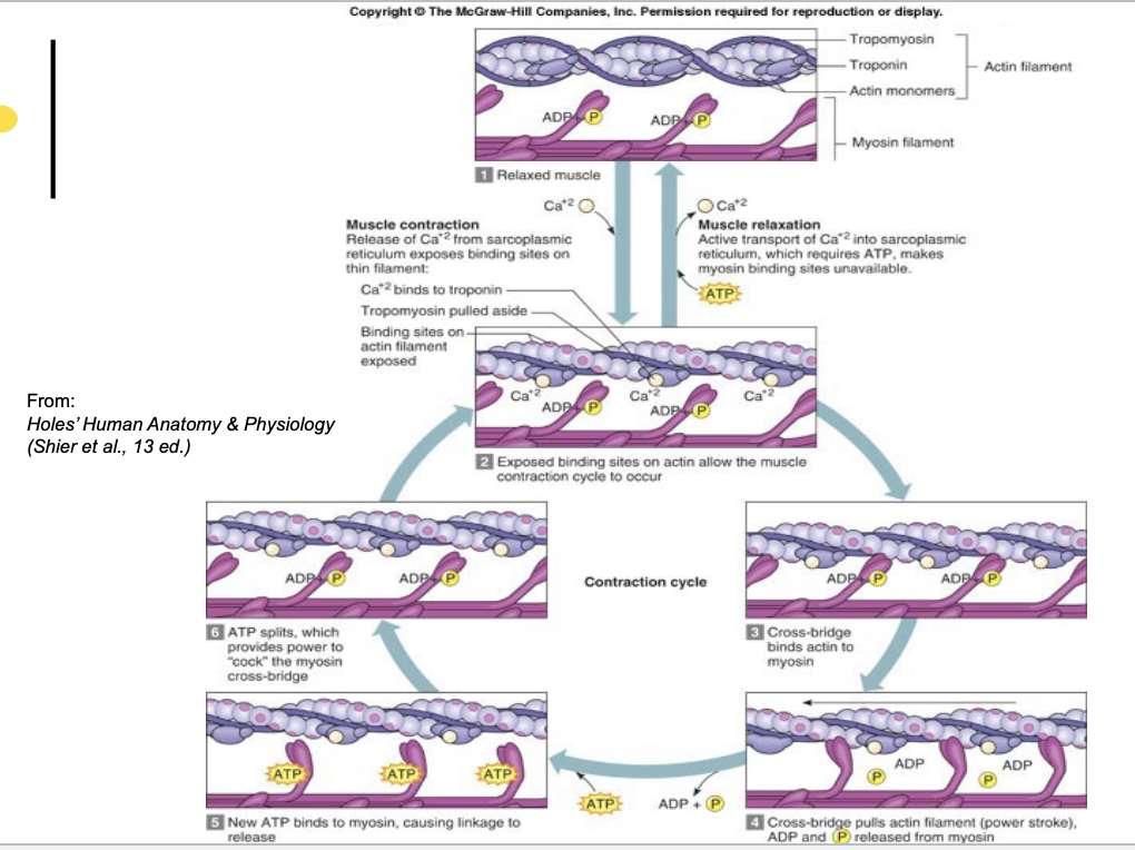
Contraction of sacromere
Actin filaments
long, unbranched polymers
thin
cytoskelton contains branched network of actin microfilaments
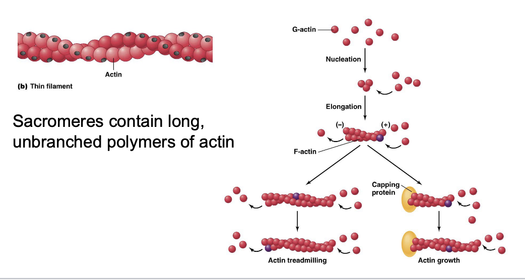
controlled polymerization of actin cause cell movement
Myosin filaments
Thick filaments
actin binding motor protein
essential light chains and regulatory light chains determine the specific properties of myosin
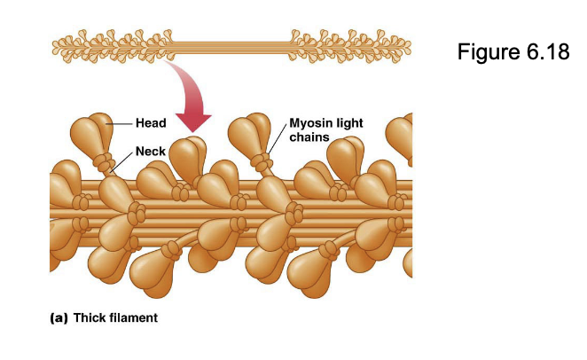
structurally divergent and carry out diverse functions
Ex: myosin V dimers move cargo by walking along actin filaments
myosin movements displays unitary displacement: the length of the step is determined by the length of myosin isoform’s neck
myosin motors catalyze ATP hydrolysis
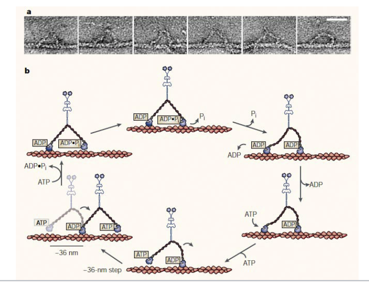
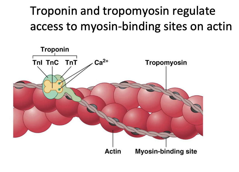
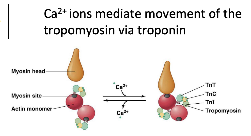
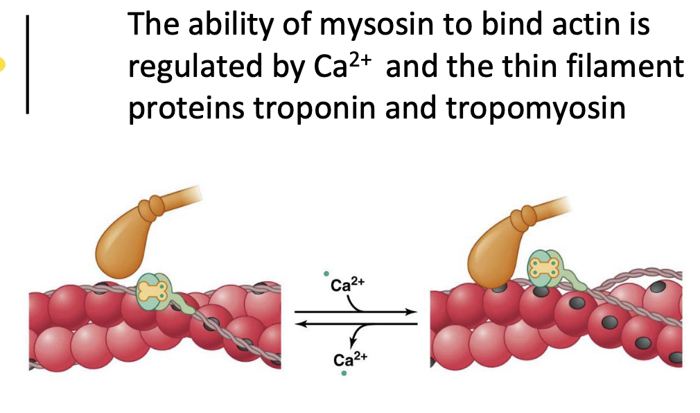
myosin heads interact with actin based thin filaments
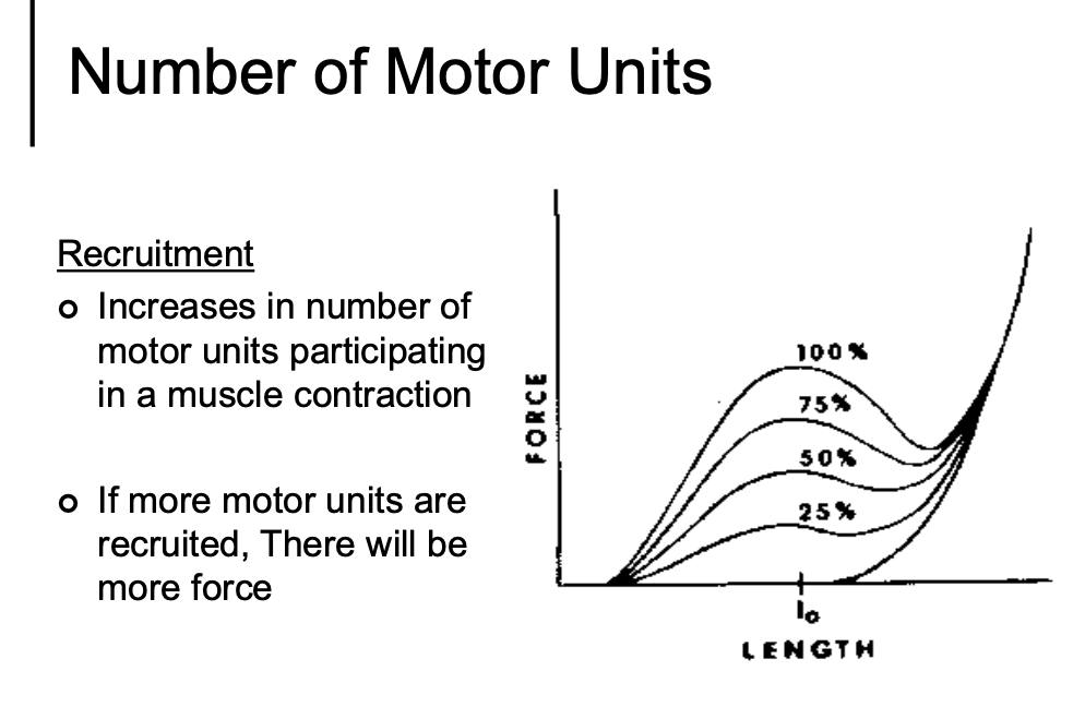
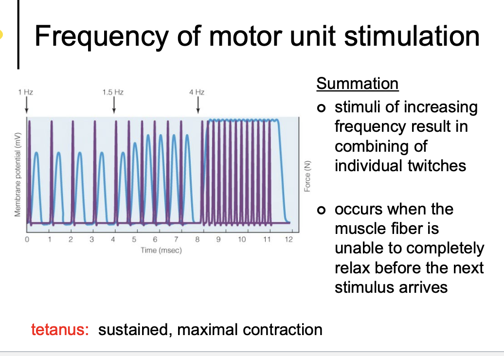
Gradation of force
the force of muscle action varies from slight to maximal based on the:
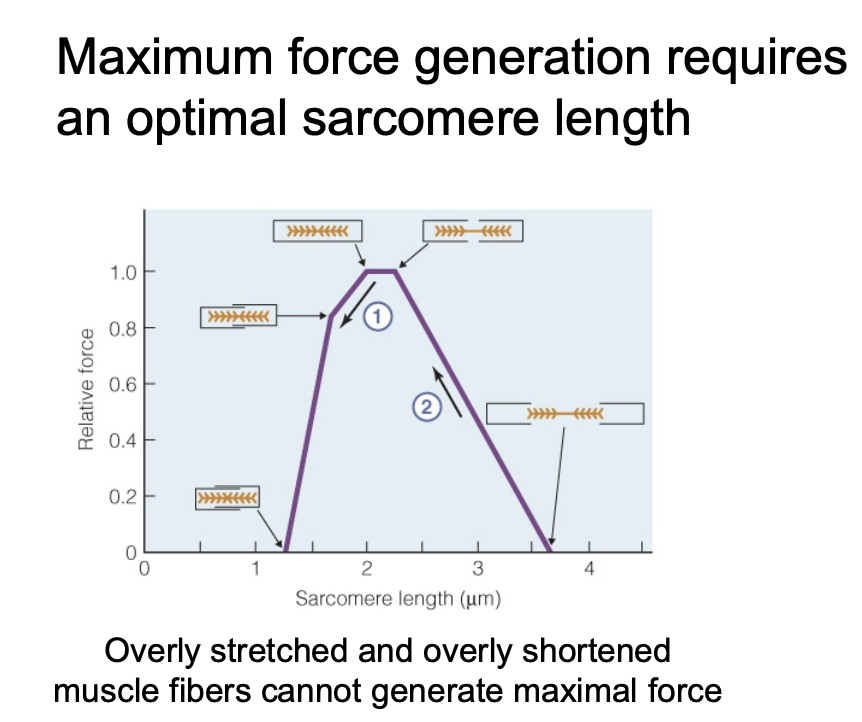
the length of muscle fiber before stimulation
the tuber of motor units recruited
the frequency of motor unit discharge
Skeletal muscle contraction
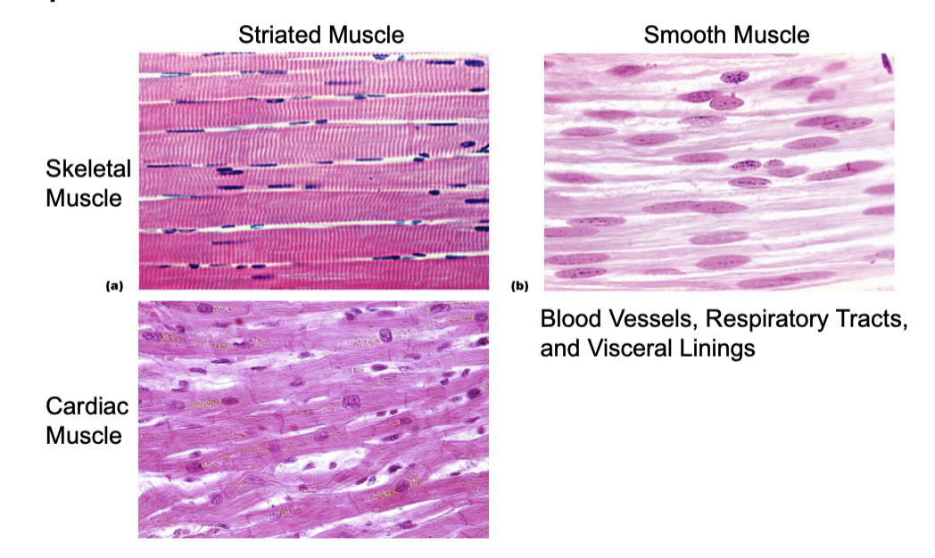
Muscles are defined by organization of their thick and thin filaments
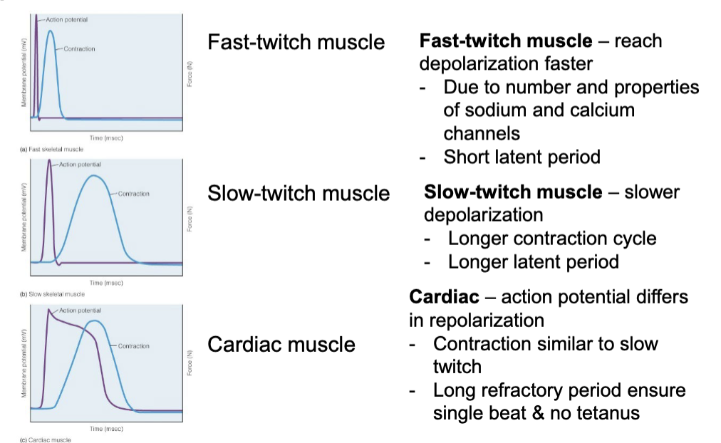
Action potentials differ in types of striated muscle
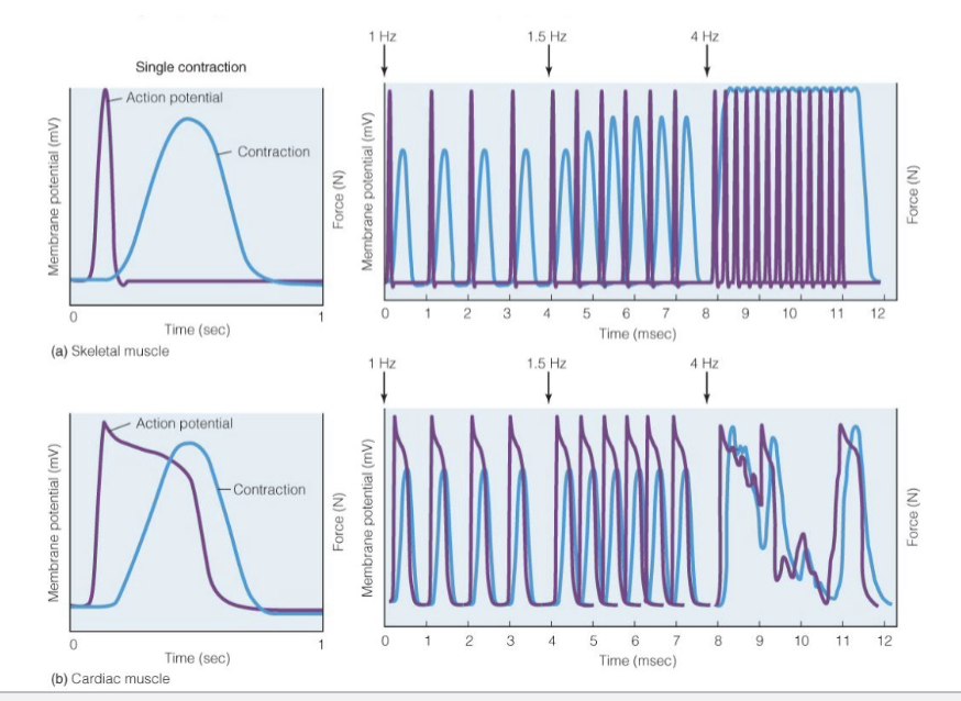
High frequency stimulation causes arrhythmic cardiac muscle contractions
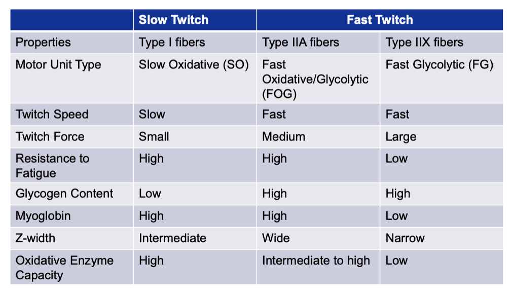
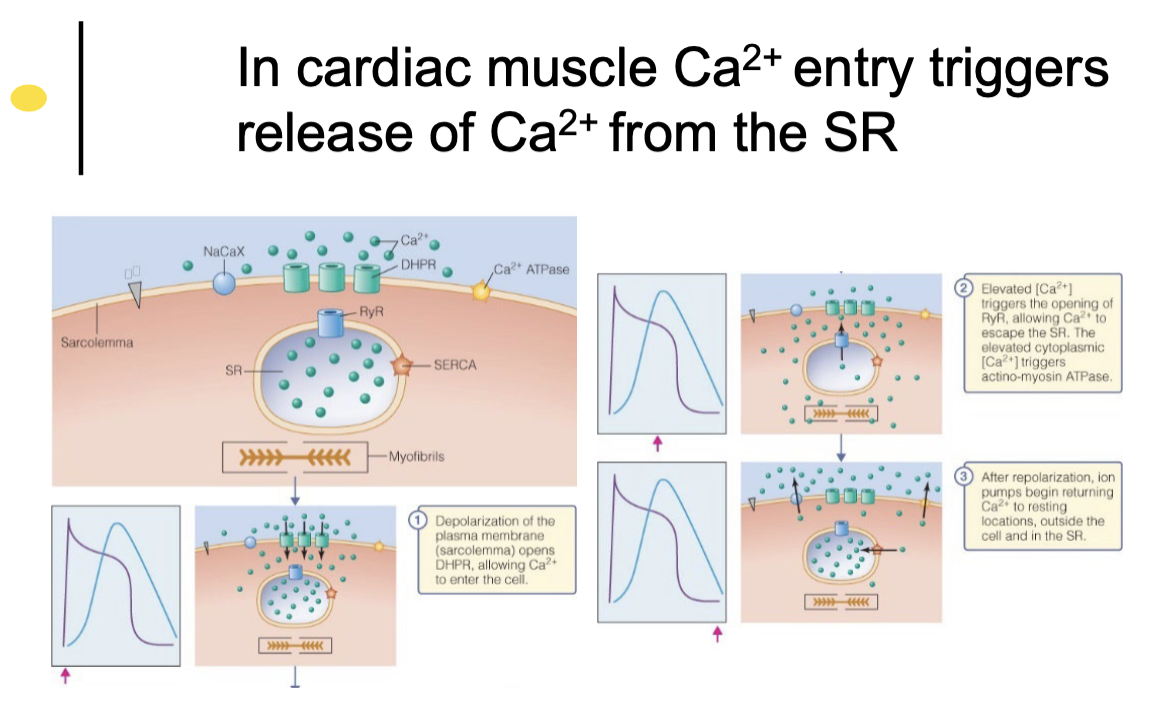
Neurotransmitters stimulate skeletal muscle, while cardiac muscle stimulates other cardiac muscle
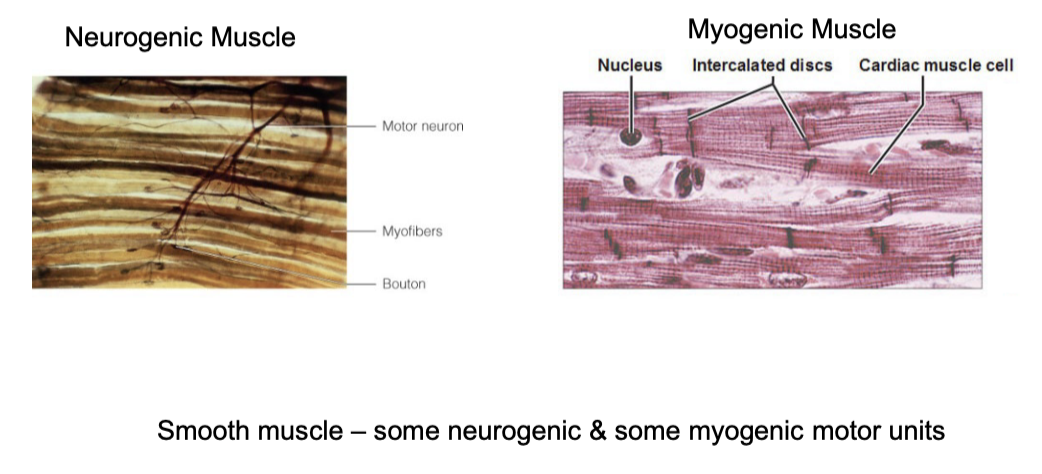
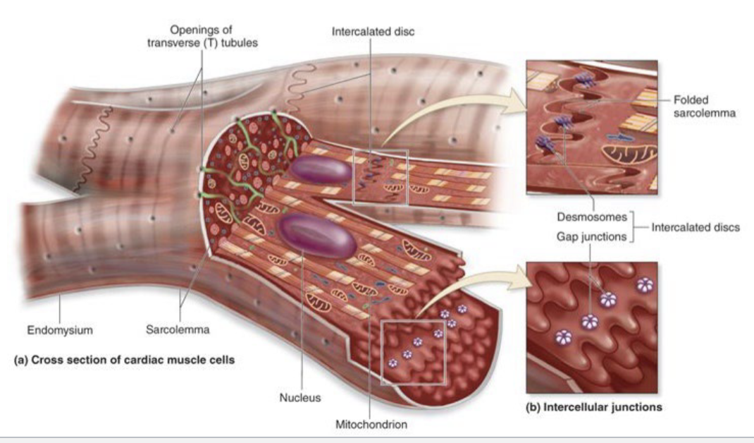
Cardiomyocytes have unique structures that distinguish them
Smooth muscle lacks organized sacromeres
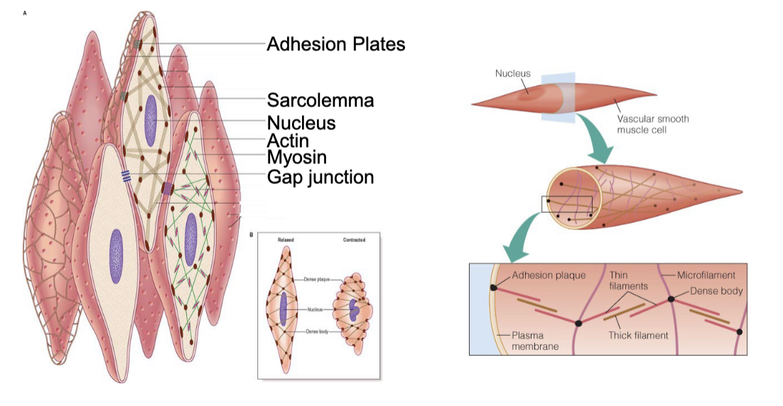
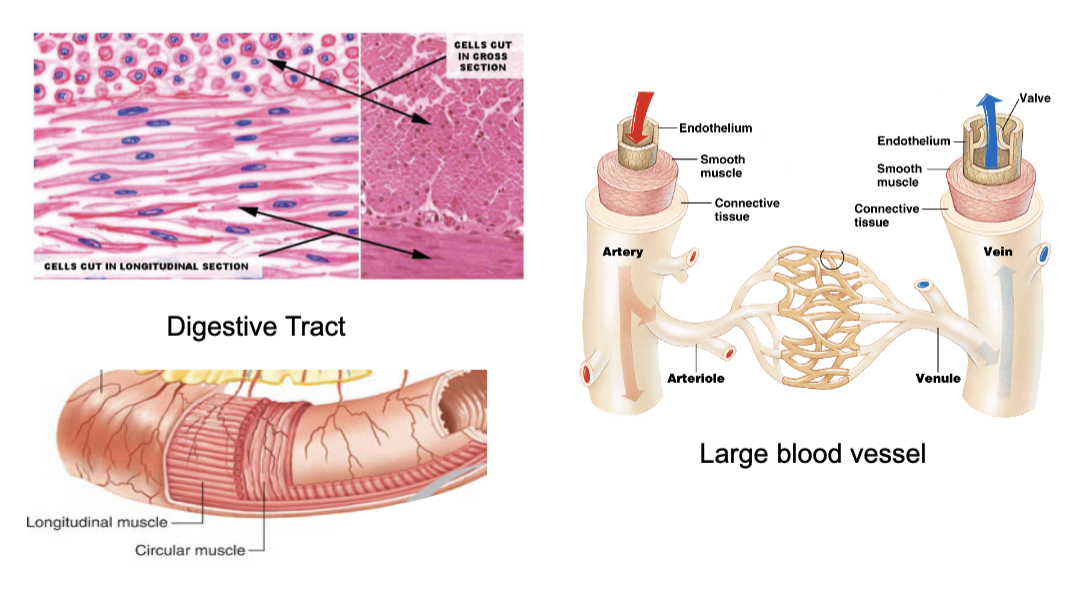
smooth muscles form circular and longitudinal layers