Male Reproductive
The primary organs in the male reproductive
system are the testes; have dual functions
produces sperm (spermatogenesis)
produces androgens (steroidogenesis)
the reproductive ducts which carry sperm from
the testes to the exterior of the body; these are
epididymis
ductus deferens
ejaculatory duct
urethra
accessory glands which produce secretions that
combine with sperm to create semen; these are
seminal vesicles
prostate gland
bulbourethral/Cowper’s glands
The male genitalia (external sex organs) are the
scrotum and penis
Testis
The male gonad or reproductive gland
contained in scrotum
Each testis measures about 3.8 cm long and 2.5
cm wide
surrounded by 3 tunics
tunica vaginalis-
the serous covering of the testis
derived from peritoneum
consists of a visceral and a parietal lamina
tunica albuginea-
the fibrous covering of the testis
extensions divide testis into 250-300
compartments called lobules
tunica vasculosa-
the vascular layer of the testis
consists of a plexus of blood vessels
clothes the inner surface of the tunica
albuginea
Lobules
each lobule contains 1 to 4 seminiferous
tubules, where sperm are produced
each tubule averages about 80 cm in length and
forms a loop
seminiferous tubules in a lobule join to form
tubulus rectus, which carries sperm into the rete
testis, a reticular network of tubules
from there, efferent ductules carry sperm out of
the testis into the epididymis,
the testis and epididymis are supplied by the
testicular artery and their veins drain into the
pampiniform plexus, which forms the bulk of the
spermatic cord
lymphatics accompany the testicular vessels
and drain into the lumbar (aortic) nodes
in between the seminiferous tubules are the
Leydig cells
Seminiferous Tubules
contains
germ cells at various stages of development
Sertoli/sustentacular cells
Sertoli cells extend from the basement
membrane of the seminiferous tubules to reach
the lumen
Sertoli cells are in constant contact with lots of
spermatogenic cells, and they send cytoplasmic
processes to surround those cells
tight junctions between adjacent Sertoli cells
near the base of the seminiferous epithelium
constitute the blood-testis barrier (BTB), a
structure that partitions seminiferous tubules
into a basal and an adluminal compartments
Leydig cells
found in interstitial supporting tissue between
the seminiferous tubules
they are almost non-existent prior to the onset
of testicular testosterone production at puberty
they produce testosterone
Spermatogenesis
The development of male gametes (spermatozoa) from spermatogonia
begins at puberty and continues throughout life
occurs in the seminiferous tubules
takes about 74 days
has three phases
proliferative/mitotic phase
meiotic phase
spermiogenic phase
Proliferative Phase
Type A spermatogonia (stem cells) which lie on
the basal lamina of the seminiferous tubules
undergo mitosis to produce more type A
spermatogonia to renew their stock and type B
spermatogonia
the type B spermatogonia undergo further
mitotic division to form primary spermatocytes
Meiotic Phase
each primary spermatocyte undergoes the first
meiosis to form two secondary spermatocytes,
each containing a haploid number of
chromosomes
spermatogonia do not separate completely after
meiosis due to incomplete cytokinesis and
remain joined by intercellular bridges
intercellular bridges are thought to facilitate
biochemical interactions allowing synchrony of
germ cell maturation
each secondary spermatocyte undergoes a
second meiosis to form two round spermatids
Spermiogenic Phase
the process that converts spermatids into mature
sperm (spermatozoa)
it has four stages, these are:
the Golgi phase
the cap phase
acrosome phase
maturation phase
as each division takes place, the daughter cell
migrates closer to the lumen of the seminiferous
tubule, so that spermatids are immediately
adjacent to the lumen
the release of sperm into the lumen of the
seminiferous tubules is called spermiation; the
remaining unnecessary cytoplasm and
organelles are removed during spermiation
Spermatozoon
It consists of:
Head
Midpiece
Tail
Head
The head contains the condensed nucleus which is capped by an apical vesicle (acrosome) filled with hydrolytic enzymes
The acrosome plays an important role in fertilization
Midpiece
it contains large helical mitochondria that generate the energy for swimming
Tail
contains microtubules and propels the sperm during motility
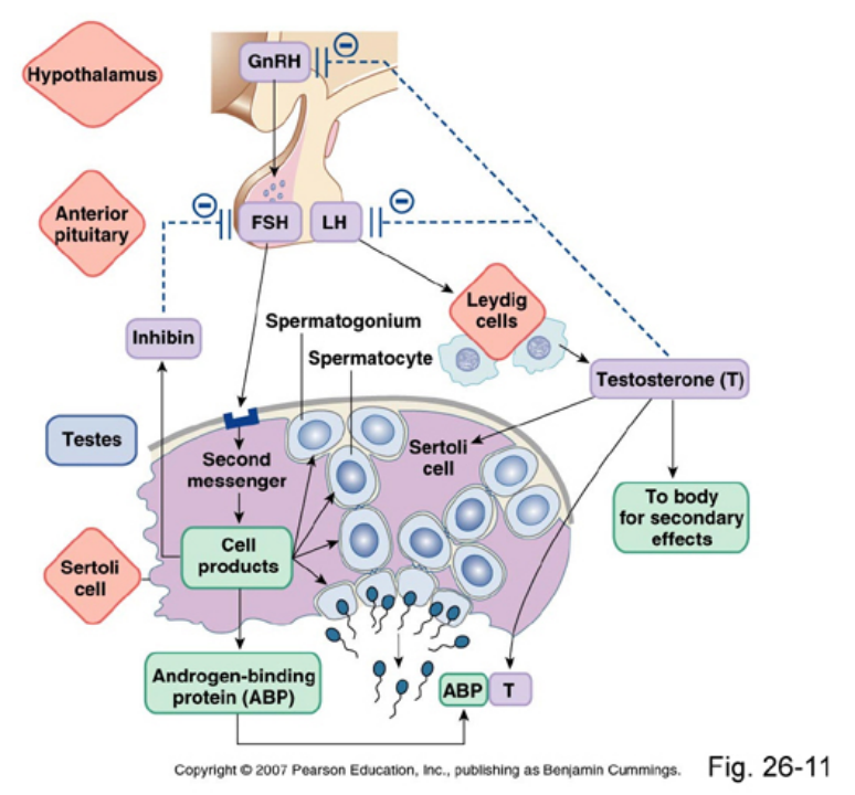
Factors Regulating Spermatogenesis
Hormonal Control of Spermatogenesis Endocrine factors secreted by the hypothalamus, pituitary gland and local regulators of testis function, as well as modulators of gene expression in the pituitary gland are involved in the regulation of spermatogenesis
both Sertoli cells (SC) and Leydig cells (LC)
regulate spermatogenesis by steroidogenesis
and growth factors production
SC stimulated by FSH produce inhibin (In),
activin (Ac) and androgen binding protein (ABP)
inhibin has negative feedback control on
pituitary and FSH secretion
inhibin also binds to Leydig cells (LC) regulating
testosterone (T) production
activin enhances FSH production
activin binds to round spermatids and
spermatogonia (SP), effecting spermatogenesis
activin also blocks testosterone synthesis in the
Leydig cells
ABP is essential to concentrating testosterone in
levels high enough to initiate and maintain
spermatogenesis, which can be 20-50 times
higher than the concentration found in blood
FSH and LH are known to influence germ cell
fate. They suppress apoptosis of germ cells
Leydig cells also produce epidermal growth
factors (EGF) which bind to spermatogonia and
spermatids regulating cell divisions
considerable amounts of the testosterone are
metabolized to estrogens by the enzyme
aromatase in Sertoli cells
this reaction is blocked by inhibin
FSH is required for the initiation of
spermatogenesis
once spermatogenesis is initiated, FSH is no
longer required; testosterone alone will maintain
active spermatogenesis
Temperature
The testes are located in a sac called the scrotum
which holds the testes away from the body since normal body temperature is too warm for sperm production
Scrotal temperature is maintained by:
the counter current heat exchanger formed by
the close association between the testicular
artery and the pampiniform plexus of veins
surrounding the vas deferens which cools the
arterial blood before entering the testicular
epithelium
testicular altitude (relative to the abdomen)
regulated by cremaster muscle which lowers or
raises testes within scrotal sac to cool or warm
the testes respectively
regulation of scrotal skin area by the dartos
muscle (smooth muscle) within the dermis of the
scrotum: increase (relaxed dartus muscle) or
decrease (contracted) surface area for heat
exchange
the scrotum is well supplied with sweat glands
which presumably aid in cooling the testes
these features maintain a testes temperature
about 3o C below the body temperature
Other Factors
Dietary deficiencies (such as vitamins B, E and
A)
anabolic steroids
metals (cadmium and lead)
x-ray exposure
dioxin
alcohol
infectious diseases
will also adversely affect the rate of spermatogenesis
Sperm Maturation and storage
Sperm from the seminiferous tubule enter the
epididymis via other ducts
the sperm are stored in the epididymis for up to
2 weeks where they mature and develop motility
as sperm transit the epididymis, they are bathed
in a specialized fluid rich in proteins, ions, and a
number of other molecules
complex interactions between spermatozoa and
epididymal fluid contribute to sperm maturation
sperm is stored in the vas deferens until
ejaculation
Sperm Capacitation
Freshly ejaculated sperm are unable or poorly
able to fertilize eggs
they must first undergo a series of changes that
give them the ability to fertilize
the acquisition of the ability to fertilize is known
as capacitation
capacitation occurs while sperm reside in the
female reproductive tract for a period of time
it is stimulated by secretions in the vagina,
uterus, and uterine tubes
it takes between 5-7 hours
it is associated with
removal of adherent seminal plasma
proteins
reorganization of plasma membrane lipids
and proteins
an influx of extracellular calcium ions
increase in cyclic AMP
decrease in intracellular pH
capacitated sperm
display hyperactivated motility
undergo the acrosome reaction
Semen
The grayish white bodily fluid ejaculated at the
time of orgasm
it contains sperm (2-3%) and secretions from
the
testis
epididymis (<5%)
seminal vesicles
prostate gland
bulbourethral glands or Cowper’s glands
the initial secretion is from the bulbourethral or
Cowper’s glands (<5%)
it is secreted just before emission of semen
it is thought to serve as a lubricant(?) for inserting the penis into the vagina
provide the mucus which provides a jelly like consistency to the semen and is important for the mobility of the sperm in the cervix and the vagina
next is the secretion from the prostate (30%)
prostatic secretion contains: citric acid, prostate-specific antigen, acid phosphatase, zinc, and proteolytic enzymes
the zinc content in the prostatic secretions is vital for stabilizing chromatin which contains the DNA in the sperms. Deficiency of zinc
can lead to lower fertility as it can render the
sperms fragile.
it is alkaline
it neutralizes any residual urine, which tends
to be acidic, and the acidity of the vaginal
secretions
liquefies coagulated semen into a viscous
fluid
the terminal portion is from the seminal vesicles
this is about 60% of total semen volume
contains:
mucus
amino acids
fructose as the main energy source for the
sperm
prostaglandins to stimulate female uterine
contractions to move the semen up into
the uterus
normal semen volume is between 1.5-5.0 ml
the pH is 7.0-8.3
a count of 15 million and above spermatozoa/ml
is considered the normal range
counts below 15 million/ml are considered low
Transport
Testosterone circulates in blood at concentrations above its aqueous solubility by binding to circulating plasma proteins
about 65% binds avidly to sex-hormone binding
globulin (SHBG)
about 33% is bound to lower-affinity, high
capacity binding sites (albumin, ⍺1-acid
glycoprotein, transcortin)
1-2% remaining non-protein bound
the “free” (non-protein bound) fraction is the
most biologically active with the loosely protein-
bound testosterone constituting a larger
“bioavailable” fraction of circulating testosterone
circulating testosterone levels demonstrate
distinct circhoral and diurnal rhythms
testosterone is secreted at adult levels during 3
epochs of male life:
transiently during the first trimester of
intrauterine life
during neonatal life
after puberty to maintain virilization
testosterone undergoes metabolism to both
bioactive metabolites and to inactivated oxidized
and conjugated metabolites for urinary and/or
biliary excretion
Action
testosterone has both androgenic and anabolic
effects
its action is initiated by the binding of
testosterone or its analogs to the androgen
receptor causing its activation
in addition, testosterone is also converted to its
bioactive metabolites, dihydrotestosterone
(DHT) and estradiol
in the majority of target cells, some testosterone
is converted into DHT by the enzyme 5-α
reductase
DHT has higher binding affinity to the androgen
receptor and 3-10-fold greater molar potency
than testosterone
DHT cannot be aromatized to estrogen, and
thus its effects are purely androgenic
testosterone and DHT do bind to the same
intracytoplasmic androgen receptor
the androgen-receptor complex binds to DNA
this results in mRNA syntheses, and
subsequently in syntheses of cytoplasmic
proteins, which lead to cell growth and other
secondary effects mediated by androgens
congenital 5-α reductase deficiency individuals
may have normal male external genitalia, often
with an unusually small penis (micropenis) and
the urethra opening on the underside of the
penis (hypospadias), ambiguous genitalia, or
normal female genitalia
these individuals have male internal genitalia
but usually have female primary sex
characteristics and may be raised as females
high concentrations of testosterone at puberty
leads to marked virilization including phallic
growth and, occasionally, masculine gender
reorientation although prostatic development
remains rudimentary