Chapter 8 - Particles in the Nucleus
8.1 Atoms, Isotopes and Radioisotopes
Earth is naturally radioactive; we are constantly exposed to background radiation.
Human senses cannot detect radiation, but our bodies are adapted to low levels. High doses can be damaging.
Radioactive atoms (radioisotopes) have beneficial uses (e.g., medicine).
The Standard Model
All matter is composed of 12 fundamental particles (fermions): 6 quarks and 6 leptons.
These are not seen in everyday situations; produced in high-energy particle accelerators.
Quarks (make up hadrons like protons & neutrons): up (u), down (d), charm (c), strange (s), top (t), bottom (b).
Leptons: electron (e⁻), muon (μ⁻), tau (τ⁻), and their neutrinos (ν_e, ν_μ, ν_τ).
Hadrons = particles made of quarks (baryons = 3 quarks; mesons = quark + antiquark).
Proton = uud (2 up + 1 down).
Neutron = udd (1 up + 2 down).
Fermions Table (masses relative to proton):
Quarks: Fractional charges (+2/3 or –1/3).
Leptons: Electron, muon, tau have charge –1; neutrinos have charge 0.

Antiparticles:
Same mass, opposite charge (or opposite quantum numbers).
Example: positron (e⁺) is antiparticle of electron.
The Structure of Atoms
Atoms consist of a tiny, dense nucleus (protons + neutrons = nucleons) surrounded by an electron cloud.
Nucleus contains >99% of atom’s mass but is extremely small (~10⁻¹² of atom’s volume).
Atomic notation: AZX
Z (atomic number) = number of protons.
A (mass number) = protons + neutrons (nucleons).
Neutrons (N) = A – Z.
Electrically neutral atom: Number of electrons = number of protons (Z).

Isotopes
Atoms of the same element (same Z) but different number of neutrons (different A).
Same chemical properties, different physical properties (e.g., density, mass).
Nuclide: Refers to a specific nucleus.
Hydrogen isotopes:
Protium (¹H): 1 proton, 0 neutrons.
Deuterium (²H): 1 proton, 1 neutron.
Tritium (³H): 1 proton, 2 neutrons.
Worked Example Highlights:
For ¹⁹⁷₇₉Au: Z=79 (protons), A=197 (nucleons), N=118, electrons=79.
Isotopes have same Z, different N and A.

Radioisotopes
Unstable isotopes that undergo spontaneous transmutation (emit radiation/particles and change into different element/isotope).
Most atoms are stable; only certain combinations of protons & neutrons are stable.
Radioactive if nucleus is unstable (naturally occurring or artificial).
Symbol: Radiation warning trefoil (Figure 8.1.11).
Periodic table: All isotopes of elements with Z > 83 (bismuth) are radioactive; many lighter elements have radioactive isotopes (shaded yellow in Figure 8.1.12).

Artificial Transmutation: How Radioisotopes Are Manufactured
Most radioisotopes (~3000) are produced artificially (only ~270 occur naturally).
Common method: Neutron absorption in a nuclear reactor.
Stable isotope + neutron → radioactive isotope.
Example: Cobalt-59 (stable) + neutron → Cobalt-60 (radioactive, used in medicine).
Produced in facilities like ANSTO (Lucas Heights, Sydney) using nuclear reactors.
Stability of Nuclei and Why Radioactive Nuclei Are Unstable
Strong nuclear force (mediated by gluons) holds nucleus together — very strong but short-range.
Electrostatic repulsion between protons tries to break nucleus apart.
Line of stability (Figure 8.1.15): Stable nuclei cluster around a curved line (N ≈ Z for light elements; N > Z for heavier elements).

For stability, neutrons help “dilute” proton repulsion.
Unstable nuclei (away from line) emit radiation (α, β⁻, β⁺) to reach stability.
Bismuth-209 is the heaviest stable isotope; heavier nuclei are unstable.
Examples:
Small balanced nuclei (e.g., carbon-12) are stable.
Large nuclei with too many protons or neutrons become unstable.

8.2 Radiation
Early researchers (Marie Curie, Ernest Rutherford, Paul Villard) discovered three types of radiation: alpha (α), beta (β), and gamma (γ).
Alpha & beta are particles emitted from the nucleus.
Gamma is high-energy electromagnetic radiation.
Radioactive decay: Process where unstable nucleus emits particles/radiation.
Alpha (α) Decay
Occurs in heavy unstable nuclei.
Emits an alpha particle (⁴₂α or ⁴₂He²⁺): 2 protons + 2 neutrons (helium nucleus).
Nuclear equation example (Uranium-238): ²³⁸₉₂U → ²³⁴₉₀Th + ⁴₂α + energy

Parent nucleus → daughter nucleus (different element = nuclear transmutation).
Atomic number decreases by 2; mass number decreases by 4.
Energy released mostly as kinetic energy of the fast-moving alpha particle.
Conservation: Atomic number (Z) and mass number (A) are conserved in nuclear reactions.
Beta (β) Decay
Beta-Minus (β⁻) Decay
Occurs when nucleus has too many neutrons.
A neutron changes into a proton + electron (β⁻) + antielectron neutrino (ν̅_e).
Example (Carbon-14): ¹⁴₆C → ¹⁴₇N + ⁻¹₀β + ν̅_e + energy

Atomic number increases by 1; mass number unchanged.
Beta particle is a high-energy electron from the nucleus.
Beta-Plus (β⁺) Decay
Occurs when nucleus has too many protons.
A proton changes into a neutron + positron (β⁺) + electron neutrino (ν_e).
Example: ¹²₆C → ¹²₅B + ¹₀β⁺ + ν_e
Atomic number decreases by 1; mass number unchanged.
Positron (β⁺) is the antiparticle of the electron (positive charge).

Gamma (γ) Decay
High-energy electromagnetic radiation (no mass, no charge).
Emitted when daughter nucleus is in an excited state after α or β decay.
Releases excess energy; nucleus rearranges to more stable state.
Example (Iodine-131 decay sequence): Beta decay → excited Xenon-131 → stable Xenon-131 + γ
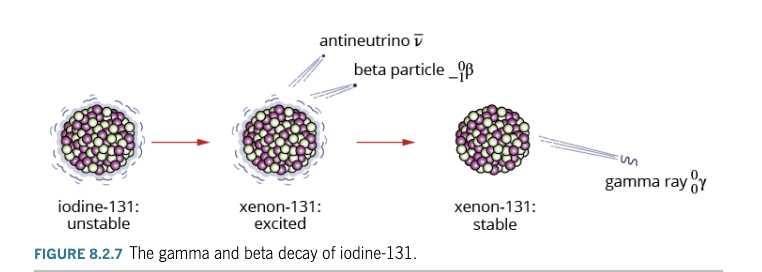
No change in atomic or mass number.
Travels at speed of light.
Conservation Laws in Nuclear Reactions

Mass-energy: Total energy (rest mass + kinetic) is conserved (E = mc²).
Charge: Total charge before = total charge after.
Momentum: Total momentum before = total after.
Atomic and mass numbers are always conserved.
Worked Example Insight: Energy differences in decays are calculated in MeV (mega-electron volts).


Range of Energies for Different Radiations
Alpha particles: 5–10 MeV (or MeV range).
Beta particles: Up to a few MeV.
Gamma rays: Typically < 1 MeV (e.g., up to 412 keV for gold-198).
Higher energy → greater speed/frequency (for gamma).
Detection of Radiation
Human senses cannot detect it.
Geiger Counter (Geiger-Müller tube):
Filled with argon gas.
High voltage between electrodes.
Radiation ionises gas → current pulse → audible click.
Used for mineral exploration, safety monitoring, medical/industrial checks.
Physics in Action: How Technetium-99m is Produced
Most widely used radioisotope in nuclear medicine (cancer diagnosis/treatment).
Produced from Molybdenum-99 (from Lucas Heights reactor) via beta-minus decay: ⁹⁹₄₂Mo → ⁹⁹ᵐ₄₃Tc + ⁻¹₀β + ν̅_e
Tc-99m then emits gamma ray: ⁹⁹ᵐ₄₃Tc → ⁹⁹₄₃Tc + ⁰₀γ
Short half-life → produced on-site in hospital generators (lead-shielded).
8.3 Properties of Alpha, Beta and Gamma Radiation
Early experiments (with radium) using a magnetic field showed three distinct types of radiation (Figure 8.3.1):
Alpha (α): Deflected one way (positively charged).
Beta (β): Deflected the opposite way (negatively charged).
Gamma (γ): Not deflected (no charge).
Differences in charge, mass, and speed.
Speed and Charge
Alpha (α) Particles (Figure 8.3.2)
Consist of 2 protons + 2 neutrons (helium nucleus, ⁴₂He²⁺).
Charge: +2.
Mass: Relatively heavy.
Speed: Slow — up to 20,000 km/s (about 0.1c or 10% speed of light).
Beta-Minus (β⁻) and Beta-Plus (β⁺) Particles (Figure 8.3.3)
β⁻: Fast-moving electrons (negative charge).
β⁺: Positrons (positive charge, antiparticle of electron).
Mass: Very light (much lighter than alpha).
Speed: Very fast — up to ~90% speed of light (0.9c).
Gamma (γ) Rays (Figure 8.3.5)
High-energy electromagnetic radiation (part of EM spectrum, Figure 8.3.4).
Charge: 0 (no charge).
Mass: None (no rest mass).
Speed: Speed of light (3.00 × 10⁸ m/s).
Penetration Ability and Ionisation
Alpha (α) Particles (Figure 8.3.6)
High ionising ability: Strongly interact with atoms, knock off electrons, create ions.
Low penetration: Travel only a few centimetres in air; stopped by paper, skin, or thin card.
Easily absorbed — become helium atoms after gaining electrons.
Beta (β) Particles (Figure 8.3.8)
Low ionising ability: Fewer interactions due to smaller charge and higher speed.
Moderate penetration: Travel a few metres in air; stopped by a few mm of aluminium or thin metal/plastic sheet.
Can pass through skin but stopped by thin metal.
Gamma (γ) Rays
Very low ionising ability: Weak interaction with matter (only ionise if direct collision).
High penetration: Travel long distances in air; penetrate through human body, aluminium sheets, and several cm of lead (thick lead or concrete needed to stop).
Summary Table 8.3.1: Comparison of Properties
Property | Alpha (α) Particle | Beta (β) Particles | Gamma (γ) Ray |
|---|---|---|---|
Mass | Heavy | Light | None |
Speed | ~10% c (slow) | ~90% c (very fast) | Speed of light (c) |
Charge | +2 | –1 or +1 | 0 |
Ionising Ability | Very high | Low | Very low |
Range in Air | A few centimetres | 1 or 2 metres | Many metres |
Penetration in Matter | ~10⁻² mm (paper/skin) | A few mm (thin aluminium) | High (thick lead/concrete) |
Physics in Action: Applications
Monitoring Thickness of Sheet Metal (Figure 8.3.10)
Beta-minus particles used.
Source under sheet; detector above.
Fewer beta particles reach detector if sheet is thicker → adjusts rollers automatically.
Gamma Knife Therapy (Figure 8.3.11)
Focused gamma beams from many directions target bra
in tumours.
High dose at tumour; surrounding tissue receives low dose.
Smoke Detectors
Use alpha emitter (e.g., Americium-241) — alpha particles ionise air; smoke reduces current, triggers alarm.
8.4 Half-Life and Decay Series
Different radioisotopes decay at different rates.
Activity: Rate of radioactive decay (measured by Geiger counter as counts per second).
Example:
Polonium-218: High initial activity, drops rapidly (almost zero in ~30 minutes).
Uranium-235: Low activity, remains almost constant over decades (Figure 8.4.1).
Half-Life (t₁/₂)
Definition: Time taken for half of the radioactive nuclei in a sample to decay.
Half-life is constant for a given radioisotope and cannot be changed by chemical reactions, heat, etc.
Determined only by nuclear instability.
Example: Polonium-218 has t₁/₂ ≈ 3.10 minutes.
Mathematical Formula:
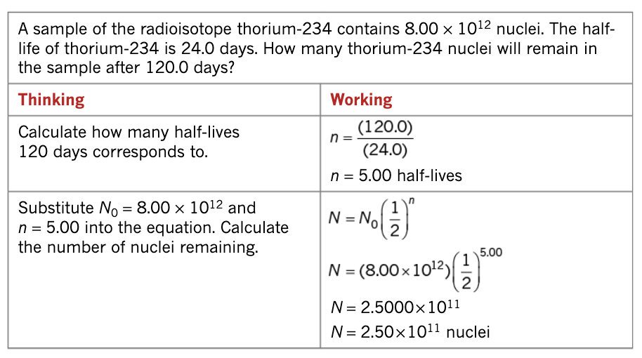
Number of nuclei remaining:

Number of half-lives:

Decay Curve: Exponential decay — activity halves
with each half-life.
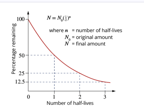
Worked Example Insight:
After each half-life, activity and number of nuclei halve (100% → 50% → 25% → 12.5%, etc.).
Activity (A)
Activity (A): Number of disintegrations per second (measured in becquerels, Bq). 1 Bq = 1 disintegration/second.
Formula:
?
Activity also follows:
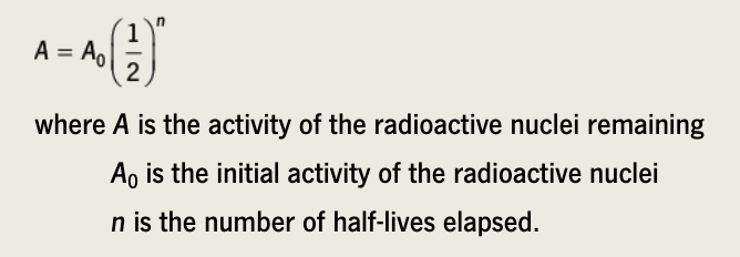
Worked Example Insight:
Technetium-99m (short half-life) activity drops rapidly — useful for medical imaging.

Common Radioisotopes and Their Applications (Table 8.4.1)

Decay Series
Decay of a radioisotope often produces another radioactive daughter nucleus → continues until a stable isotope is reached.
Decay series: Sequence of decays (Figure 8.4.4 shows Uranium-238 series).

Uranium-238 series ends at stable Lead-206.
Earth is old enough (4.5 billion years) that only 4 natural decay series remain active (Uranium, Actinium, Thorium, Neptunium).
Applications:
Rock dating: Measure proportions of radioactive elements and daughters in rocks.
Extension: Radiocarbon Dating33
Uses Carbon-14 (t₁/₂ = 5730 years, β⁻ decay to Nitrogen-14).
Living organisms have constant C-14/C-12 ratio (from atmosphere).
After death, C-14 decays and is not replaced → ratio decreases.
Compare C-14/C-12 ratio to estimate age (accurate up to ~60,000 years).
Examples: Shroud of Turin (medieval), mammoth fossils.
8.5 Radiation Doses and Effects on Humans
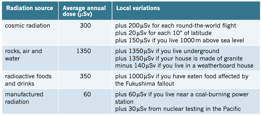
We are constantly exposed to low-level background radiation (natural sources).
High exposure from α, β, γ sources (medicine, industry, nuclear power) can be harmful.
Monitoring is essential for workers (radiologists, nuclear plant staff) and patients (cancer treatment).
Dosimeters measure personal exposure

Ionising Radiation
Ionising radiation: High-energy EM radiation (frequency > 2.00 × 10¹⁶ Hz) that can remove electrons from atoms, creating ions (Figure 8.5.3).
Includes: Gamma rays, X-rays, and some UV.
Non-ionising radiation: Lower energy (radio waves, microwaves, visible light, infrared, UV-A) — generally harmless in everyday amounts (Figure 8.5.2).
Effects of Ionisation:
Breaks chemical bonds in molecules (e.g., water → ions).
Can damage cells, cause cancer, or lead to radiation sickness.
Measuring Radiation Exposure
Absorbed Dose (AD)
Energy absorbed per kilogram of tissue.
Formula:

Unit: Gray (Gy) → 1 Gy = 1 J/kg.

Dose Equivalent (DE)

Accounts for the type of radiation (different biological damage).
Formula: DE = AD × QF (QF = quality factor / weighting factor).
Unit: Sievert (Sv) or millisievert (mSv), microSievert (μSv).

Quality Factors (Table 8.5.1):
α particles: 20 (highly damaging due to high charge, mass, low speed).
Neutrons (10 keV): 10.
β particles, γ rays, X-rays: 1.
Background Radiation
Average annual exposure in Australia: ~1.5 mSv.
Sources (Figure 8.5.4 & 8.5.6):
Natural (~90%): Cosmic rays, rocks/soil, food/drink, radon gas, natural radioactivity in air.
Artificial: Medical (X-rays), nuclear power, etc.
Background radiation is low and generally not harmful.
Effect of Radiation Exposure on Humans
Symptoms: Nausea, fatigue, hair loss, reduced white blood cells, cancer risk.
Rapidly dividing cells (cancer, bone marrow) are most vulnerable.

Medical Applications of Radiation
Cancer treatment: Targeted radiation (e.g., cobalt-60) damages cancer cells more than healthy tissue (Figure 8.5.8).
Radioactive tracers (Table 8.5.5): Short half-life isotopes injected/ingested to image organs.

Scintigraphy: Gamma camera detects tracer emissions to create images (Figure 8.5.10).
Typical Medical Exposures (Table 8.5.4):
Chest X-ray: 30 μSv.
CT scan: 5000 μSv.
Radiotherapy course: Very high (targeted).
Physics in Action: Scintigraphy
Uses radioactive tracers with same chemical properties as stable isotopes.
Tracer emits gamma rays → detected to map organ function.
Dosimeters (Figure 8.5.5):
Thermoluminescent dosimeters (TLDs) used by workers for accurate monitoring.