Chapter 17.5 - Eyesight Pathway & Transduction
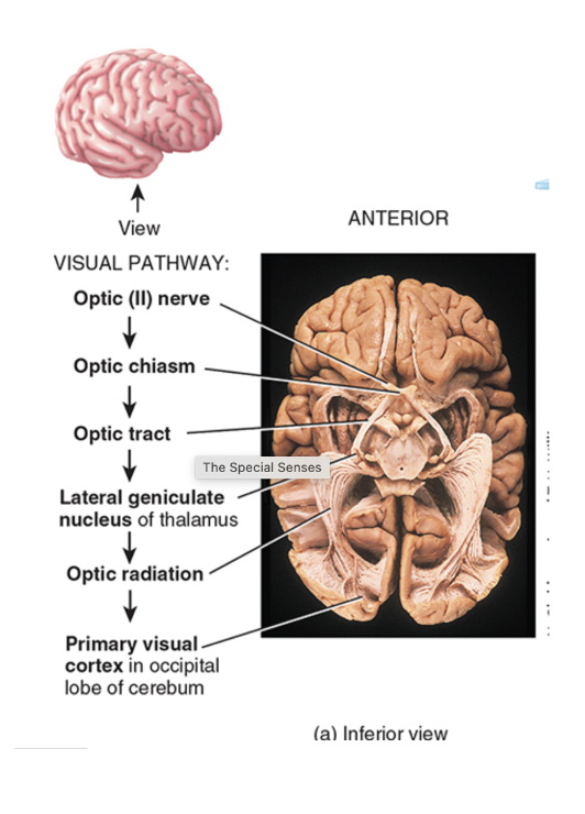
What is the visual pathway?
Axons of retinal ganglion cells exit eyeball at optic chiasm and form optic nerve on each side
Axons from the temporal half of each retina at the optic chiasm go directly to the thalamus on the same side
Axons from the nasal half of each retina cross the optic chiasm and go to the opposite thalamus
Axon branches project to the midbrain - pupil constriction and head and eye movement coordination
Axon branches extend to hypothalamus which regulates sleep and other activities in response to light/darkness
Axons project from thalamus to occipital lobe of cerebrum on same side

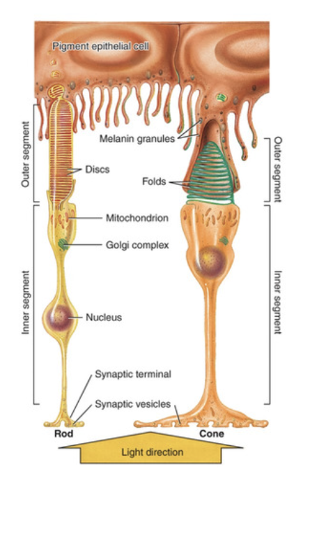
Photoreceptors and Photopigments
located in pigmented layer of retina
rods: cylindrical or rod-shaped and contain photopigment rhodopsin
cones: tapered or cone-shaped and contain photopigments (blue, green, and red)
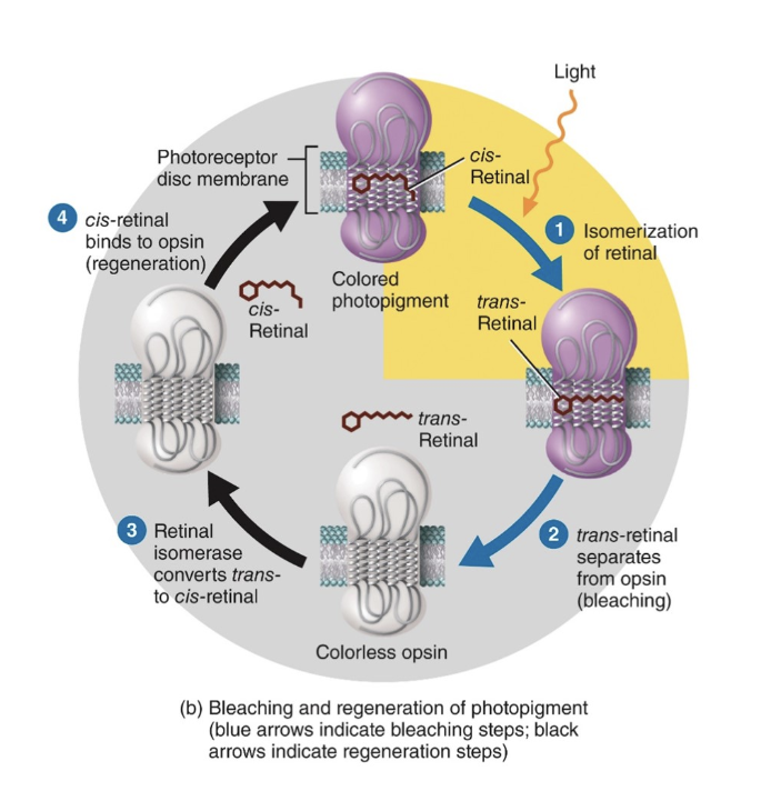
What are all photopigments made up of?
Part 1: glycoprotein known as opsin
Part 2: retinal molecule (derivative of vitamin A which has carotene in it and absorbs light)
How does phototransduction occur?
occurs in outer segment of a photoreceptor
activation by a light stimulus causes a hyperpolarizing receptor potential NOT A DEPOLARIZATION POTENTIAL
retinal has 2 forms: cis and trans
trans: retinal form separated from opsin (bleaching)
cis: retinal binds to opsin again (regeneration)
Steps of phototransduction in DARKNESS
Darkness: cis-retinal form of photopigment
Darkness: high cGMP in the cytosol of photoreceptor outer segment
Darkness: cGMP binds and opens cation channels so Na+ enters cell
Darkness: photoreceptor depolarizes
Depolarization spreads to synaptic terminal which has Ca+ channels in its membrane - Ca+ enters cell
Steps of Phototransduction in LIGHT
cis-retinal converts to trans-retinal
G protein called transducin is activated
Transducin activates cGMP phosphodiesterase
cGMP is broken down
Lower cGMP means reduced Na+ inflow
Decreases Na+ inflow causes a hyperpolarizing potential - more negative and much closer to resting potential of -70 mV
Hyperpolarization spreads causing decrease in Ca+ entry
Decreased Ca+ = decreases of the inhibitory neurotransmitter