Parasympathetic Nervous System
Overview:
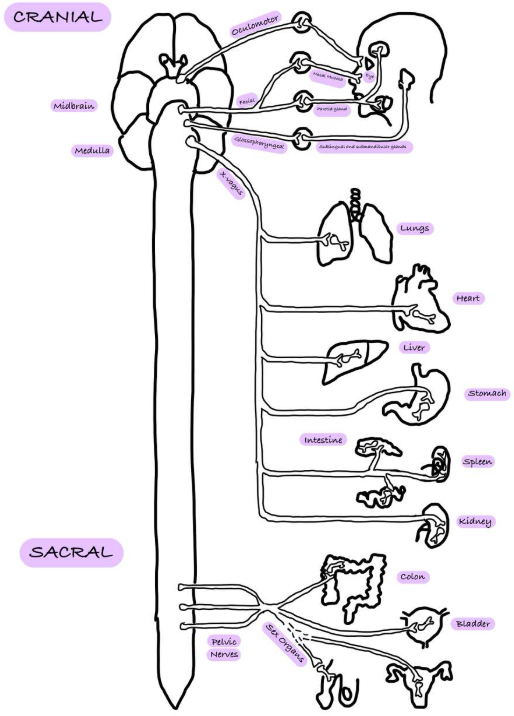
Cranial Outflow:
Oculomotor (III) - smooth eye muscles.
Facial (VII) - lacrimal/salivary glands.
Glossopharyngeal (IX) - salivary glands (specifically parotid gland).
Vagus (X) - postganglionic fibres usually in a target organ, such as the heart.
Sacral Outflow:
Sacral nerves form pelvic plexuses containing scattered ganglia.
They innervate distal parts of the intestine, bladder, ureter and reproductive organs.
Neuroeffector Pathway:
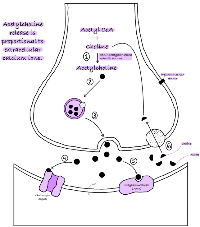
Acetylcholine is the neurotransmitter involved in this pathway.
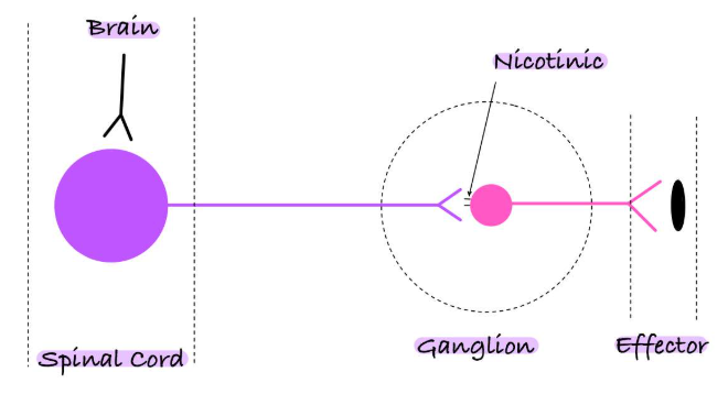
Cholinergic Synapses and Receptors:
Receptors:
Nicotinic - at ganglia; ionotropic (ion channel receptors regulate Na+ and K+ transport) - 2 ACh bind to the a-subunit proteins.
Muscarinic - at autonomic target tissues; metabotropic - G-protein coupled to secondary messenger alters K+ and Ca2+ ions.
Synapses:
ACh acts postsynaptically on nicotinic receptors at ganglionic synapses.
ACh acts postjunctionally on muscarinic receptors at the effector cell.
Parasympathetic Drug Action:
Parasympathomimetic Drugs - cholinomimetic agonists, which increase parasympathetic action.
Parasympatholytic Drugs - muscarinic antagonists, which decrease parasympathetic action.
Cholinesterase Inhibitors (ACHEIs) increase parasympathetic action.
Ganglionic Blocking Drugs - nicotinic antagonists, which decrease both sympathetic and parasympathetic action.
Co-transmission:
During low frequency stimulation, ACh is released.
During high frequency stimulation, both ACh and VIP (vasoactive intestinal polypeptide) are released.
This is co-transmission because multiple neurotransmitters (ACh and VIP) are released from the same nerve terminal, but in an activity-dependent manner.
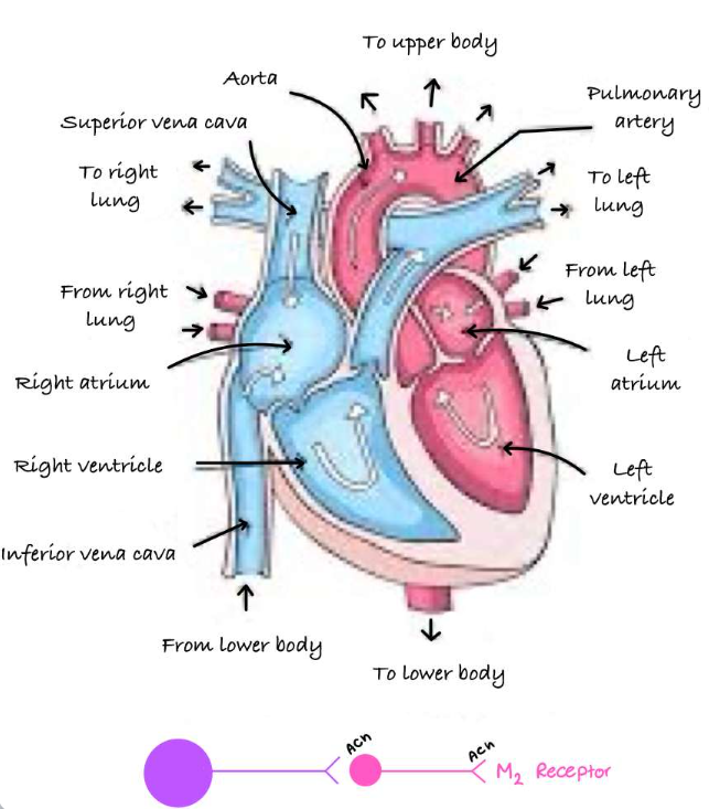
Control of the Heart:
Postganglionic nerves release ACh which acts at M2 muscarinic receptors to decrease heart function.
Sinoatrial Node - decreases heart rate.
Atrial Muscle - decreases contractility.
Atrioventricular Node - decreases rate of conduction of electrical impulses.
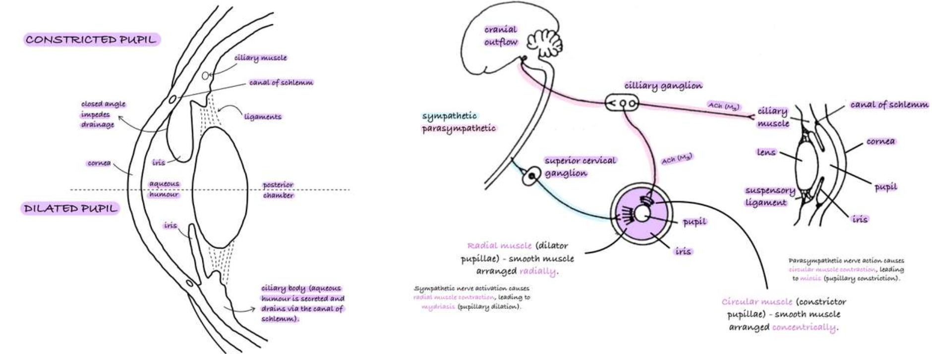
Control of Pupil Diameter:
Relaxation of ciliary muscle causes:
Suspensory ligaments to contract.
Flattened lens.
It allows for focus at long distance.
Contraction of ciliary muscle causes:
Suspensory ligaments to relax.
Enlarged lens.
It allows for focus at near distances (accommodation).
Cycloplegia is the paralysis of ciliary muscle, leading to a loss of accommodation.
Clinical Modulation of Pupil Diameter:
Mydriatic Drugs
Muscarinic antagonists which cause mydriasis (loss of drive to constrictor pupillae) and cycloplegia.
Antiglaucoma and Miotic Drugs
Muscarinic agonists which cause:
→ Pressure in the eye to build up due to the production of aqueous humour, and a lack of drainage through the canal of Schlemm.
→ Pupil dilation - the iris occludes the canal.
→ Miosis, causing an increase in outflow, and a decrease in intraocular pressure.