Urinary
Urinary Elimination Overview
Basics of the Urinary System
Major Components:
Kidneys
Ureters
Bladder
Urethra
Developmental Considerations
Children and Micturition:
Babies exhibit a micturition reflex.
Toilet training typically begins at age 2 to 3; children should be able to hold urine for two hours and communicate the need to use the bathroom.
Enuresis: Bedwetting that occurs past the age of bladder control (usually beyond 5–6 years).
Effects of Aging:
Nocturia: Increased frequency of urination at night.
Increased involuntary urination due to physical problems, such as neuromuscular issues or altered cognitive function.
Urinary Retention and Stasis: Often linked to urinary tract infections (UTIs)—adults may show confusion as a symptom of UTI.
Factors Affecting Voiding
Developmental Considerations: Age-related factors in voicing patterns.
Food and Fluid Intake:
Fluid reabsorption due to dehydration can lead to concentrated urine.
Fluid overload results in diluted urine excretion.
Body Positioning:
Men typically void sitting upright.
Psychological Variables:
Factors such as anxiety, depression, and embarrassment can hinder urination.
Activity and Muscle Tone:
Kegel exercises are important for strengthening pelvic floor muscles.
Pathological Conditions:
Congenital diseases, diabetes, renal failure, electrolyte imbalances, and medications (e.g., diuretics) can affect urination.
Urinary Terminology - Definitions
Micturition or Voiding:
The process of emptying the bladder; controlled by brain and spinal reflexes.
Urinary Incontinence:
Involuntary loss or leakage of urine; can be classified as stress, urge, overflow, or functional incontinence.
Urinary Retention:
Inability to completely empty the bladder, causing urine accumulation and potential distention.
Autonomic Bladder:
A condition in which the bladder operates reflexively without voluntary control, often due to spinal cord injury or neurological disease.
Common Urinary Symptoms & Abnormal Findings
Dysuria: Painful or difficult urination, commonly associated with UTIs.
Polyuria: Excessive urine output (>2,500–3,000 mL in 24 hours); may indicate conditions like diabetes.
Nocturia: Frequent urination at night; common in older adults or patients with chronic heart failure (CHF), diabetes, or an enlarged prostate.
Enuresis: Involuntary urination past typical bladder control ages.
Pyuria: Presence of pus or white blood cells in urine, indicating infection; urine may appear cloudy.
Urgency: Sudden strong urge to urinate, regardless of bladder fullness.
Proteinuria: Protein in urine indicating potential kidney dysfunction (may cause foamy urine).
Oliguria: Decreased urine output (<400 mL in 24 hours); potential indication of dehydration or renal failure.
Glycosuria: Glucose in urine, often seen in uncontrolled diabetes mellitus when blood glucose exceeds renal threshold (~180 mg/dL).
Frequency: Increased urinary frequency, often with small volumes, potentially due to UTI, anxiety, or pregnancy.
Anuria: Absence of urine production (<100 mL in 24 hours); indicates severe renal failure or urinary blockage.
Types of Urinary Incontinence
Transient: Sudden onset, lasting <6 months.
Mixed: Exhibits characteristics of two or more types of incontinence.
Overflow: Caused by bladder overdistention.
Functional: Results from external factors impacting the bladder.
Reflex: Bladder empties without the sensation of urgency.
Total: Continuous leakage with no control over urination.
Stress: Involuntary leakage associated with increased abdominal pressure.
Diseases Associated With Renal Problems
Congenital Conditions: Abnormalities in urinary tract development.
Polycystic Kidney Disease: Genetic disorder affecting kidney function.
UTIs: Infections, particularly common in females (Catheter-Associated UTI (CAUTI) in hospital settings).
Urinary Calculi: Kidney stones leading to urinary obstruction.
Hypertension and Diabetes: Both can cause significant renal damage over time.
Gout: Caused by high uric acid levels, can result in kidney damage.
Connective Tissue Disorders: Such as lupus, which can have renal involvement.
Effects of Medications on Urine Production & Elimination
Diuretics: Prevent water and certain electrolyte reabsorption in renal tubules, leading to increased urination. (pee alot)
Cholinergic Medications: (e.g., atropine) stimulate detrusor muscle contraction, leading to urinary retention.
CNS Depressants: Analgesics, antianxiety agents, and tranquilizers may decrease the urge to void.
Medications Affecting Urine Color
Anticoagulants: May turn urine red.
Diuretics: Produce pale yellow urine.
Pyridium: Causes urine to appear orange.
Antidepressants (e.g., amitriptyline) and B-complex vitamins: May turn urine green or blue-green.
Levodopa: Can cause brown or black urine.
Nursing Process for Urinary Elimination
Assessment:
Collect data on voiding patterns, frequency, and history of issues.
Observe nocturia and inspect urine appearance and odor.
Record intake and output (normal output around 30 mL/hr).
Physical Examination:
Palpate bladder for distention, inspecting urethral meatus for signs of infection or discharge.
Correlation of Findings: Match assessments with diagnostic test results.
Exploring Voiding Issues:
Document duration, severity, and patient perception of urinary issues.
Physical Assessment of Urinary Functioning
Kidney Evaluation:
Detailed palpation usually conducted by advanced practitioners.
Bladder Assessment:
Palpate or use bedside scanners; fullness can be checked between the symphysis pubis and umbilicus.
Urethral Inspection: Look for signs of infection or discharge.
Skin Assessment: Evaluate color, texture, hydration, and color of excreted wastes.
Urine Evaluation: Check for color, clarity, odor, and presence of sediment.
Characteristics of Urine
Color and Smell:
Dark urine indicates concentration; lighter is normal.
Ammonia smell could indicate infection.
pH Levels:
Normal range is 5–6 (4.5–8); low pH may suggest infection, diet alterations; high pH may indicate renal issues.
Urine density should range from 1.015 – 1.025; unusually high density signals dehydration.
HIGH PATIENTS IS DEHYRADTED AND LOW PATIENT IS OVERLOAD
Constituents of Urine:
Various organic and inorganic substances present in urine.
Measuring Urine Output
Procedure for Measurement:
Have the patient void into designated containers.
Use gloves and measure accurately at eye level.
Record the output; discard unless a specimen is needed.
Let patient pee alittle bit into the toilets before collecting the uri
Urine Specimens Collection Techniques
Types of Specimens:
Routine urinalysis, clean-catch or midstream specimens, sterile specimens from an indwelling catheter, and 24-hour urine collections.
Specimens in infants/children may require special techniques like point-of-care testing.
collect urines for 24 hrs and keep on ice
Question: A urine speciurinement from a patient with an indwelling catheter should be obtained from the collection receptacle, (False, the urines can be collected from the patient’s catheter itself
Care for Incontinent Patients
Scheduled toileting and measuring absorbent products can help manage leaks effectively.
Nursing Diagnoses Related to Urinary Functioning
Possible Diagnoses:
Urinary incontinence, Urinary retention;
Patterns alteration, anxiety, social isolation due to urinary issues, and caregiver role strain.
Planned Patient Goals
The patient will maintain enough urine production for fluid, electrolyte, and acid-base balance through various interventions like bladder scheduling and medication management.
Interventions for Normal Voiding Habits
Maintain a schedule, ensure privacy, and follow appropriate hygiene measures.
Strengthening of muscle tone through Kegel exercises.
Prompt response to urges to void (go to the bathroom) and increasing fluid intake as appropriate based on diagnosis.
Maintaining normal voiding habits
Promoting fluid intake depending on diagnosis
limiting sodium intake
Respond ASAP to the urge to void
Drink 8-10 glasses of water
Monitor caffeine, alcohol, or medication
schedules that promote voiding and may
interfere with sleep
Strengthening muscle tone stress incontinence
involuntary loss of urine related to increased
abdominal pressure. Kegel exercises
Assisting with toileting have a schedule and be
patient provide privacy relieve anxiety and
discomfort
Hygiene promote proper perineal care helps for
bedridden patients and immobilized patient
Patients at Risk for UTIs
At-Risk Group:
Sexually active women, those using diaphragms for contraception, and those with indwelling catheters or diabetes.
Education on hygiene practices is crucial (e.g., wipe front to back).
postmenopausal women
older patients confusion burning on urination
medical term:
bacteriuria: bacteria in the urine
bacteremia: bacteria in the bloodstream
pyelonephritis: kidney infection
cystitis: bladder infection
Reasons for Catheterization
Primary Reasons:
Relieve urinary retention, obtain sterile specimens, accurately measure output, aid in wound healing, and provide comfort in palliative care.
Emptying the bladder before, during, or after select surgical procedures and
before certain diagnostic examinations. Pregnant women may get before c
section
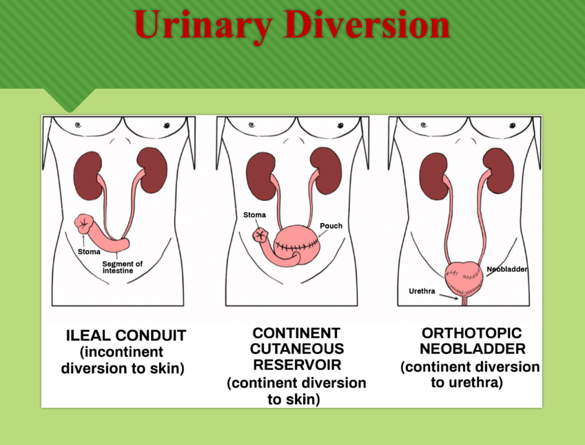
A urinary diversion may be performed when bladder has
been removed, dysfunction or neurogenic disfunction with
frequent UTI’s
Ureterostomy or ileal conduit is a permanent incontinent
urinary diversion created by transplanting the ureters into a
closed off portion of the intestinal ileum and bringing the
other end out onto the abdominal wall forming a stoma.
Urine collected through a pouch.
2 types Continent urinary reservoir you are in control of this
ostomy
Orthotopic neo-bladder uses a ileal pouch to replace the
bladder
Urinary Diversion and Care Information
Indications for a Urinary Diversion:
Conditions necessitating bladder removal or severe dysfunctions.
An ileal conduit allows urine to be collected in a pouch.
Two main types: continent urinary reservoir and orthotopic neobladder allow for bladder replacement.
Patient Education for Urinary Diversion
Instruction on care management, self-care behaviors, potential fears, and concerns is critical for those undergoing urinary diversion.
Explain reason for diversion and
rationale for treatment
Demonstrate effective self-care
behaviors
Describe follow-up care and support
resources
Report where supplies may be
obtained in the community
Verbalize related fears and concerns
Demonstrate a positive body image