P1: Introduction to Radiology
Imaging Diagnostics
- x-ray
- Radiodiagnostics
- skiagraphy
- single picture e.g. new radiogram every hour to track contrast from administration to GIT
- sciascopy
- see real-time motion e.g. peristaltic action of GIT
- Radiotherapy
- used for therapy of tumour
- Ultrasonography
- CT
- MRI
- Gamma camera (PET scan)
- Non-ioninsing
- ultraviolet
- light
- infrared radiation
- microwaves
- radiowaves
- Ionising (mutagen; dematches DNA)
- X-ray
- CT
- Gamma camera
Primary Radiation: radiation generated in the focus anode X-ray tubes, called the primary radiation
Extra-focal Radiation: radiation produced outside the focus
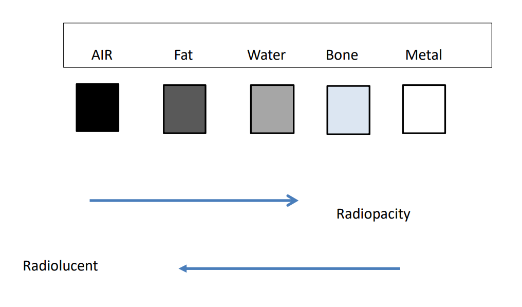
Air: nothing absorbs electrons, so all reach the cassette → black
Fat: very little of the electrons are absorbed → dark grey
Soft Tissue/Fluid: more electrons are absorbed, around 1/2 reach cassette → medium grey
Bone: most of the electrons are absorbed, very few reach cassette → white
Metal/Positive Contrast Medium: all electrons are absorbed, none reach the cassette → super-white
Radiographic Report:
- analysis of radiogram
- position e.g. lateral recumbency
- species e.g. dog, cat, rabbit, snake etc
- sex e.g. female or male
- description of pathology
- location
- size/measurement
- count e.g. stones in urinary bladder
- density e.g. radiolucent as fat
- shape
- structures
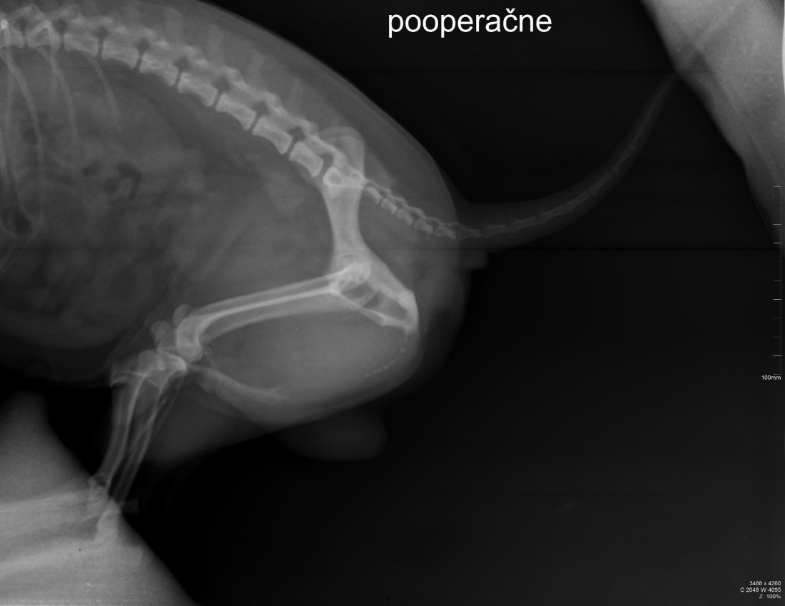
e.g.
lateral recumbency
dog (square vertebrae)
male (os penis)
flexura urethralis
particles with radiopacity of bone
8-9
irregular shape
urinary stones
- X-ray tube (cathode [emission of electrons], anode [
- mA - total dose of electrons
- kV
- time
1. very low doses of electrons (mA, kV) or time
1. not powerful enough for penetration 2. radiogram is white (underexposed) 2. high doses of electrons (mA, kV) or time
1. too powerful for thinner areas e.g. carpus, better for thorax 2. radiogram is black (overexposed)
1. cannot see a contrast between bone and soft tissue
- Patient
- parallel with cassettes
- minimum 2x radiograms
1. e.g. thorax laterolateral & ventrodorsal or dorsoventral 2. must be able to see multiple perspectives to identify pathology
- two types of patient
1. emergency
1. no preparation 1. 2. normal
1. preparation
1. sedation, anaesthesia 2. starved, no faeces in intestines
- Distance
- x-ray tube → cassette: 80-100 cm
- patient → cassette: must be touching cassette if distance is less than 10 cm or on a secondary grid (eliminates secondary radiation) if more than 10 cm
Evaluation of long bone must include proximal and distal joints
In the Slovak Republic:
- The owner restrains the animal whilst taking radiograms
- Male or Female?
- Age? (must be over 18)
- Are you healthy? (cancer, thyroid issues etc.)
- Are you pregnant? (if female)
- MUST SIGN BOOK SAYING THEY UNDERSTAND THE RISKS
- Personal Protection Equipment
- Metal dress
- Metal throat cover
- Wrist/arm protector
- Goggles (if necessary)