Lecture 16 – Anatomy of Hearing.docx
Lecture 16 – Anatomy of Hearing
19.03.24
- 1. The external ear
- Everyday use of the word “ear” means technically the external ear
- External ear consists of
- auricle (pinna)
- Meatus (ear canal)
- Tympanic membrane (ear drum)
- Task of the auricle: localize the sound source (important: evolution)
- Tympanic membrane works like a loudspeaker or microphone membrane
- External auditory meatus
- External ear canal
- 7 mm in diameter and 2.5 cm long
- This generates resonane frequencies at 3400 Hz
- From an acoustic point of view: ear canal is a filter than amplifies frequencies between 2 kHz and 5 kHz
- Terminates at the tympanic membrane
- Two-third of ear canal housed in bone (osseus portion)
- One-third of ear canal composed of cartilageous parts
- Resonating cavity that contributes to hearing
- Determine resonant frequency
- Outer third-line with hair cells and cerum (ear wax) – protects by trapping dirt and insects
- Tympanic membrane
 Also known as eardrum
- Separates the middle ear from the outer ear
- Oval shaped, 10 mm in diameter
- Thin three-layered sheets of tissue
- Landmakrs
- Umbo – point of attachment for maleus, middle ear bone – location is cone of light (reflects light from otoscope)
- Responsible for initiating mechanical impedance-matching process of middle ear
- First layer: outer (cuticular) layer
- Second layer: intermittent (fibrous) layer
- Third layer: inner (mucous) layer
- 2. The middle ear
- The middle ear consists of the tympanic cavity
- This cavity contains the smallest moving bones of the human body – the ossicles
- Malleus (hammer): touches the tympanic membrane and transmits to
- The incus (anvil) which transmits to the
- Stapes (stirrup) which transmits to the internal ear (oval window)
- Malleus and stapes are attached to muscles (may attenuate to transmission of sound by these bones
- This cavity contains the smallest moving bones of the human body – the ossicles
- The stapes connects directly to the internal ear through the oval window 🡪 transmission of stapes movement to the lymphatic fluid inside the internal ear
- The middle ear consists of the tympanic cavity
- Ossicles
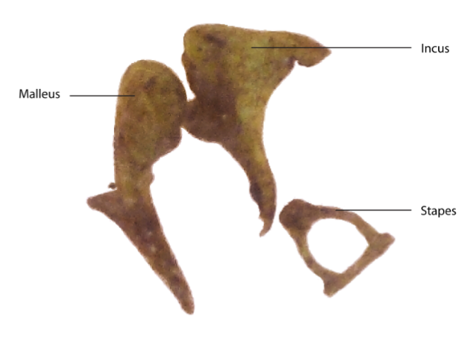 Malleus
 - Largest of the ossicles (9 mm long and weighs only 25 mg)
- Provides point of attachment with tympanic membrane
- Bulk of bone is the head or caput
- Incus
- Shaped like an anvil
- Weight 30 mg and is around 7 mm long
- Provides intermediate link of ossicular chain
- Incus and malleus articulate by means of a saddle joint
- Stapes (stirrup)
- Third bone of ossicular chain
- Weights 4 mg with an area of 3.5 mm^2
- Helps to transmit sound vibrations from eardrum to oval window
- Articulation of the incus and stapes of ball and socket type
- Ossicular chain is held in place by ligaments
- Tympanic muscles
- Muscles of middle ear attached to ossicles
- Smallest muscles of human body
- Stapedius muscle
- Imbedded in posterior wall of middle ear
- Pulls stapes posteriorly
- Tensor tympani
 Pulls malleus anterior and medial
- Stapedius muscle
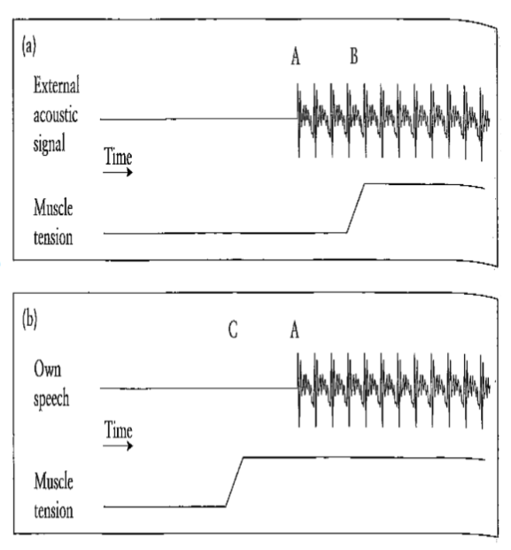
- Sound attenuation in the middle ear
- The middle ear performs a kind of “volume control” 🡪 muscles of malleus can be tensed, resulting in a low frequency damping
- It must be activated though by neural impulses 🡪 in order to be activated as noise control, the noise has to be processed by the internal ear 🡪 damage could have been occurred already
- Additionally, these muscles are activated just before a person starts to speak 🡪 damping mechanism to protect against own voice
- Pressure increase in the middle ear
- Sound waves are mechanically transmitted by ossicles of the middle ear to the internal ear, which is filled with watery liquid
- Ossicles perform conversion of pressure changes from an elastic medium (air) to pressure changes of an incompressible liquid (water)
- Ossicles function like a cone: from large surface (tympanic membrane) to a smaller surface (stapes)
- This leads to a pressure increase 🡪 pressure variations at the interal ear are about 20 times stronger than original air pressure variation
- This pressure increase is necessary to generate the necessary activation of the liquid (otherwise reflection would occur)
- Pressure equalization in the tympanic cavity
- The middle ear is not completely airtight, a connection with the eustachian tube allows for pressure equalization (e.g. meteorological pressure changes)
- The Eustachian tube leads from the middle ear to the nasopharynx
- Without pressure equalization, the meteorological changes would “push” the ear membrane inwards 🡪 feeling of “pressure on the ear” (felt e.g. when going downhill, or in an airplane, or fast elevator)
- The middle ear is not completely airtight, a connection with the eustachian tube allows for pressure equalization (e.g. meteorological pressure changes)
- 3. The internal ear
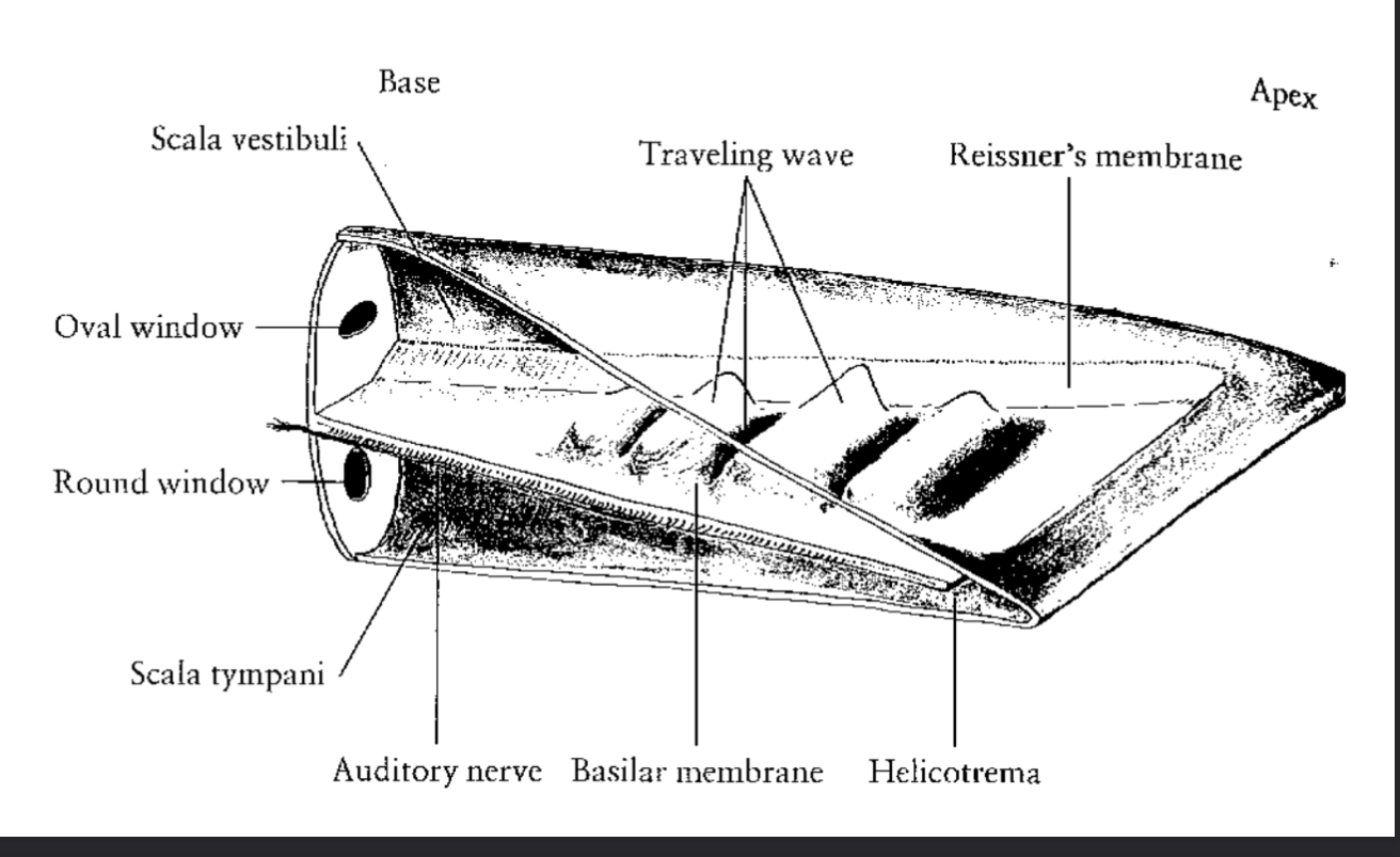
- Cochlea: part of the inner ear relevant for hearing 🡪 sound waves are transformed into neural impulses
- Is shaped like a snail shell
- Contains two passages, separated by basilar membrane
- Upper: scala vestibuli
- Lower: scala tympani
- These two passages meet at the apex (the tip) in the helicotrema
- Scala vestibuli connects to middle ear (stapes) through the oval window
- Cochlea: part of the inner ear relevant for hearing 🡪 sound waves are transformed into neural impulses
- The “uncoiled” cochlea
- Physiology of hearing
- The pressure waves from the middle ear (stapes) reach the cochlea through the oval window 🡪 longitudinal pressure waves are generated in internal ear fluid through scala vestibuli to the apex
- These pressure waves return via scala tympani to the round window
- Round window serves as pressure release, since the fluids are incompressible