w3: tissue level of organisation
Video1: epithelial tissue
Structural organisation of the human body
Atoms > molecules > macromolecules > organelle > cell > tissue > organ > organ system > organism
Types of Tissues
Four main types of tissues:
Epithelial Tissue: Lines surfaces, provides protection.
Connective Tissue: Connects/supports structures.
Muscle Tissue: Includes skeletal, cardiac, and smooth muscle.
Nervous Tissue: Communicates messages throughout the body.
Epithelial Tissue Functions
Protection: Forms barriers (e.g., skin).
Selective permeability: Controls the passage of substances.
Secretion and absorption: Vital for bodily functions (lungs, intestines).
Filtration and sensory reception.
The Embryonic Origin of Tissues
Starting point: Fertilised Zygote → Blastocyst
A fertilised egg develops into a blastocyst, which then differentiates into 3 primary germ layers
The 3 Germ Layers:
Ectoderm (outer layer)
Forms the exoskeleton (skin, nails, hair)
Also gives rise to neurons and epithelial cells
Mesoderm (middle layer)
Develops into organs
Produces red blood cells and cardiomyocytes (heart muscle cells)
Endoderm (inner layer)
Forms the inner lining of organs
Gives rise to liver cells and pancreas cells
Key concept:
Every tissue in the body can be traced back to one of these three layers
This process is called gastrulation, when the blastocyst reorganises into the three distinct germ layers
Structure of Epithelial Tissue
All epithelial tissues share certain characteristics:
Highly cellular: Dense packing with minimal intercellular substance
Attached to a basement membrane for anchorage.
Have cellular polarity:
Apical surface faces the exterior/interior lumen.
Exterior = faces outside the bodye.g. skin surface, exposed to the outside world
Interior lumen = faces the inside of a hollow organ or tube e.g. the inside of your intestines, blood vessels, bladder, airways
The basal surface connects to the basement membrane.
Lateral surfaces are adjacent to neighbouring cells.
Epithelial tissue is avascular: It relies on the underlying connective tissue for nutrients. (lacks direct blood supply)
Basement Membrane
Key role in anchoring/gluing epithelial tissues to connective tissues.
Semi-permeable: controls the exchange of nutrients and waste products
Structure separates the epithelium from the connective tissue directly
Thin (20-100 nanometers), made up of fine granular fibrous proteins (collagen) and glycans (proteoglycans).
Contains two layers: the basal lamina (epithelial) and the reticular lamina (connective).
Video2: lining epithelium
Types of Epithelial Tissues
Two Main Types: Lining epithelia and glandular epithelia.
Lining Epithelia
Definition: Epithelial membranes covering internal organs and cavities, and surfaces exposed to the external environment.
Function: Serve as protective barriers.
Classification Criteria
Number of Layers:
Simple Epithelium: One layer of cells.
Stratified Epithelium: More than one layer (2 to 100+ layers).
Cell Shape:
Squamous: Flat and wide.
Cuboidal: Cube-like, equal height and width.
Columnar: Tall and skinny.
Simple Epithelia Types
Simple Squamous:
Location: Blood vessels, lungs, lining of the heart, lymphatic vessels
Function: allows materials to pass through by diffusion and filtration, and secretes lubricating substance
Simple Cuboidal:
Location: ducts and secretory portions of small glands and kidney tubules
Function: secretes and absorbs
Simple Columnar:
Location: bronchi, uterine tubes, uterus (smooth nonciliated tissues) in the digestive tract, bladder
Function: absorbs and secretes mucous enzymes
Pseudostratified Columnar:
Structure: Appears stratified but is a simple epithelium with cilia and goblet cells.
Location: Lines trachea and upper respiratory tract
Function: secretes mucus and ciliated tissue moves mucus
Stratified Epithelia Types
Stratified Squamous:
Location: esophagus, mouth and vagina
Function: protects against abrasion
Stratified Cuboidal:
Location: Sweat glands, salivary glands, and mammary glands
Function: protective tissue
Stratified Columnar:
Location: Male urethra and ducts of some glands
Function: secretes and protects
Transitional Epithelium:
Structure: Changes shape between squamous and cuboidal.
Location: Bladder, urethra, and ureters
Function: allows urinary organs to expand and stretch
Specialised Epithelial Tissues
In addition to classical simple/stratified epithelia, there are 4 specialised types: Endothelium, Mesothelium, Respiratory epithelium, and Transitional epithelium.
Endothelium (simple squamous epithelium):
Structure
Single layer of thin, flat cells
Smooth, continuous lining
Sits on a basement membrane facing the lumen
Function
Diffusion and gas exchange
Location
Lines cardiovascular and lymphatic vessels (e.g. capillaries, veins, arteries)
Mesothelium (simple squamous epithelium):
Structure
Thin layer with lubricating cells
Supported by dense connective tissue beneath
Function
Protection of organs
Supports movement (reduces friction between organs)
Location
Lines organs and body cavities
Specifically: pleura (lungs), peritoneum (abdominal cavity)
Keratinised Stratified Squamous:
Structure
Anucleate (dead) cells in the superficial/outer layers
Keratinised surface
Tough and resistant to tearing
Function
Protection
Forms an impermeable layer
Location
Skin
Non-Keratinised Stratified Squamous:
Structure
Nucleated squamous cells in superficial layers (cells are still living)
Upper layers protect the underlying tissues
Sits on a basement membrane above the lamina propria
Function
Protection
Location
Moist surfaces where tubular systems open to the outside
Lining of mouth, esophagus, upper nostrils, vagina
Respiratory Epithelium (mucociliary escalator):
Structure
Pseudostratified appearance
Columnar and ciliated cells
Contains goblet cells that secrete mucus
Function
Air filtration via the mucociliary escalator:
Traps pathogens and dust in mucus
Cilia move mucus up and out, away from the lungs
Location
Lines the entire respiratory tract
Nasal passages through to the bronchi
Transitional Epithelium:
Structure
Stretched state: appears as stratified squamous
Relaxed state: appears as stratified cuboidal
Function
Acts as an osmotic barrier (regulate the passage of water and solutes)
Allows for contraction and expansion of the organ
Location
Lining of ureters and bladder
Video3: Glandular epithelia
Glandular Epithelium
Glandular epithelium produces secretions (e.g., mucus, sebum, milk, gastric juices)
A lumen is a hollow channel within a tubular structure, such as blood vessels or the gastrointestinal tract
Glandular cells may be unicellular (e.g., goblet cells) or aggregate to form multicellular glands.
Glandular epithelial cells grow down into the connective tissue and form glands
Epithelial glands form by invagination/infolding
Classification of Glands
Two primary types of glands:
Exocrine Glands: Secrete substances through ducts; either into the limen or onto the surface of the epithelium (sweat glands, salivary glands, mammary glands, pancreas)
Endocrine Glands: Secrete hormones into the extracellular space > transported by bloodstream; long-distance (e.g., adrenal, pituitary, thyroid, pancreas).
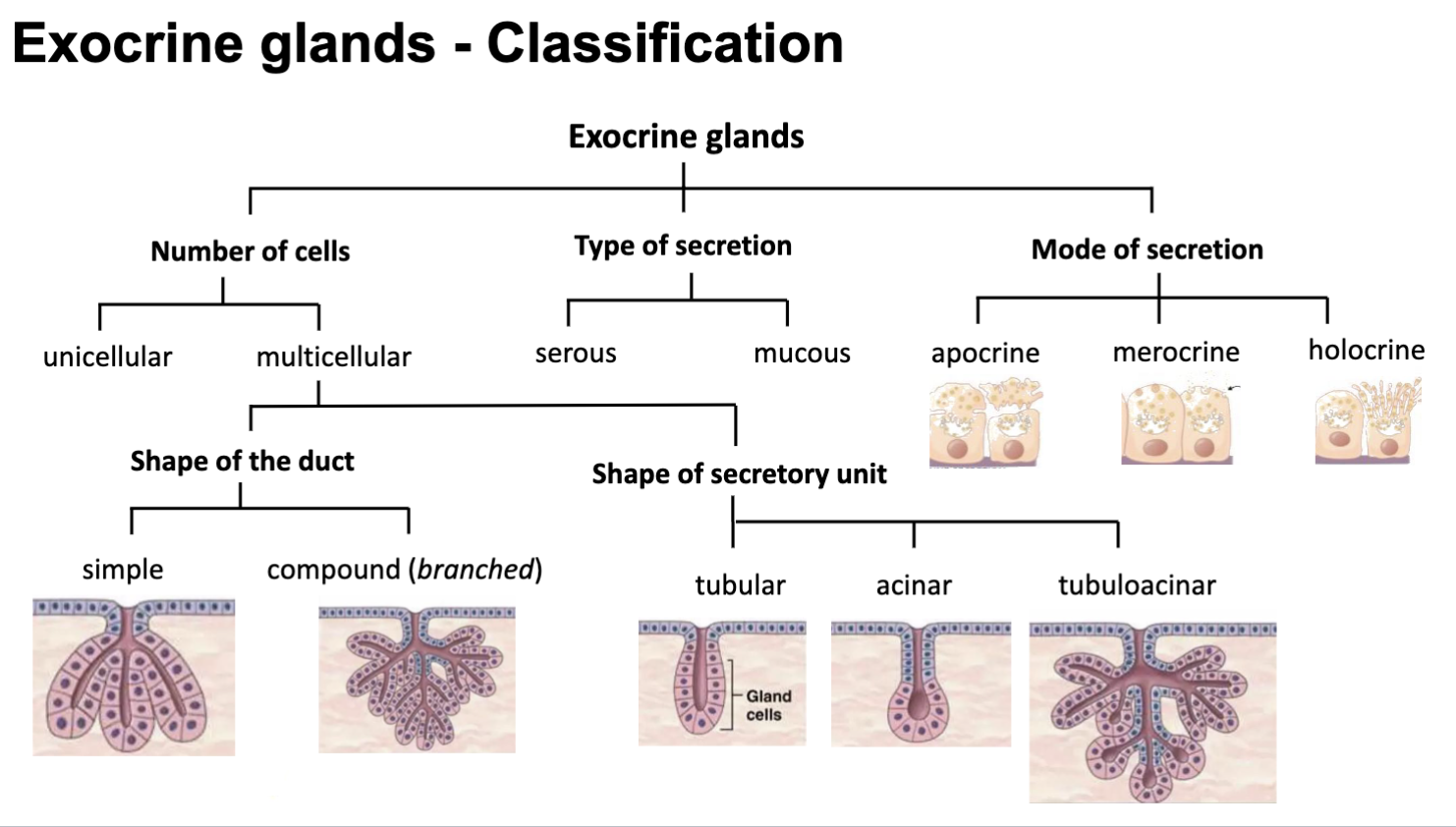
Exocrine Glands Classification
Exocrine Glands — Classification
Number of cells
Unicellular — gland made up of a single secretory cell (e.g. goblet cells)
Multicellular — gland made up of many cells working together
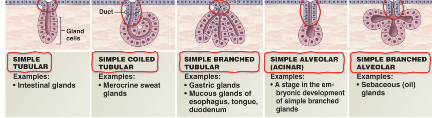
Shape of the duct (multicellular only)
Simple — single, unbranched duct leading to the surface (can be tubular, coiled tubular, branched tubular, simple acinar or simple branched alveolar
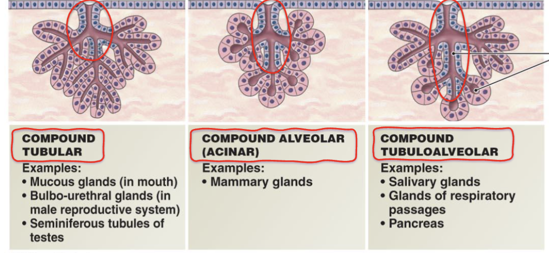
Compound — duct is branched, allowing secretions from multiple units to drain through one opening (can be compound tubular, compound alveolar, compound tubuloalveolar)
Type of secretion
Serous — produces a thin, watery, protein-rich fluid (e.g. digestive enzymes)
Mucous — produces thick, viscous mucus for lubrication and protection
Shape of the secretory unit
Tubular — secretory unit is a straight or coiled tube shape
Acinar — secretory unit is rounded/flask-shaped (also called alveolar)
Tubuloacinar — combination of both tubular and acinar components in the one gland
Mode of secretion
Apocrine — part of the cell itself is pinched off and released along with the secretion (e.g. mammary glands)
Merocrine — secretion is released via exocytosis (vesicles)
Holocrine — the cell ruptures (e.g. sebaceous/oil glands)
Video4: cell junctions
Overview of Epithelial Tissues and Cell Junctions
Epithelial tissues perform various roles: protection, control the passage of substances, filtration and sensory reception.
Tight packing of epithelial cells resembles a brick wall, minimising intercellular space.
Structural integrity relies on cell junctions, which connect the plasma membranes of adjacent cells.
Functions of Cell Junctions
Because epithelial cells are packed so tightly together, they need special connections between them called cell junctions, which physically link neighbouring cells together and help control what passes between them
Connect cells and surrounding structures
Withstand mechanical stress
Facilitate communication
Types of Cell Junctions
Tight Junctions (occluding junction)
Eliminate free space between cell membranes
located apically (near the top)
form a waterproof seal
function as a strict control of substance passage, forces substances to pass through receptors on cell membranes
Found in areas like the blood-brain barrier, stomach, and bladder.
Adherens Junctions
joining of actin filaments of adjacent cells (cytoskeletal enforcement)
located midway
forms a belt
functions: regulate cell structure, cell adhesion and cell division
Typically found in epithelial tissues and blood vessels.
Desmosomes
Connect adjacent cells through protein fibres (joining of cytoskeletal protein filaments)
form locaslised spot junctions
function: cell adhesion and provides mechanical strength and stability
Found in the skin, heart muscle (myocardium), and bladder.
Differentiate from adherens junctions: localised spot junctions with deeper cellular attachment.
Hemidesmosomes
Half a desmosome; connects cells to the underlying basement membrane instead of to each other.
Located on the basal surface, anchoring cells to connective tissues.
located on epidermis of the skin
Gap Junctions
aligned channel protein pores that connect adjacent cells allowing cytoplasmic communication.
Permit small molecules, nutrients, and signalling molecules to pass.
faciliates cell communication and allows sheets of cells to function in unison
Workshop
What type of Epithelial cell lines the trachea: Pseudo stratified
Goblet cells make mucus that catches dust and dirt particles
Cilia help to move substances across the cell surfaces, moving crap out of the respiratory pathway; cilia also trap dust/dirt particles from moving down the respiratory airways
Cellular adaptation under ongoing stress is called metaplasia
cellular adaptation?
Overview of the Integumentary System
Skin is the largest organ of the body.
Functions of Skin
Protection: Shields against UV radiation, pathogens, and physical injury.
Immunity: First line of defence against infections.
Sensation: The largest sensory organ, it detects pressure, temperature, and pain.
Thermoregulation: Maintains core body temperature (~37°C).
Water Balance & excretion: Regulates hydration and excretes metabolic wastes (urea, uric acid, ammonium).
Vitamin D Production: Activates vitamin D through UV light for biochemistry and musculoskeletal health.
Structure of Skin
Composed of three layers:
Epidermis:
Stratified squamous keratinised epithelium tissue, providing a waterproof barrier.
Dermis:
Dense connective tissue layer supporting the epidermis.
Hypodermis:
Adipose tissue that cushions, insulates, and stores energy.
Layers of the Epidermis
statified squamous keratinised epithelium
composed of keratinocytes (specialised epithelial cells that form the protective layer
Epidermis is avascular, keratinised, attached to connective tissue via basement membrane, semipermeable
Composed of five distinct layers, from outermost to innermost:
Stratum Corneum: Dead, flat keratinised cells; protective. No nuclei, no organelles. Forms waterproof barrier. Desquamation = cells shed
Stratum Lucidum: Only in thick skin (palms/soles); keratinocytes filled with eleidin (keratin).
Stratum Granulosum: Keratinocytes secreting/accumulating keratin granules as they migrate towards the surface; thin granular layer.
Stratum Spinosum: Thick layer with desmosome microfilaments; provides strength and flexibility. Multiple layers of keratinocytes tighly held together
Stratum Basale: Innermost layer; continuously reproduces keratinocytes (also called stratum germinativum). Basal keratinocytes = stem cells.
Skin Types Comparison
Thin Skin:
Covers most of the body, no stratum lucidum, thinner stratum corneum.
Thick Skin:
Present on palms and soles, thicker stratum corneum, contains stratum lucidum, no hair follicles.
Cell Types in the Epidermis
Keratinocytes
Comprise ~90% of epidermal cells.
Develop in stratum basale and mature as they ascend.
Key roles:
Provide protection, structure and regeneration of epidermis.
Produce keratin and lipids for a waterproof barrier.
Aid in calcium regulation by enabling UVB absorption and vitamin D activation.
Langerhans Cells
Specialised antigen-presenting dendritic cells of the immune system.
Derived from bone marrow; found mostly in stratum spinosum.
Functions:
Provide first-line immune defence against pathogens.
Monitor environment for threats; process and present antigens to initiate immune response.
originate in bone marrow > travel to skin via blood > ingest foreign antigens > present foreign antigens on cells surface > migrate to lymph nodes and warn immune system of invaders
Melanocytes
Responsible for melanin production, which gives skin its colour.
Located in the stratum basale
Melanin is secreted by melanocytes and then packaged into melanosomes
Melanosomes are ingested by keratinocytes (endocytosis)
Keratinocytes move to apex of cell to protect nucleus from UV
Melanin is produced in response to UV exposure, packaged in melanosomes, and distributed to keratinocytes.
Variations in skin colour relate to melanocyte activity, not quantity.
Merkel Cells
Found in stratum basale; function as mechanoreceptors (sensory receptors that gather information about the environment).
Merkel cells are modified epidermal cells that detect changes in pressure and stretch; relay sensory information to the nervous system.
Concentrated in highly sensitive areas (fingertips, lips); crucial for touch perception.
Cells of the Epidermis
Keratinocyte
Specialised epithelial cell found throughout the entire epidermis
Produces keratin and lipids to form a waterproof barrier
Helps activate Vitamin D
Langerhans Cell
Immune cell located in the stratum spinosum
Acts as the skin's first line of immune defence by detecting and presenting foreign antigens
Melanocyte
Melanin-producing cell located in the stratum basale (deepest layer)
Gives skin its pigmentation
Protects DNA from UV damage
Merkel Cell
Sensory cell also located in the stratum basale
Detects touch, stretch and pressure on the skin
Quick memory tip: think of each cell by its job — Keratinocytes = waterproofing, Langerhans = immune guard, Melanocytes = sun protection, Merkel = touch sensor.
Connection Between Epidermis and Dermis
Junction between epidermis and dermis is corrugated, not straight.
Features epidermal ridges (downward extensions) and dermal papillae (upward extensions) - increases surface area and structural integrity.
The epidermis (outer skin layer) depends on the dermis (underlying connective tissue) for support and nourishment, while the dermis relies on the epidermis for protection against environmental factors and overall skin integrity.
Junction contributes to fingerprint formation.
Dermis Structure
Divided into two layers:
Papillary Dermis:
Thin, loose connective tissue below the epidermis.
Loose CT - Rich in cells and ground substance; contains blood vessels (nourish the epidermis).
Reticular Dermis:
Dense irregular connective tissue contains thick collagen fibres for strength and support.
Both layers support epidermis, contribute to thermoregulation and sensation
Hypodermis
Innermost skin layer primarily composed of adipose tissue (fat cells).
Functions include energy storage, insulation, and cushioning of tissues beneath.
Ancillary Structures of the Skin
Include hair follicles, arrector pili muscles, sebaceous glands, nerves, and sweat glands.
Function to maintain skin integrity, respond to stimuli, regulate temperature, enhance health.
Hair Follicles
Originate mostly in the dermis (occasionally hypodermis); absent in thick skin areas (palms, soles).
Associated with sebaceous glands (secrete oil) and arrector pili muscles (control hair position).
Involved in touch sensation and thermoregulation.
Thermoregulation Mechanism
Cold Environment:
Blood vessels in the dermis constrict to prioritise blood flow to vital organs.
Arrector pili muscles contract, causing goosebumps and hair to stand straight to trap heat.
Hot Environment:
Blood vessels dilate, increasing blood flow towards the skin surface for heat exchange.
Sweating facilitates heat loss through evaporation.
Arrector pili muscles relax allowing heat to escape
Sebaceous Glands
Located in dermis, connected to hair follicles.
Produce sebum (oil) regulated by hormones, active from puberty.
Functions include skin lubrication, moisture, and antimicrobial properties (combat bacteria).
Sweat Glands — Two Types
Merocrine (Eccrine)
Duct opens directly onto the skin surface
Found all over the body, especially palms, soles of feet and forehead
Smaller — 30-40um in diameter
Produces watery secretion (water and salt/NaCl)
Function: thermoregulation (cools the body down)
Apocrine
Duct opens onto a hair follicle
Found in densely hairy areas e.g. armpits
Larger — 80-100um in diameter
Produces thick, viscous secretion (proteins and fatty acids)
Function: associated with body odour when the secretion is broken down by bacteria on the skin
Quick memory tip: Merocrine = cooling sweat (watery, everywhere), Apocrine = smelly sweat (thick, hairy areas)
Nerves in the Skin
High density of nerve endings; skin serves as the body's contact with the environment.
Different nerve fibres detect temperature, touch, pain, and pressure, relaying information to the nervous system.
Types of Tissues
Human body consists of four tissue types: epithelial, connective, muscle, and nervous.
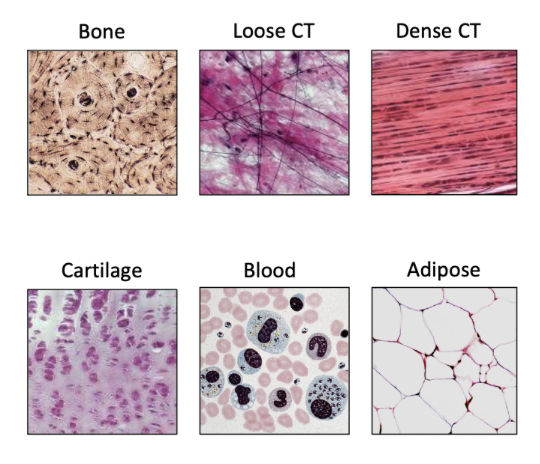
Connective Tissue Overview
Connective tissues vary in form (e.g., bone, cartilage, fat, blood).
Structures like tendons and ligaments facilitate movement.
Loose connective tissues under the skin provide support.
Key components of all connective tissue: (1) cells (variation in types and numbers), (2) fibres (collagen, elastin, reticulin), (3) amorphous ground substance (gel-like, fills spaces).
Together, fibres and ground substance form the extracellular matrix.
Extracellular Matrix
Surrounds cells; properties vary across connective tissues.
Example: blood matrix is fluid; bone matrix is solid and calcified.
Functions of Connective Tissue
Provide support and protection for organs.
Connect tissues and organs while allowing flexibility for movement.
Facilitate communication between cells and tissues for homeostasis (molecule messenger, passing of nutrients, gases and waste)
Store energy and cushion internal organs (e.g., adipose tissue), assist in defence (immune cell transport), and repair tissue damage (adipose tissue).
Mechanical support: the skeleton protects organs; the skin acts as a defence barrier.
Examples of connective tissue for each type:
Bone (supportive CT): provides structural support, facilitates movement by serving as attachment points for muscles, and houses bone marrow, which produces blood cells.
Cartilage (supportive CT): Provides flexibility and cushioning at joints.
Dense regular CT: Found in tendons and ligaments, it resists pulling forces in one direction.
Dense Irregular CT: found in the dermis, it provides strength and structural support while allowing flexibility in multiple directions.
Blood (fluid CT): Transports nutrients and waste throughout the body.
Adipose CT: Stores energy in the form of fat, provides insulation, and cushions organs.
Classification of Connective Tissue
Fluid Connective Tissues: blood and lymph (transport nutrients, oxygen, waste).
Supportive Connective Tissues: cartilage and bones (physical support).
Connective Tissues Proper: subdivided into:
Loose connective tissues (fewer fibres, more ground substance).
Dense connective tissues (more fibres, categorised as regular or irregular).
Specialised Connective Tissues: adipose tissue (insulation, energy storage).
Connective Tissue Proper
Subdivided into loose and dense categories based on structural properties:
Loose Connective Tissue: high ground substance, supports organs, nourishes cells, provides passage for immune cells. Located underlying epithelial tissue, covering blood vessels and nerves, fascia between muscles and pleural and pericardial sacs
Dense Regular Connective Tissue: fibres in parallel, strong tensile strength, found in tendons and ligaments.
Dense Irregular Connective Tissue: random fibre arrangement, provides multidirectional strength, found in dermis and around organs.
Summary of Dense Connective Tissues
Dense Regular: strong, unidirectional support.
Dense Irregular: multidirectional support, physical protection from injury.
Loose Connective (areolar tissue, adipose tissue & reticular tissue): support for organs, nourishment, and a passageway for blood vessels and nerves.
All connective tissue contains, extracellular matric(solid, liquid, syrup/viscous), fibres and cells