Body Log - Flashcards making:
Describe the composition of cartilage in terms of its cells and extracellular components
Classify the 3 major types of cartilage and describe the structural differences between them
Relate the different anatomical distribution of the 3 major types of cartilage to their different functions
Recognise the following in photomicrograph and discuss their anatomical locations and salient histological features in relation to their function: Hyaline cartilage (trachea), Hyaline articular cartilage, elastic cartilage, fibrocartilage
Outline the cause and morphological features of achondroplasia
Explain how bone is a living tissue and how it is formed and how it undergoes remodelling
Describe the microstructure of compact (cortical) and spongy (cancellous/ trabecular) bone
Recognise compact bone (including its cells, lacunae, canaliculi, Haversian and Volkmann’s canals, lamellae and periosteum) and trabecular bone from photomicrographs
Explain the importance of vitamin D in normal bone stability
Describe how the morphology and/ or mechanical properties of bone can change in disease
Describe the genetic basis and phenotypical changes in Osteogenesis imperfecta
Outline the consequences of four different and contrasting diseases for the affected individual: Rickets different to Osteomalacia and the difference between the two conditions and explain the importance of diet and behavioural factors in their prevention
For Osteoporosis: Describe the radiological and structural changes which occur + List the most common risk factors and explain the importance of osteoporosis as a risk factor for fractures in elderly
Explain how bone is a living tissue, how it is formed and how it undergoes remodelling
Understand the key role water plays as a structural and functional component
Appreciate how limited nutrient diffusion affects chondrocyte metabolism and ECM maintenance
Briefly outline the differences in appositional and interstitial growth in Hyaline cartilage growth
Describe how Hyaline cartilage acts as a template to long bone growth in endochondral ossification
Describe Epiphyseal Growth plate zonation - their roles in new growth for developing bone
Briefly outline the general features of Achondroplasia and how it results in dwarfism
How is bone a living tissue?
Highly vascular (good blood supply)
Innervated (supplied by nerves)
Metabolically active capable of growth, remodelling and repair
Bone stores calcium and phosphorus and serves as a reservoir for these essential minerals
How is Bone formed? Brief overview:
Ossification occurs in two main ways: Intramembranous and endochondral

Intramembranous:
Bone is going to directly form from the mesenchyme with osteoblasts forming osteoid. Examples of this is most bones in skull and facial bones form
Endochondral:
Where mesenchyme cells first form cartilage which serves as a template, this cartilage gets replaced by bone. KEY POINT: The cartilage does not turn into bone, it gets replaced by bone. Examples of this is most of the long bones: humerus, femur, vertebrae and ribs
Detailed process of Intramembranous bone formation: We dont need to know?


Mesenchyme is embryonic connective tissue containing mesenchymal stem cells. Mesenchymal cells condense, meaning cells become closely packed together.
Mesenchymal stem cells differentiate into osteoblasts, osteoblasts are bone-forming cells
Osteoblasts secrete osteoid, which is unmineralized bone matrix. At this point mainly contains Type 1 collagen and ground substance
Osteoblasts then release calcium salts in the form hydroxyapatite crystals, which are deposited into osteoid
As the osteoblasts are producing bone matrix around themselves, some osteoblasts become completely surrounded by the matrix. So, the osteoblasts that become trapped in their own matrix are called osteocytes. Therefore, the little space containing the osteocyte is called a lacuna.
Osteocytes aren’t completely isolated inside their lacunae, they have long thin cytoplasmic processes that extend through tiny channels called canaliculi (which allow communication and exchange nutrients/ waste to neighbouring cells)
An area where bone formation has started is called an ossification centre, where multiple areas begin producing bone, which englarge and fuse together
The newly formed bone matrix then becomes organised into rods and plates of bone called trabeculae, which forms a network, producing cancellous (spongy) bone
The spaces between the trabeculae contain haematopoietic tissue, which is the marrow
The surrounding mesenchyme forms the periosteum, which is connective tissue covering around the extrernal surface of the bone. While the inside of the bone (surfaces facing the marrow spaces) is the endosteum
New bone that is first produced is called woven bone, where its collagen fibres are arranged relatively irregularly, so is not as mechanically strong as mature bone
Woven bone is remodelled into lamellar bone, where collagen is organised into layers called lamellae (more organised and mechanically stronger)
Towards outside, underneath periosteum → compact bone develops // Towards inside → cancellous (spongy) bone remains
Detailed process of Endochondral bone formation: We dont need to know
Mesenchyme cells first form chondroblasts, which synthesize the cartilage matrix, the type 2 collagen and ground substance.
The cartilage forms in the shape of the bone that’s going to replace it. This is hyaline cartilage and it has those chondrocytes in lacunae. It is surrounded by the perichondrium, this is the framework for the formation of bone.
Some of the chondrocytes in the centre swell (hypertrophy, where size of cell increases, but not the number) and develops vacuoles, these chondrocytes compress the matrix, and the matrix starts calcifying.
Part of the perichondrium near the middle of the shaft becomes vascularized, that starts changing the perichondrium to the periosteum, because these cells go from being chondrogenic cells to becoming osteogenic cells.
They form osteoblasts which synthesize the bony matrix. The presence of bone doesn’t allow the cartilage cells to get nourishement, the hypertrophied cartilage cells compress the surrounding cartilage matrix. They synthesize type X collagen, osteocalcin, alkaline phosphotase. These hypertrophied cells are needed for calcification. The calcification of the cartilage matrix reduces nutrition to those cells as well, so they die. That creates spaces in the matrix lined with calcification.
The osteoclasts, thats out bones digesting cells form spaces in the bone collar, this allows blood vessels along with osteoprogenitor cells and haematopoietic cells to reach the area where chondrocytes have died.
The osteoprogenitor cells differentiate into osteoblasts, 7:57
Bones formed from the mesoderm, for embryonic development. The mesoderm forms the mesenchyme and the mesenchymal stem cells, which can differentiate into different kinds of cells:
chondroblasts which can form chondrocytes (cartilage cells)
osteoblasts which can form osteocytes (bone cells)
There are three kinds of cartilage:
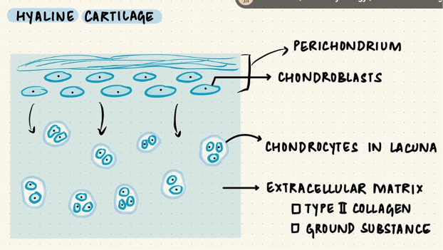
Hyaline: contains cells in extracellular matrix. These cells are called chondrocytes, which are located in spaces called the lacunae (either singly or in groups). The matrix consists of collagen, mainly type 2 collagen. Also, a well-hydrated ground substance which are synthesized by the chondroblasts. Chondroblasts are located in the perichondrium, which is the connective tissue layer that covers cartilage
Elastic:
Fibrocartilage:
Bone has osteoblasts, which develop from mesenchymal stem cells called Osteoprogenitor cells. These synthesize the bone matrix which is then calcified. Bone matrix contains type 1 collagen and ground substance, which gets mineralized with the addition of ions like calcium hydroxyapatite to form the calcified bone matrix.

During this process, some of the osteoblasts get trapped in the matrix and differentiate into oesteocytes, which are also located in spaces. One in each lacuna. Bone also has osteoclasts, which are multi-nucleated giant cells that resorb bone. They digest the bone matrix.

Osteoblasts form bone
Osteocytes maintain it
Osteoclasts destroy it
Bone is ditributed in compact and cancellous bone.

Compact bone = dense
Cancellous bone = spongy
Rods and plates forming trabeculae
Spaces between the trabeculae are filled with bone marrow
The structure of bone: (labelled parts)

In long bone:
Diaphysis: Central portion of the shaft, the diaphysis has medullary cavity filled with marrow
Epiphysis: The ends of the bones
Metaphyses: The expanded ends of the diaphysis
Periosteum: The surface of compact bone covered with connective tissue called the periosteum
Contains inner cellular layer, containing osteoprogenitor cells, osteoblasts and osteoclasts
The marrow cavities are lined by endosteum which also has these cells
How is bone remodelled?
Bone remodelling is a lifelong process, where old bone is replaced by new bone.
Remodelling: Maintains bone strength, repairs microdamage and helps regulate calcium levels
The cycle begins with osteoclast (bone-resorbing cells), which resorbs bone over a few weeks. Mononuclear cells then