Marieb - Chapter 7, 5.2b
Vertebral column
Introduction
axial support of the body
from the skull to the pelvis where it transmits the weight
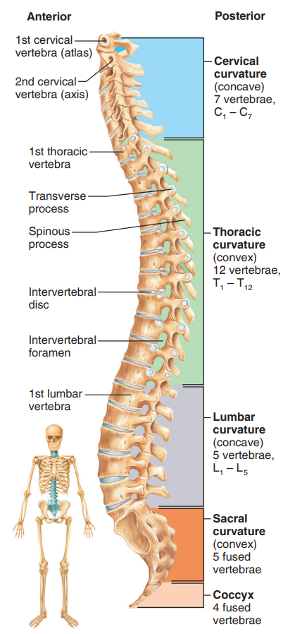
26 bones, between them an intervertebral disc, the spinal cord in the center, and surrounded by ligaments
Sacrum + coccyx + 7 CV + 12 TV + 5 LV
Note:
primary curvatures → TV + LV (they are present when we are born)
secondary curvatures → CV (they develop after birth)
Intervertebral discs
cushion, shock absorber
herniation due to aging or due to twisting forces
Structure patterns of the vertebrae: what they have in common
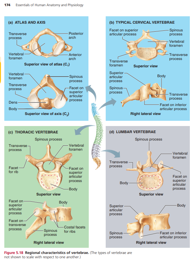
Body → facing the anterior part of the column, bears weight
Vertebral arch → arch formed from the joining of all posterior extensions, (laminae and pedicles) from the vertebral body
Vertebral foramen → canal through which the spinal cord passes
Transverse process → 2 lateral projections from the vertebral arch
Spinous process → single projection arising from the posterior aspect of the vertebral arch
Superior + inferior articular process → projections at the lateral of the vertebral foramen, allow a vertebrae to form joints with other vertebrae
Cervical vertebrae (C1 - C7)
C1 → atlas has no body, transverse process contains large depression to accommodate the occipital condyles of the skull (YES joint)
C2 → axis, pivot function, large upright process (dens)
The joint between C1 and C2 allows to do “NO”
Typical CV: C3 to C7
small, light
spinous process divided in 2 branches
transverse processes contain foramina (openings) for arteries
Thoracic vertebrae (T1 -T12)
they are all typical
larger than CV
only vertebrae to articulate with ribs (2 costal facets that articulate with them on the side, they receive the head of the ribs)
The transverse process articulates with tubercles of ribs
The spinous process is sharply downward
Lumbar vertebrae (L1 - L5)
the biggest type
short spinous process
Sacrum
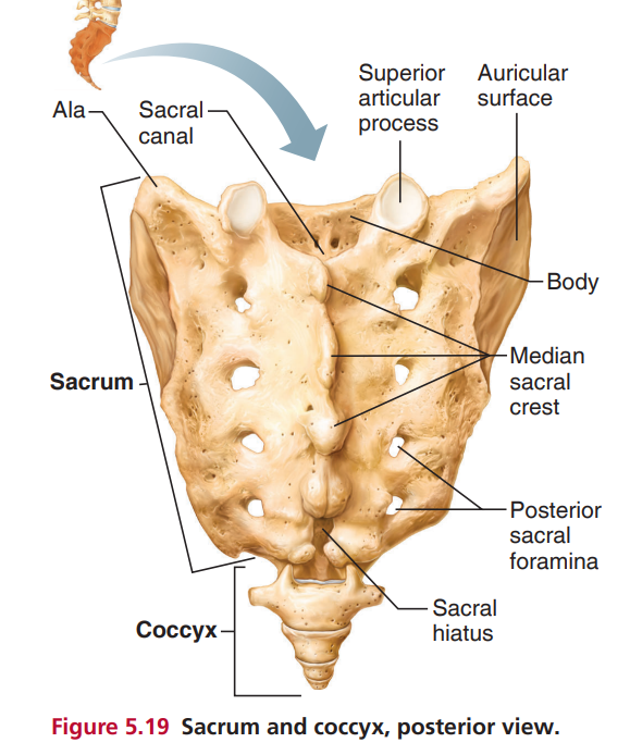
formed by 5 vertebrae fused
articulates above with L5 and inferiorly with coccyx
Each wing articulates with hip bone → Sacroiliac joint
the sacrum formed the posterior wall of the pelvis
posterior surface → median sacral crest
there are some longitudinal openings → posterior sacral formaina
the vertebral canal continues inside the sacrum with → sacral canal and it end with the sacral hiatus (large inferior foramina)
Coccyx
fusion of 3 to 5 bones
“tailbone“