Ophthalmic Lasers Part 2
Canaloplasty
microcatheter, placed in Schlemm’s canal to enlarge drainage canal
Tube shunt implant surgery
implanting shunt and aqueous drains through a valve into a filtering bleb
Enhancing fluid outflow: GATT, Kahook Tabectome, iStent (20% reduction)
Microtrabeculectomy (shunt fluid outside eye): XEN Gel stent, Preserflow
Excimer laser Trabeculotomy (ab interno) - MIGS
Characteristics (excimer)
short UV (193 um) cold laser
precision, no collateral tissue damage, nanosecond pulses
Method of action
enhances aqueous outflow (0.5 mm holes)
through TM including inner wall of schlemm’s canal
Indications: pts on max medical therapy, refractory POAG, long term lowering of IOP
can be performed with lensectomy
maintains integrity of TM and schlemm’s canal
Contraindications
narrow angles, iris neo
inadequate view of TM on gonio
advanced VF defects (scotoma within 10 degrees of fixation)
Procedure
1.2 mm limbal incision made temporally + nasally
viscoelastic used to protect endothelium
excimer laser tip contacts TM and makes 8 spots (anterior trabecula), equally spaced at 500 um
Laser cyclophotocoagulation surgery
Methods: transscleral, transpupillary, endolaser (TTE)
Mean IOP dropped by 50.3 %
Indications:
later stage glaucoma (meds not effective), failure of other filtering surgeries, painful blind eye, alleviation of pain in neovascular glaucoma
Procedure
Nd:YAG laser with scleral endoscopic probe (50-80 micropulses, 0.5ms duration)
destruction of ciliary epithelium to REDUCE aqueous production
retrobulbar anesthesia: bupivicane, lidocaine
Complications
eye pain, photophobia
RD, hypotony, phthisis
loss of BCVA, macular edema, scleral thinning
inflammation
Anterior vitreolysis (aka laser floater removal)
Three P’s: photocoagulation, photodisruption, photoactivation
Photocoagulation (Excimer laser)
laser absorbed by RPE (chromophore) and produces heat to denature proteins
thermal energy - coagulative necrosis
outer retinal layers more affected!
Photodisruption (Femtosecond laser)
energy released in short time and small retinal area
acoustic wave disrupts tissue
Photoactivation (PDT)
chemical
PDT uses IV-injected green dye called Vertporfin (chemically inert) but activated by light and destroys neovascular tissue
used for wet AMD
Types of lasers
Focal laser treatments: macula edema due to focal retinal vessel leakage
GRID laser treatments: macular edema due to DIFFUSE leakage
panretinal laser treatment (PRP): treat PDR (uses ruby argon lasers) - 500 um sized
PanRetinal Photocoagulation
Grading of laser vascular coagulation (PRP)
MINIMAL visible constriction of vessel
total constriction and SPASM of vessel
total constriction of vessel WITH coagulation of surrounding tissue
total constriction, CHARRING vessel, COAGULATION of surrounding tissue
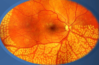
Complications
painful during/ immediately after t/x
inflammatory events
retinal architecture distorted
disrupts normal retinal connectivity
decreased VF (especially in periphery)
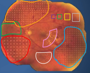
Pattern scanning laser (PASCAL)
Nd: YAG solid state laser (56 spots in 0.5 secs)
delivers multiple laser spots with shorter pulse duration in preset pattern
Less energy needed
less choroidal heating
less patient discomfort than classic PRP woohoo!
predetermined pattern helps to reduce retinal injury than just random firing of laser
Retinal detachment lasers
Causes/ risks of Retinal detachments
axial myopia
lattice degeneration (30% associated w/ rheg RD)
trauma
cataract surgery
PVD/ traction (10-20% risk)
idiopathic
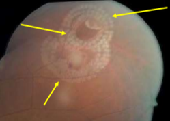
Goals
generate firm chorio-retinal adhesion surrounding the retinal break
counter vitreoretinal traction
prevent liquefied vitreous from passing into subretinal space
Healing
8 hours post op - RPE 50% weaker
18 hours post op - RPE improved to 100%
5 days post op*** - maximal strength achieved!!
Complications
laser hitting macula
choroidal effusion, angle closure glaucoma, ERM
Ant seg burns, hemorrhage, CNVM, new retinal breaks
Follow ups
BIO about 3 weeks after treatment
retreatment needed if: scarring incomplete, retinal break or subretinal fluid present