Cell-Cell Interactions PP notes
Types of tissues in animals
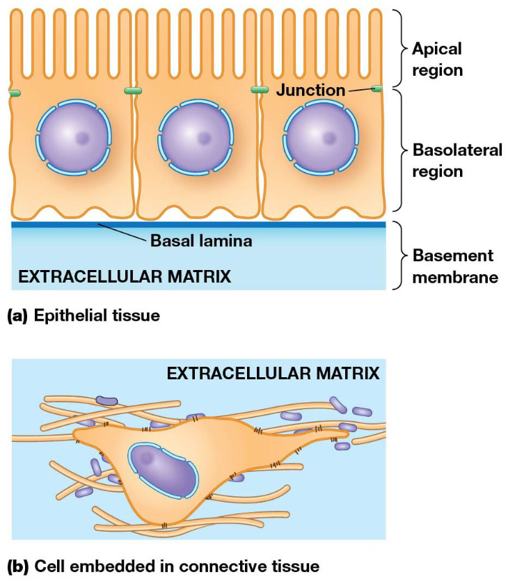
Epithelium: sheets/layers of cells with polarized functional domains, apical and basal
lines organs
outer layer of skin
Connective tissue: loosely organized fiber/gel in which cells are attached to each other
cartilage, bone, blood
Adhesive Junctions
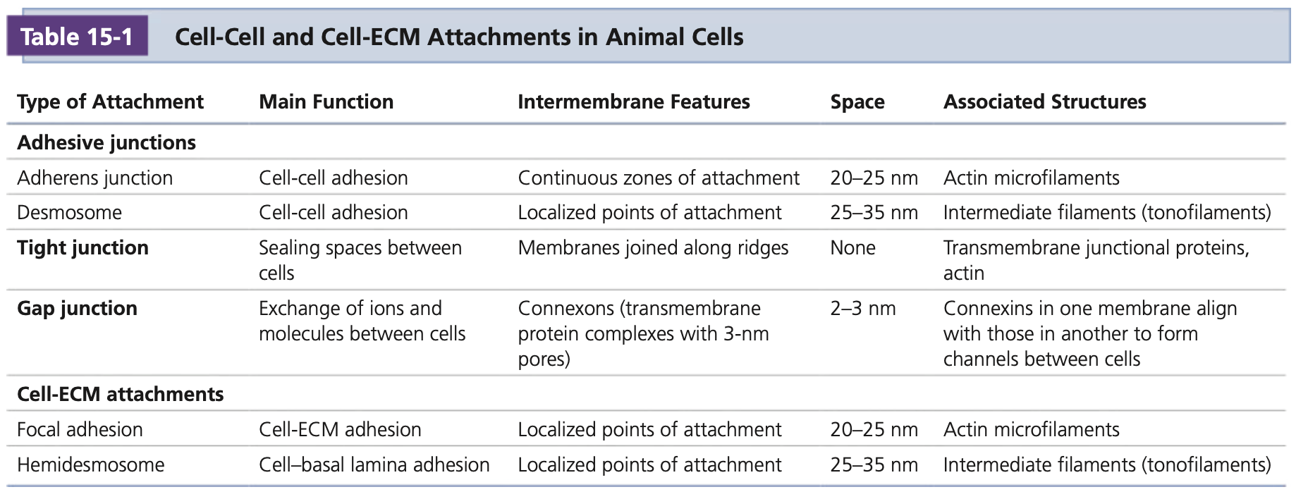
Adhesive junctions anchor the cytoskeleton to the cell surface, relying on specialized adhesion proteins which can interact with the other extracellular proteins on neighboring cells
Homophillic interactions involve cells with identical receptors interacting with each other, while heterophilic interactions have cells with different receptors. Many receptors also interact with the cytoskeleton via linker proteins.
Cells assemble/disassemble in response to cellular events
Adhesion proteins are recycled through endocytic and exocytic pathways
Adhesion proteins are sites of assembling signaling complexes and cytoskeletal structures
Cell adhesion is coordinated with cell signaling, movement, proliferation, and survival
Adherens Junctions
Cadherin-mediated junctions that interact with actin
Prominent in epithelial cells
Cadherins
repeats in extracellular domain
transmembrane domain
varying cytosolic ends
Structure diagram
E-Cadherin
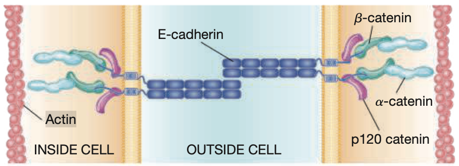
5 repeats on extracellular domain, two E-cadherin molecules can interact
associate in pairs in a calcium dependent manner
cytosolic portions interact with cytoskeleton
Beta-Catenin and Alpha-catenin
B-catenin binds to cytosolic tail of cadherin
bound by a-catenin, which recruits actin to the junction
a-catenin stretches and binding sites become available
vinculin can bind to sites and strengthen connection to F-actin
p120 Catenin
binds to cytoplasmic tail of cadherins near the plasma membrane
regulates stability of cadherins and how quickly they are endocytosed
Epithelial-mesenchymal transition (EMT)
breakdown of epihelium into loosely organized mesenchyme cells
accompanied by changes in cadhein expression
occurs in cancer cells which metastasize
Desmosomes
button-like points of strong adhesion between adjacent cells in tissue
structure diagram
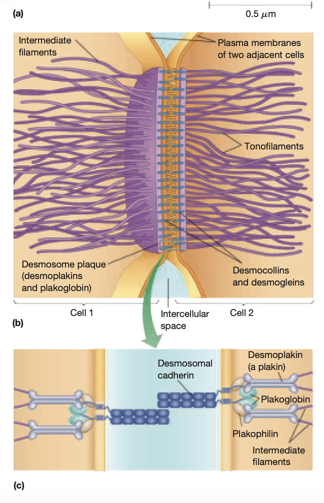
provide structural integrity
abundant in cells under mechanical stress
Desmosome core: extracellular space between two connected cells
desmosomal cadherins are desmocollins and desmogleins
linker proteins bind to domains and link to cytoskeleton
Plakoglbin binds desmocolin and desmoplakin
desmoplakin attaches intermediate filaments
p120ctn family is plakophilin can bind to cadherins and desmoplankin
thick plaque with linker proteins and intermediate filaments located between the membrane of adjoined cells
Lectins
carbohydrate-binding proteins
promote cell-cell adhesion by binding to sugars at outer cell surface
cand bind to carbs on two different cells, linking them together
Cell Adhesion Molecules (CAMs)
CAMs are members of the immunoglobulin super family (IgSF)
well-organized loops like immunoglobulins in their extacellular domains
homophilic interactions occur via domains
ex. Neural cell adhesion molecule, N-CAM
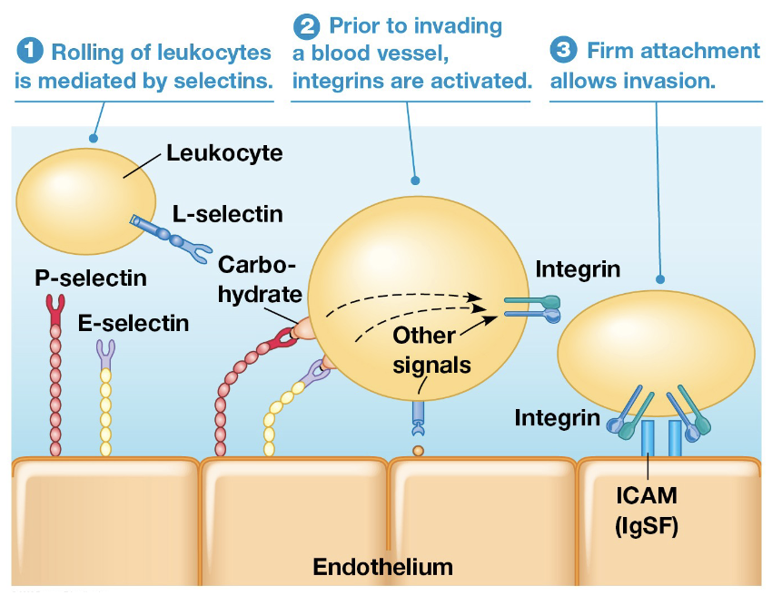
Leukocyte Adhesion
Leukocytes interact with platelets or blood vessels during inflammation
reactions are mediated by selectins
L-selectin - leukocytes
P-selectin - platelets
E-selectin - endothelial cells of blood vessels
stable adhesions at site of inflammation are mediated by integrin and ICAM
Tight Junctions
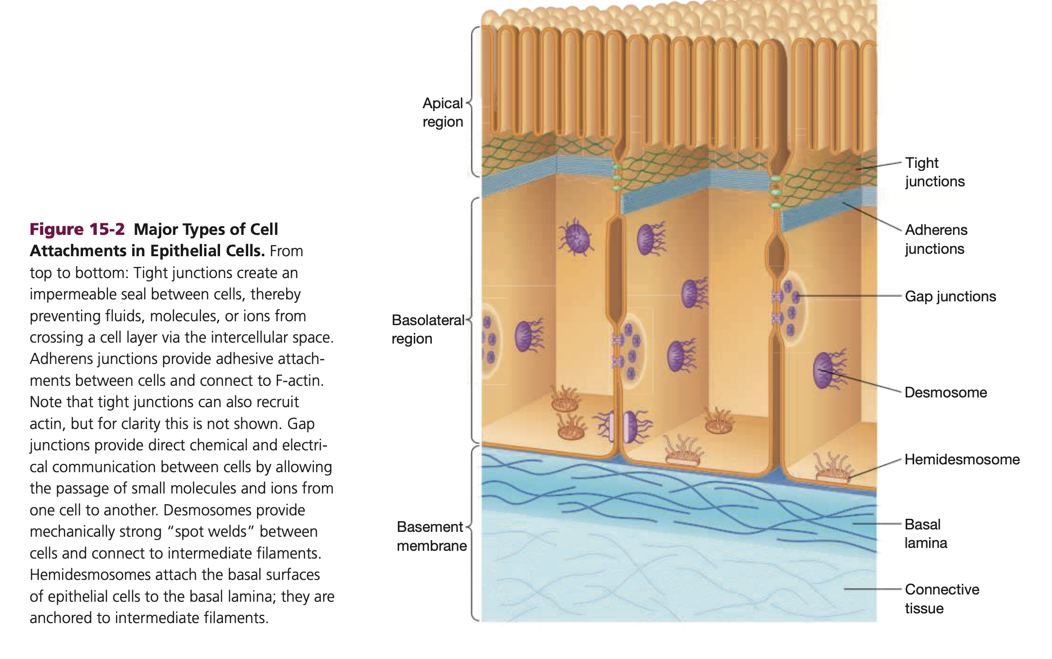
Tight junctions form a seal between two cells, leaving no space between their plasma membranes. They form a continuous belt around the apical (top) ends of lateral surfaces on each cell, and molecules cross the cell layer by passing through the cells
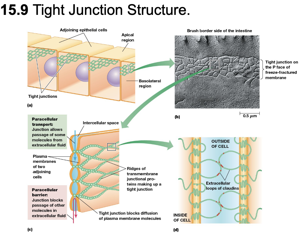
Tight junctions have several transmembrane proteins such as occludin and junctional adhesion molecules (JAMs), as well a claudins.
Function of Claudins
proteins with 4-membrane spanning domains
interlock in adjacent cells to form tight seal
large extracellular loop forms ion-selective pores to allow passage of specific ions
called paracellular transport
Tight junctions block lateral movement of lipids and proteins in the plasma membrane
lipids blocked only in outer monolayer
integral membrane proteins completely blocked
Gap Junctions
A gap junction is a region where the plasma membranes of cells are in contact, with a very small gap in between. The gap includes small passages between the cells, allowing small molecules and ions to pass directly from one cell to another
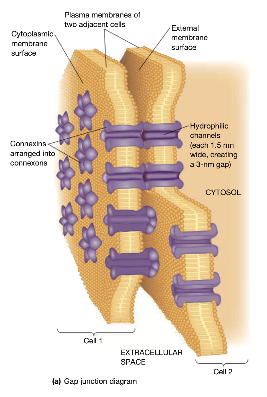
In a gap junction, the plasma membranes of adjacent cells are connected by hollow cylinders called connexons. Each connexon has 6 subunits of connexin proteins, forming a circle. (innexin and invertebrates). Every connexon has a channel about 3nm wide, only allowing small molecules to pass through.
Extracellular Matrix (ECM)
The extracellular matrix in animal cells is crucial for tissue structure and function, as it takes on several different roles:
Bone consists of a rigid ECM containing a small number of interspersed cells
Cartilage is tissue mostly made of matrix materials more flexible than bones
Connective tissue which surrounds glands/blood vessels is gelatinous, containing interspersed fibroblast cells
ECM consists 3 classes of molecules:
Structural proteins
collagens, elastins
strength & flexibility
Protein-polysaccharide complexes
proteoglycans
provide matrix
Adhesive glycoproteins
fibronectins, lamins
allow cells to attach to matrix
Collagens
most abundant in ECM in animals
secreted by cells in connective tissues, such as fibroblasts
rigid triple helix of intertwined polypeptides
high glycine and rare amino acids
glycine spacing important for helix
Collagen fibers are composed of fibrils
fibrils made of many collagen molecules, twisted into chains of three
In ER lumen, 3 chains form triple helix procollagen
short, nonhelical sequences at ends
nonhelical ends cleaved by procollagen peptidase outside the cell
spontaneously associate into fibrils, fibers
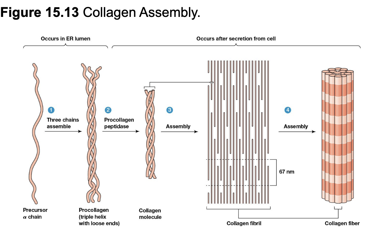
stability of fibrils reinforced by hydrogen bonds between amino acids (hydroxylysine and hydroxyproline)
form crosslinks between/within collagen molecules
Elastins
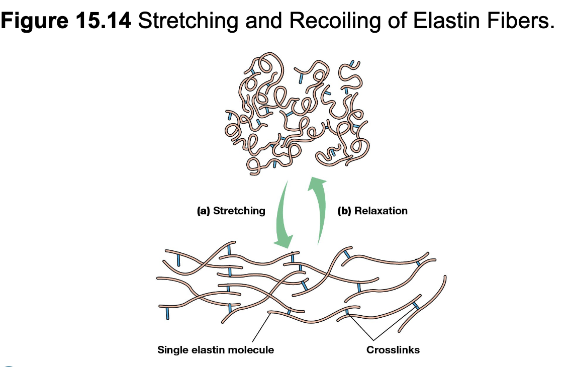
provide elasticity to ECM by fibers composed of elastins
rich in glycine and proline, crosslinked by bonds between lysine residues
tension causes network to stretch
release of tension causes molecules to relax
Collagen and elastin fibers are combined in a gel-like network of proteoglycans, glucoproteins, and glycosamionglycans attached to a protein
Glycosaminoglycans (GAGs) are large carbohydrates with repeating disaccharide units
most common types are chondroitin sulfate, keratan sulfate, and hyaluronate
repeating unit has one amino sugar
N-acetylglucosamine (GlcNAc)
N-acetylgalactosamine (GalNAc)
other sugar is sugar or sugar acid
galactose (Gal)
glucuronate (GlcUA)
amino sugar has 1+ sulfate attached
hydrophillic, attract water and cations
forms gelatinous matrix where collagen and elastin are embedded
Proteoglycans
GAGs in ECM covalently bound to proteins form proteoglycans
each has number of GAG chains attached along length of a core protein
vary in size depending on core protein and length of GAG chains
integral components of plasma membrane
core polypeptides embedded in membrane
covalently linked to membrane phospholipids
Hyaluronate
occurs both as backbone of cartilage proteoglycans and a free molecule
lubricating properties
most abundant where friction needs to be reduces, ex. joints
Fibronectins
family closely related glycoproteins in ECM
RNA transcribed from fibronectin gene processed to produce many varients
can be soluble in blood/body fluids, insoluble fibrils in ECM, and intermediate associated with cell surfaces
two large suvunits linked by C-terminal, 2 disulfide bonds
folded into rodlike domains
several domains bind 1+ ECM macromolecules (collagen, heparin, fibrin)
other domains recognize & bind cell surface receptors via RGD (arg-gly-asp) sequence
acts as bridging molecule between cells and ECM
cell migration
soluble form is plasma fibronectin
promotes blood clotting
Basal Lamina
thin sheet of specialized extracellular material
underlines epthelial cells
surround muscle cells, fat cells, and schwann cells
major adhesive glycoprotein is laminins
structural support and permeability barrier
contain type IV collagen, proteogluycans, laminins, and nidogen
can alter properties of basal lamina by secreting enzymes in it
metalloproteinases (MMPs) degrade ECM locally, allowing cells to pass through (require metal cofactors)
Laminin
3 long polypeptides, α, β, and γ
disulfide bonds hold PP together in shape of cross
several domains, such as binding sites for type IV collagen, heparin, heparin sulfate and entactin
receptors on cell surfaces
Integrins
large family of cell surface receptors
bind to fibronectins or laminins
integrate the cytoskeleton with ECM
consist of 2 large transmembrane polypeptides, α & β
differ in binding specificity and subunit size
extracellular parts of subunits form binding sites
specificity depends on alpha unit
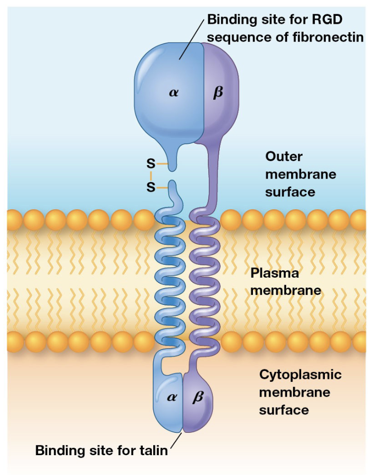
many types of subunits, resulting in many types of integrin heterodimers
recognize RGD sequences in ECM glycoproteins
tails interact with cytosolic proteins that link integrins to cytoskeleton
focal adhesions attach migraotory and non-epithelial cells (fibroblasts) to ECM
contain clustered integrins
interact with bundles of actin filaments
interact with signaling pathways
MAP kinase activation induces integrin clustering
act as receptors for intracellular signaling
Hemidesmosomes
found in epithelial cells
contain alpha-6-beta-4 integrin
attached to keratin, an intermediate filament
linker proteins form a dense plaque to connect integrins to cytoskeleton
plankin family are linker proteins
pectin attaches keratin to integrins
BPAG2 and its plakin, BPAG1, bridge keratin and laminin
Anochorage-dependent growth requires cells to be attached to a substratum, which involves the activation of intracellular pathways following integrin clustering.
A costamere is an attachment structure at the surface of striated muscle which align with the Z discs, containing the protein dystrophin.
Dyrstrophin mutation causes muscle disease
acts as a spring
links to laminin, connecting costaneres to ECN
interacts with a complex including integral membrane proteins