Reading Notes - Chapter 2
2.1 - electrical signals are the vocabulary of the nervous system
Ions → electrically charged molecules
Anions → negatively charged ions
Cations → positively charged ions
Intracellular fluid → watery solution found within cells
Extracellular fluid → fluid in the spaces between cells
Cell membrane → lipid bilayer that encloses a cell
In a neuron at rest, of the many ions, most of them are anions
Then all of the ions are dissolved in the intracellular fluid in the cell and the extracellular fluid outside the cell membrane
Microelectrode → small electrode used to record electrical potentials inside living cells
Resting potential → the difference in electrical potential across the membrane of a nerve cell at rest (neuron at rest exhibits about -50 to -80 mV)
Millivolts (mV) → a thousandth of a volt
Insert microelectrode into interior of a neuron → place electrode in the extracellular fluid → find that the inside of the neuron is more negative than the fluid around it
Ion channel → (important type of membrane-spanning protein) a pore in the cell membrane that permits the passage of certain ions through the membrane when the channel is open
Some ion channels stay open all the time
Diffusion → tendency for molecules of a substance to spread from regions of high concentration to regions of low concentration
Electrostatic pressure → the tendency of charged molecules or ions to move toward areas with the opposite charge
Charged particles exert electrical force on one another: like charges repel and opposite charges attract
Ex.: positively charged cations (like K+) are attracted to the negatively charged interior of the cell and anions are repelled by the cell interior and then tend to exit to the extracellular fluid
Sodium-potassium pumps → energetically expensive mechanisms that pushes sodium ions out of a cell and potassium ions in (pump 3 Na+ ions out for every K+ ions pumped in → leads to buildup of K+ ions inside cell and reduces Na+ inside cell) → K+ ions can leave interior moving down their concentration gradient and causing buildup of negative charges inside cell → exert electrostatic pressure to pull positively charged K+ ions back inside → reach the equilibrium potential
Equilibrium potential → point at which the movement of ions across the cell membrane is balanced, as the electrostatic pressure pulling ions in one direction if offset by the diffusion force pushing them in the opposite direction
Cell’s resting potential is about -65 mV


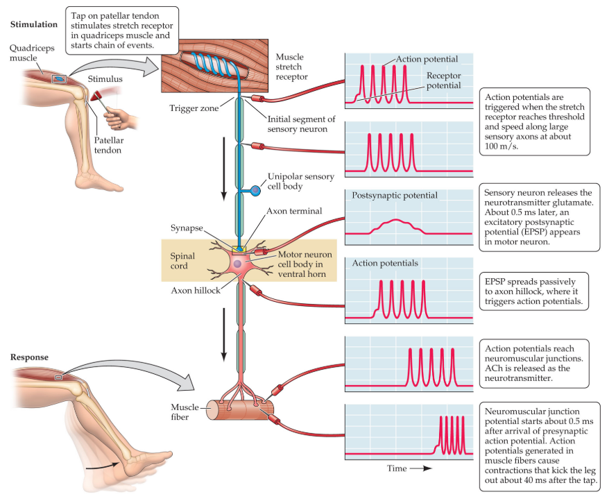
Axon hillock → cone-shaped region where the axon emerges from the cell body
Action potentials are changes in resting membrane potential that happens in initial segment of the axon just after the axon hillock → moves rapidly down the axon
Two concepts to understand how action potentials are triggered:
Hyperpolarization → increase in membrane potential (when the neuron becomes EVEN MORE NEGATIVE on the inside, relative to the outside)
Ex.: if neuron’s resting potential is -65mV, hyperpolarization makes it even farther from zero, maybe -70mV
Depolarization → decrease in membrane potential
Ex.: if neuron’s resting potential is -65mV, depolarization to roughly -50mV makes the inside of the neuron more like the outside (closer to zero)
Local potentials → electrical potential that is initiated by stimulation at a specific site, is a graded response that spreads passively across the cell membrane and decreases in strength with time and distance
Threshold → the stimulus intensity that is just adequate to trigger an action potential in an axon
Action potential → sudden and brief response (0.5 to 2.0 millisecond)
Also called spike
Rapid reversal of the membrane potential that momentarily makes the inside of a neuron positive with respect to the outside
All-or-none property → the condition that the size (amplitude) of the action potential is independent of the size of the stimulus
Afterpotentials → the positive or negative change in membrane potential that may follow an action potential
Voltage-gated Na+ channel → a Na+ selective channel that opens or closes in response to changes in the voltage of the local membrane potential
Mediates the action potential
Refractory → unresponsive
Two phases of refractoriness:
Absolute refractory phase → brief period immediately following the production of an action potential
No amount of stimulation can induce another action potential because the voltage-gated Na+ channels can’t respond
Relative refractory phase → period after absolute phase with reduced sensitivity
Only strong stimulation can depolarize the axon to threshold to produce another action potential
Conduct velocity → speed at which an action potential is propagated along the length of an axon
Saltatory conduction → form of conduction that is characteristic of myelinated axons in which the action potential jumps from one node of Ranvier to the next
Multiple sclerosis (MS) → disorder characterized by widespread degeneration of myelin (“many scars”)


Postsynaptic potentials → a local potential that is initiated by stimulation at a synapse, can vary in amplitude, and spreads passively across the cell membrane, decreasing in strength with time and distance
Excitatory postsynaptic potential (EPSP) → a depolarizing potential in a neuron that is normally caused by synaptic excitation. EPSPs increase the probability that the postsynaptic neuron will fire an action potential
Inhibitory postsynaptic potential (IPSP) → a hyperpolarizing potential in a neuron. IPSPs decrease the probability that the postsynaptic neuron will fire an action potential


Spatial summation → summation of postsynaptic potential that reach the axon hillock from different locations across the cell body. If this summation reaches threshold, an action potential is triggered
Temporal summation → summation of postsynaptic potentials that reach the axon hillock at different times. The closer in time the potentials occur, the greater the summation


2.2 - Synaptic Transmission Requires a Sequence of Events
Steps in transmission at a chemical synapse
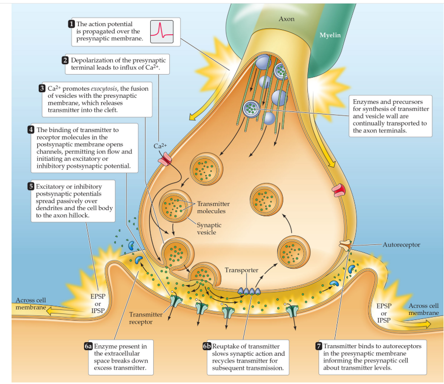
synaptic vesicles → small, spherical structure that contains molecules of neurotransmitter
synaptic cleft → space between the presynaptic and postsynaptic cells at a synapse
synaptic delay → brief delay between the arrival of an action potential at the axon terminal and the creation of postsynaptic potential
ligand → a substance that binds to receptor molecules, such as a neurotransmitter or drug that binds to postsynaptic receptors
acetylcholine (ACh) → neurotransmitter that is produced and released by parasympathetic postganglionic neurons, by motor neurons, and by many neurons in the brain
neurotransmitter receptor → specialized protein, embedded in the cell membrane, that selectively senses and reacts to molecules of a corresponding neurotransmitter
curare → neurotoxin that causes paralysis by blocking acetylcholine receptors in muscle
bungarotoxin → a neurotoxin isolated from the venom of the many-banded krait that selectively blocks acetylcholine receptors
agonists → substance that mimics or boosts the actions of a transmitter or other signaling molecule
antagonist → 1. a substance that blocks or attenuates the actions of a transmitter or other signaling molecule. 2. a muscle that counteracts the effect of another muscle
cholinergic → referring to cells that use acetylcholine as their synaptic transmitter
transporters → specialized membrane component that returns transmitter molecules to the presynaptic neuron for reuse
Neural circuits underlie reflexes
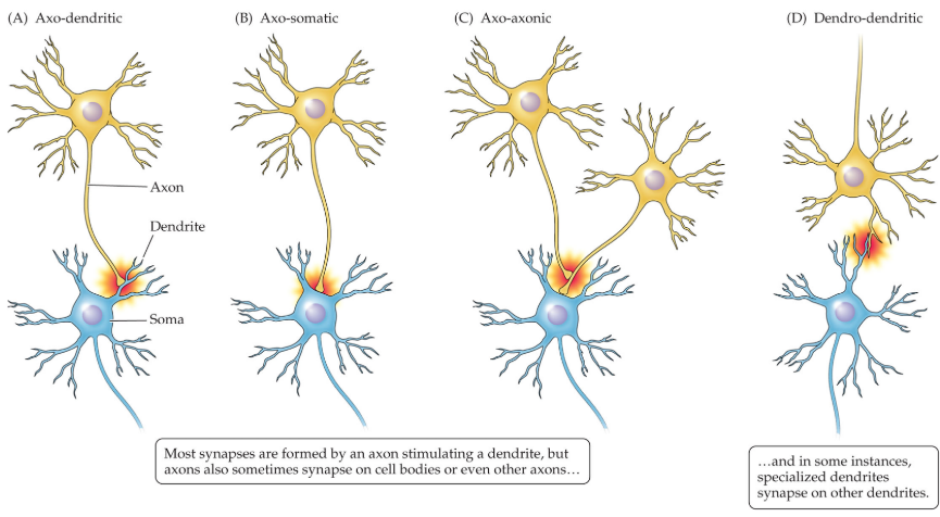
axo-dendritic synapses → synapse at which a presynaptic axon terminal synapses onto a dendrite of the postsynaptic neuron, either via a dendritic spine or directly onto the dendrite itself
axo-somatic synapses → synapse at which a presynaptic axon terminal synapses onto the cell body (soma) of the postsynaptic neuron
axo-axonic synapses → synapse at which a presynaptic axon terminal synapses onto the axon terminal to another neuron
dendro-dendritic synapses → synapse at which a synaptic connection forms between the dendrites of two neurons
knee-jerk reflex → variant of the stretch reflex in which stretching of the tendon beneath the knee leads to an upward kick of the leg