Central Nervous System
Central Nervous System (CNS)
The Brain
The Brain: The anatomy of the brain & spinal cord can seem more abstract than most of the gross anatomy that we study in the other organ systems. The brain is a complex mass of nerve, connective, & epithelial tissue. It is divided into 4 Anatomical & Functional Regions:
1. Cerebrum: Largest region & occupies most of the space in the cranial cavity; split into 2 cerebral hemispheres (left & right) & each hemisphere are made up of lobes.
2. Diencephalon: Pair of oval-shaped structures deep into the cerebrum; potruding laterally in the lateral ventricles, then meets medially encapsulating the 3rd ventricle, which is located superior to the brainstem's midbrain; it contains multiple nuclei (deep mass of gray matter surrounded by white matter); dubbed as the gateway to the cerebral cortex (plural: cortexes or corteces).
3. Cerebellum: Second largest region of the brain; located inferior to the cerebrum but posterior to the brainstem. It consists of a left & right cerebellar hemispheres.
4. Brainstem: A long structure that extends inferiorly from the diencephalon; 10 of the 12 cranial nerves originate from here; it is made up 3 sections/parts/regions: the superior midbrain, the middle round-shaped pons, & inferior medulla oblongata.
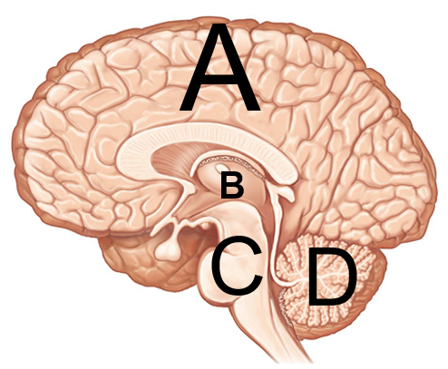
Figure 4.16 4 Anatomical & Functional Divisions of the Brain: (A) Cerebrum, (B) Diencephalon, (C) Brainstem, (D) Cerebellum
Fissures, Sulci, & Gyri: External features of the cerebellum described as grooves & folds.
Fissures: Deeper grooves/furrows, usually separating bigger parts of the brain.
Sulci: (singular: sulcus) Shallow grooves found in the cerebrum.
Gyri: (singular: gyrus) Folds found in the cerebrum; think of a gyro sandwich which is folded.
Note: It takes two gyri to form a sulcus & 2 sulci are found on each side of a gyrus.
Cranial Gray & White Matters:
Gray Matter: Located superficial in the cerebrum & cerebellum; they are mainly composed of neuron cell bodies, dendrites & synapses.
Cerebellar & Cerebral Cortexes:Superficial layer on both the cerebrum & cerebellum.
Nuclei: Deeper masses of gray matter surrounded by white matter.
White Matter: Lies deep to the gray matter & is mainly composed of bundles of axons/tracts.
Cranial Meninges: The brain & spinal cord are surrounded by a set of 3 connective tissue membranes known as meninges (singular: meninx). The 3 layers from superficial to deep are the:
1. Dura Mater: Tough layer protecting the brain; made up of 2 layers:
Periosteal Layer: Outer layer surrounding the cranial bones, equivalent to periosteum.
Meningeal Layer: Inner layer facing the other meninges; only part of dura that extends to the vertebral canal; its surrounds & supports the dural sinuses that drain deoxygenated blood from the brain; below are 3 structures formed from this layer, which separate parts of the brain:
Falx Cerebri: Separates each cerebral hemisphere; located in longitudinal fissure.
Tentorium Cerebelli: Separates cerebrum from cerebellum; located in transverse fissure.
Falx Cerebelli: Separates each cerebellar hemisphere; located inferior to the cerebellum.
Subdural Space: A potential space below the dura mater & above the arachnoid mater; potential space means it can be opened if needed, for issues such as a brain bleed.
Dural Sinuses: Spaces formed by the layers of the dura mater that collects deoxygenated blood & CSF circulated through the brain; an example is the superior sagittal sinus, which is located superior to the falx cerebri.
2. Arachnoid Mater: Second layer resembling a spider web; have small parts called arachnoid granulations.
Arachnoid granulations: Tree-like protrusions piercing through the dura mater, which reabsorbs CSF into the dural sinuses.
Subarachnoid Space: An actual space below the arachnoid mater where CSF flows.
3. Pia mater: Very thin & delicate layer that closely follows every contour of the brain even the sulci.
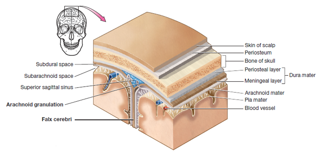
4.17 Cross-section of the cranial meninges & its associated structures
Ventricles: The brain contains a system of fluid filled cavities (spaces) called ventricles, which are located in different parts of the brain. It is lined with a spongy mass of capillaries called the choroid plexus. There are 4 ventricles & other pathways that connect each ventricles, below are their location in the brain:
Lateral Ventricles: (aka 1st & 2nd ) Forms an arc with an anterior, posterior, & inferior horns; located within the left & right cerebral hemispheres.
Interventricular foramen: Located inferior to the anterior horn; connects the lateral ventricles to the 3rd ventricle.
3rd Ventricle: Located below the corpus callosum & within the medial part of the thalamus.
Cerebral Aqueduct: Located in the midbrain; connects the 3rd ventricle to the 4th ventricle.
4th Ventricle: Located between the pons & cerebellum.
Central Canal & Apertures: Located by the medulla oblongata; drains CSF into the subarachnoid space.
Subarachnoid Space: Space that surrounds the brain & spinal cord, located just below the arachnoid mater.
Arachnoid Granulations: Reabsorbs CSF into the dural sinuses.
Cerebrospinal Fluid: Clear & colorless fluid flowing in the ventricles. It's derived from plasma & serves to protect the brain by allowing it to float in the insulating fluid of the CSF. About 500 mL/day of CSF is produced by ependymal cells lining the choroid plexus found in each ventricle & pathways; the CSF functions include:
Bouyancy: It allows the brain to attain considerable size without being impaired by its own weight.
Protection: It protects the brain from striking the cranium when the head is jolted; if the jolt is severe it could lead to traumatic brain injuries or concussions.
Chemical Stability: It rinses metabolic wastes from the nervous tissue & regulates its chemical environment.
CSF Flow in the Ventricles:
1) Left & right lateral ventricles
2) Interventricular foramen
3) 3rd ventricle
4) Cerebral aqueduct
5) 4th ventricle
6) Out of the apertures & central canal
7) To the subarachnoid space that surrounds the brain & spinal cord
8) Reabsorbed by arachnoid granulations into the dural sinuses
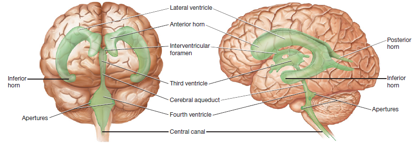
Figure 4.18 Anterior & left lateral views of the ventricles
Cerebrum
Lobes:
Frontal Lobe: Responsible for emotion, mood, memory, & aggression.
Broca Area: Motor language area for speech or sign language; located superficial & anterior on the left hemisphere by the frontal lobe; a little bit anterior to the precentral gyrus
.
Parietal Lobe: Responsible for sensory reception & integration of taste.
Temporal Lobe: Responsible for hearing, smell, learning, visual recognition, & emotional behavior.
Wernicke Area: Recognition of spoken & written language; located superficial & posterior on the left hemisphere by the temporal lobe & lateral sulcus.
Occipital Lobe: The principal visual center of the brain.
Insula: Plays a role in pain & empathy; it is a small mass of cerebral cortex located deep to the lateral sulcus.
Sulci:
Central sulcus: Shallow groove found between the frontal & parietal lobes.
Lateral sulcus: Shallow groove found between the frontal & temporal lobes.
Parieto-occipital sulcus: Shallow groove found between the parietal & occipital lobes.
Fissures
Transverse Fissure: Deep groove found between the cerebellum and cerebrum.
Longitudinal Fissure: Deep groove found between each cerebral hemisphere.
Gyri
Precentral Gyrus: Immediate fold anterior to the central sulcus & located in the frontal lobe. Also known as the primary motor cortex. Signals sent from here results in muscle contractions.
Postcentral Gyrus: Immediate fold posterior to the central sulcus & located in the parietal lobe. Also known as the primary somatosensory cortex. Awareness of stimulation occurs here.
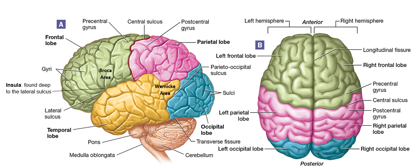
Figure 4.19 Left lateral & superior views of the brain
White Matter:
Corpus Callosum: Thick C-shape structure made up of nerve axons/tracts that connect the cerebral hemispheres to each other. Its C-shape is seen better on the lateral view.
Fornix: Thin C-shape structure located inferior to the corpus callosum. Its C-shape is seen better on the lateral view.
Gray Matter:
Basal Nuclei: Striped masses of gray matter buried deep in the white matter lateral to the thalamus; involved in motor control.
Caudate Nucleus: Superior of the three & medial; close to the lateral ventricles; tail-like shape from the lateral view.
Putamen: Inferior & lateral to globus pallidus; lens shape from the lateral view.
Globus Pallidus: Inferior & medial to putamen; lens shape from the lateral view.
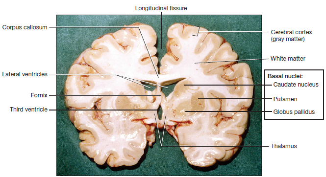
Figure 4.20 Coronal cut, anterior view of a cadaver brain: cerebellum & diencephalon
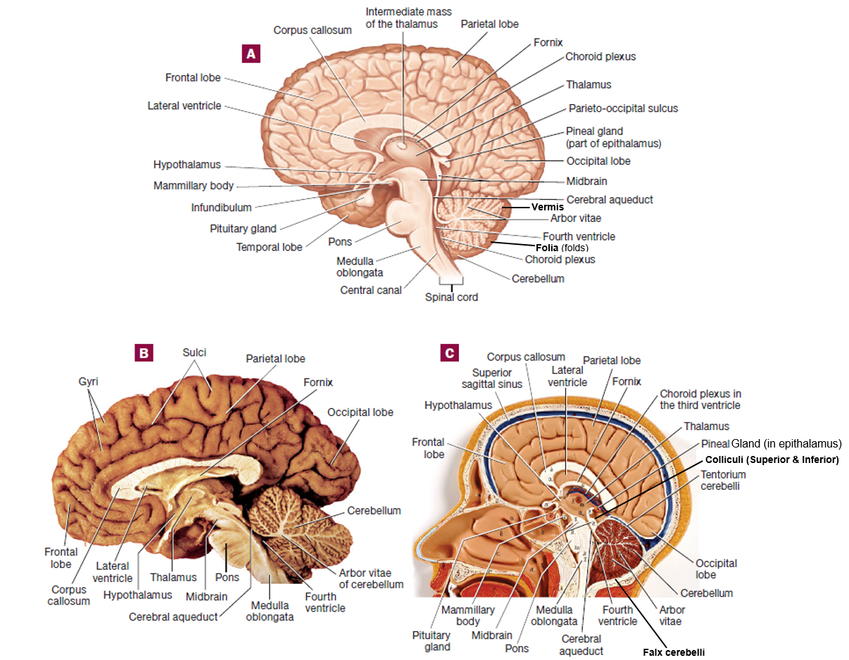
Figure 4.21 Left lateral view of the brain & midsagittal cut: (A) illustration, (B) cadaver, (C) plastic model
Cerebellum
Cerebellar Cortex: Central control point for muscle coordination.
Arbor vitae: White matter of the cerebellum; translates to"tree of life."
Folia: Folds found within the cerebellum; it is the equivalent of the gyri in the cerebrum.
Vermis: Medial structure that connects each cerebellar hemisphere. Seen better on posterior view.
Tentorium Cerebelli: Separates cerebrum from cerebellum; located in transverse fissure.
Falx Cerebelli: Separates each cerebellar hemisphere; membrane located inferior to cerebellum.
Diencephalon
Thalamus: (plural: thalami) Gateway to the cerebral cortex, filters information & relay only a small portion of it to the cerebral cortex; made up of at least 23 nuclei.
Hypothalamus: Located anterior & inferior to the thalamus & optic chiasm (part of the optic nerve); it is connected to the pituitary gland (master gland) via a stalk called infundibulum
; it rests on the hypophyseal fossa; it is the major control center of the autonomic nervous & endocrine systems.
Intermediate Mass: Conjoins the thalami medially.
Epithalamus: Located posterior & superior to the thalamus; found in it is the pineal gland, which is responsible for releasing the hormone melatonin, which has a role in regulating sleep.
Mammillary Bodies: A pair of round structures located posterior to the pituitary gland.
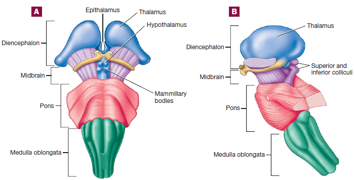
Figure 4.22 Diencephalon & Brainstem (A) Anterior view; (B) Left lateral view.
Brainstem
Note: 10 of the 12 cranial nerves originate in the brainstem; we'll learn about cranial nerves later
Midbrain: Associated with controlling our awareness to pain, collaborates in fine motor control; located inferior to the thalamus & superior to the pons; contains 4 bulges called colliculi (singular: colliculus), which are split into 2 top pairs called superior colliculi (involved with control of extrinsic eye muscles) & 2 bottom pairs called inferior colliculi (involved in relaying signals from the inner ear to the thalamus); 2 cranial nerves originate here.
Pons: Responsible for relaying signals hearing, equilibrium, taste, eye movements, swallowing, bladder control, & posture; located above the medulla oblongata & below the midbrain; round-shaped; 4 cranial nerves originate here.
Medulla Oblongata: Cardiac, respiratory, & vasomotor center; once it passes through the foramen magnum, it becomes the spinal cord; located inferior to the pons; 4 cranial nerves originate here.
The Spinal Cord
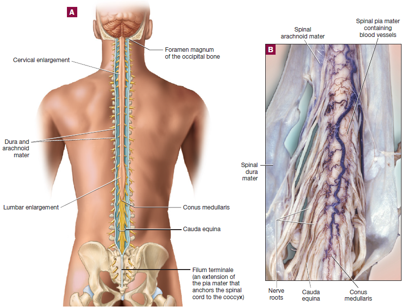
Figure 4.24 Posterior view of the spinal cord: (A) illustration, (B) close-up of inferior portion of a cadaver's spinal cord
The Spinal Cord
: As mentioned before, the medulla oblongata becomes the spinal cord after passing through the foramen magnum.
Conus Medullaris: The spinal cord then extends to the region of L1 & L2 vertebrae & thins down in this cone-shaped region.
Cauda Equina: A bundle of nerve roots that resemble a horse’s tail fill the rest of the vertebral column from L2 to S4 vertebrae, since the spinal cord stops at the L1 & L2 vertebrae. It innervates the pelvic organs & lower extremity.
Grooves: the spinal cord has an anterior deep groove called anterior median fissure & a posterior shallow groove called the posterior median sulcus.
Central Canal: A pathway traveling within the spinal cord, located in the midline & center of the spinal cord where CSF flows as well. Closed in adults, but open in children.
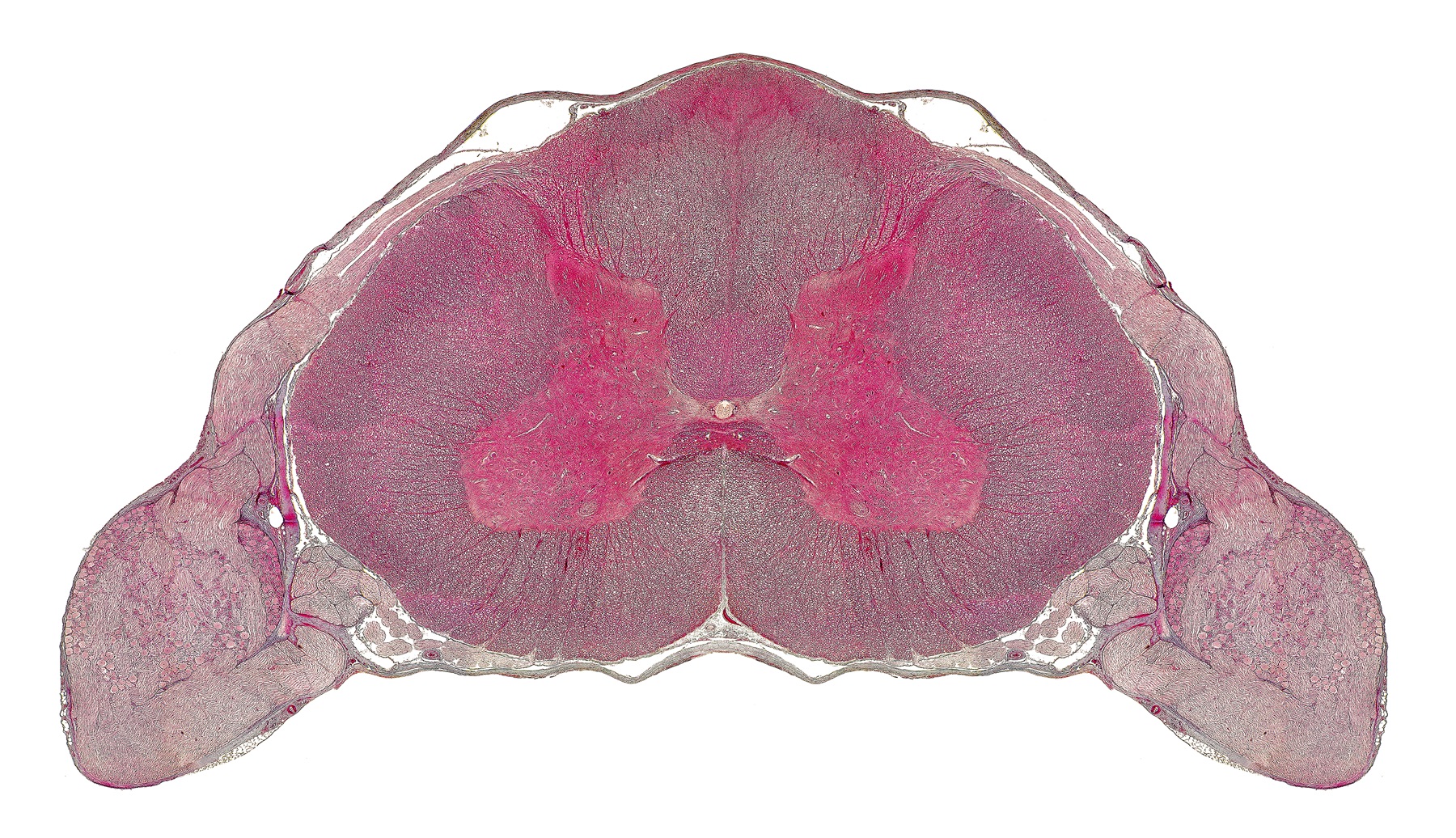
Spinal Gray Matter: The spinal gray matter is a butterfly-shaped core. Take note that the gray matter in the spinal cord is deep, unlike in the brain where it is located superficial.
Horns
Anterior Horn: The anterior portion, known as the anterior (ventral) horn, is made up of cell bodies of the motor neurons of the spinal cord, also referred to as lower motor neurons. The axons of these nerves exit via the anterior (ventral) spinal nerve roots.
Posterior Horn: The posterior (dorsal) horn receive axons from the dorsal root ganglia (singular: ganglion), which house the cell bodies of the sensory neurons & are housed just outside the cord.
Lateral Horn: In the thoracic & lumbar regions only, a lateral horn is present & contains cell bodies of the sympathetic division (fight or flight) of the autonomic nervous system (motor nervous system that controls glands, cardiac muscle, & smooth muscle).
Gray Commissure: Found in the midline; connects left & right portions of spinal gray matter.
Enlargements: These are thicker/wider sections in the spinal cord & there are 2 of them:
Cervical Enlargement: Gives rise to the nerves of the upper limbs.
Lumbar Enlargement: Gives rise to the lumbar & pelvic nerves.
Spinal White Matter: The remaining spinal cord that surrounds the spinal gray matter is known as the spinal white matter. Take note that white matter in the brain is deep, unlike the white matter of the spinal cord which is located superficial.
This contains groups of axons carrying information up the cord via the ascending fibers or down the cord via the descending fibers.
These fibers are grouped in columns called funiculi (singular: funiculus). Which are divided into 3 sections called anterior, posterior, & lateral funiculi.
The funiculi are divided up into tracts, which contain axons that have the same functional role & are defined primarily by the structures they connect.
Spinal Nerve Roots: Each spinal nerve arises from 2 points of root attachments to the spinal cord:
Anterior/Ventral Spinal Nerve Roots: Formed by 6-8 anterior/ventral spinal nerve rootlets, carries motor axons & efferent signals away from the spinal cord. The motor nerves' neurosoma/cell bodies are found within the spinal cord's gray matter.
Posterior/Dorsal Spinal Nerve Roots: Formed by 6-8 posterior/dorsal spinal nerve rootlets, carries sensory axons & afferent signals towards the spinal cord.
Posterior/Dorsal Root Ganglion: (plural: ganglia) It contains the neurosoma/cell body of the sensory nerves traveling to the spinal cord. It is easy to find since it has a bulbous/swollen appearance. Seeing this ganglion is a good landmark to use & confirm that you are at the posterior side of the spinal cord.
Spinal Nerves: Formed by the fusion of the anterior & posterior spinal nerve roots. It contains both sensory & motor axons. Therefore, it is a mixed nerve that takes efferent signals away from the spinal cord & to the body by the way of the anterior spinal nerve roots, executing a motor action. While, it carries sensory signals sensed by the body back to the spinal cord via the posterior spinal nerve roots & root ganglion.
Spinal Meninges: They (dura, arachnoid, & pia mater) are organized the same way as the cranium
There are surrounding fibers called the denticulate ligaments (extensions of the pia mater that extends to the dura mater) which anchors the spinal cord & limit its side-to-side movements.
The spinal meninges are an extension of the cranial meninges. However, in the spinal cord the dura mater is not adherent to the vertebrae the way it is in the skull, so there is space between the dura & spinal column known as the epidural space. It is filled with loose connective tissue, adipose tissue, & blood vessels.
Again, the major difference is the epidural space which exists in the spinal column but not in the cranial cavity. The spinal cord also has subdural & subarachnoid spaces.
Filum Terminale: It is an extension of the pia mater that anchors the spinal cord to the coccyx vertebra.
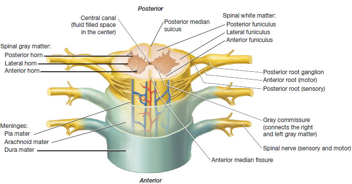
Figure 4.25 Cross-section of the spinal cord (including tags of gray & white matter structures)

Figure 4.26: Cross-section of spinal cord with cervical vertebrae (includes rootlets & spaces)