ESSENTIAL CONCEPTS OF RADIOLOGIC SCIENCE
I. Nature of Our Surroundings
Everything in the universe—what we see, touch, breathe—is composed of matter and influenced by energy. Radiology studies how energy (especially X-rays) interacts with matter (the human body).
II. Matter and Energy
Matter
Anything that has mass and occupies space.
Smallest unit: atom
States: solid, liquid, gas
In radiology, matter = patient tissues, equipment parts, shielding, etc.
Energy
The capacity to do work or cause change.
Forms:
Mechanical (movement)
Chemical
Electrical
Thermal
Nuclear
Electromagnetic (this includes X-rays)
Energy can convert from one form to another—this is what allows x-ray production.
III. Sources of Ionizing Radiation
Ionizing radiation = radiation with enough energy to remove electrons from atoms.
Sources:
Natural
Cosmic rays (from space)
Terrestrial radiation (radionuclides in soil)
Internal radionuclides (K-40 in the body)
Radon gas (the largest source)
Man-made
X-ray machines
Nuclear medicine
Consumer products (smoke detectors, older TVs)
IV. Discovery of X-rays
Discovered by Wilhelm Conrad Roentgen on November 8, 1895.
Observed fluorescence from a barium screen while experimenting with cathode rays.
He called them “X” because their nature was unknown.
This discovery began the field of diagnostic imaging.
V. Development of Modern Radiology
Key improvements:
Coolidge tube (1913) – stable x-ray tube design
CT (1970s)
MRI (1980s)
Digital radiography
Ultrasound advancements
Fluoroscopy modernization
Modern radiology shifted from static films to digital, faster, safer imaging with lower dose.
VI. Reports of Radiation Injury
Early radiologists did not use protection. Reported injuries:
Skin burns
Hair loss
Blood disorders
Cancers
Amputations
These injuries led to the establishment of radiation safety standards.
VII. RADIATION PROTECTION
Modern radiation safety focuses on protecting patients because even low doses from routine X-rays can cause small but real long-term risks.
Fetuses (especially 1st trimester) are very sensitive to radiation.
As a student/technologist, you must learn to operate systems safely and avoid becoming too comfortable or complacent, because this leads to unnecessary exposure.
Always follow ALARA
ALARA = As Low As Reasonably Achievable
→ Reduce exposure by controlling:
Time (shortest exposure possible)
Distance (stay far from the source)
Shielding (use protective barriers)
II. RADIATION PROTECTION DEVICES
1. Filtration
Metal filters (aluminum/copper) in the x-ray tube absorb low-energy x-rays.
These weak X-rays do not contribute to imaging but add to the patient's dose.
2. Collimation
Narrows the X-ray beam to the area of interest only.
Benefits:
Reduces radiation to nearby tissues.
Reduces scatter → better image contrast.
Light-locating collimators are most common.
3. Intensifying Screens
Films are placed between screens that emit light when exposed to X-rays.
Reduce patient dose by 95%, because light, not X-rays, exposes most of the film.
4. Protective Apparel
Aprons and gloves made of lead-impregnated material.
Used in fluoroscopy and some radiographic exams.
5. Gonadal Shielding
Protects ovaries/testes.
Used when:
Patient is of reproductive age.
Gonads are near the primary beam.
Shield does NOT interfere with the exam.
6. Protective Barriers
Control booth walls and windows are lead-lined.
Technologists remain behind them during exposures.
Important Patient Considerations
Avoid pelvic/abdominal x-rays during 1st trimester, unless necessary.
Avoid repeat exposures (they double the patient dose).
Use mechanical immobilizers; technologists should NEVER hold patients.
III. TEN COMMANDMENTS OF RADIATION PROTECTION
Understand time, distance, and shielding.
Avoid becoming complacent.
Never stand in the primary beam.
Always wear protective apparel when needed.
Wear your radiation monitor at the collar, outside the apron.
Never hold a patient; use devices or a family member.
Holders must wear an apron and gloves.
Use gonadal shielding when appropriate.
Avoid pelvic imaging in pregnancy (especially first trimester).
Collimate to the smallest field size.
VIII. Standard Units of Measurement
Physics uses three base quantities:
Mass (kg)
Length (m)
Time (s)
Everything else is derived from them.
Derived Quantities
Volume → m³
Density → kg/m³
Velocity → m/s
Acceleration → m/s²
Special Quantities of Radiologic Science
Quantity | Traditional Unit | SI Unit |
|---|---|---|
Exposure | C/kg | Air kerma (Gy_a) |
Dose | J/kg | Gray (Gy_t) |
Effective Dose | J/kg | Sievert (Sv) |
Radioactivity | s⁻¹ | Becquerel (Bq) |
IX. BASIC MECHANICS
Mechanics: study of objects at rest (statics) and in motion (dynamics)
Velocity (v)
How fast an object moves.
Formula:
v=dtv = \frac{d}{t}v=td

Example:
A ball travels 60 m in 4 s.
v=15 m/sv = 15\ \text{m/s}v=15 m/s
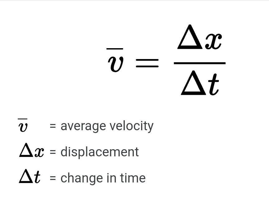
Average Velocity
vˉ=vo+vf2\bar{v}=\frac{v_o + v_f}{2}vˉ=2vo+vf
Acceleration (a)
Rate of change of velocity.
a=vf−vota = \frac{v_f - v_o}{t}a=tvf−vo

Example:
A dragster accelerates from 0 to 80 m/s in 10 s.
a=8 m/s2a = 8\ \text{m/s}^2a=8 m/s2
NEWTON’S THREE LAWS OF MOTION
Law 1: Law of Inertia
An object stays at rest or moves in a straight line unless acted on by a force.
Law 2: Law of Force
F=maF = m aF=ma
Force is measured in newtons (N).
Law 3: Action–Reaction
Every action has an equal and opposite reaction.
WEIGHT
Weight is a force caused by gravity pulling on mass.
Formula:
Wt=mgWt = mgWt=mg
Momentum
Mass × velocity.
Work
Force × distance.
W = Fd
Power
Work per unit time.
P = W/t
Energy
Ability to do work.
Measured in Joules.
Heat
Energy is transferred due to a temperature difference.
Methods:
Conduction
Convection
Radiation
(Heat principles matter for X-ray tube cooling.)
X. Terminology for Radiologic Science
Common Numeric Prefixes
Milli- 10⁻³
Micro- 10⁻⁶
Kilo- 10³
Mega- 10⁶
Giga- 10⁹
Radiologic Units (again for clarity)
Gray (Gy) – absorbed dose
Sievert (Sv) – biological effect
Becquerel (Bq) – radioactivity
Air Kerma (Gyₐ) – exposure
XI. The Diagnostic Imaging Team
Radiologist
A medical doctor interprets images.
Radiologic Technologist (RadTech)
Performs imaging procedures safely and accurately.
Radiology Nurse
Assists with patient care, contrast administration.
Medical Physicist
Oversees equipment quality and radiation safety.
Radiographer Assistant / Support Staff
Helps with positioning, workflow, and clerical tasks.
Team goal: accurate diagnosis with minimal radiation dose.
IMPORTANT HISTORICAL DATES IN RADIOLOGY
(Key highlights only)
1895 – Roentgen discovers X-rays
1901 – Roentgen receives the first Nobel Prize
1913 – Coolidge hot-filament x-ray tube
1921 – Potter-Bucky grid
1928 – Roentgen defined
1948 – Image intensifier
1973 – First CT system developed
1973 – First MRI image
1982 – PACS introduced
1990 – Helical CT
2000s–2020s – Rapid advancements in CT, DR, PET, MRI