Lect. 4 Female Anatomy
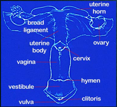
An Overview of the Female Reproductive Tract
Mammals are the only group to have a true uterus, point of connection between thye mother and her fetus(s) providing the fetus with nutrients
Species of insects, fish, and reptiles hatch eggs inside the body ovoviviparous (producing young by means of eggs which are hatched within the body of the parent)
Mammals are the only group of animals which direct “feed” nutrients from the mother’s body to the fetus
Mammals are only species to use their own bodies to Lactate - The secretion of milk products from mammary glands to provide nutrition for newborn offspring
Why should the producer care?
Everything we do with a female animal is dependent on her anatomy
Reduction of economic drain
Increase production and profits
Selection of the most desirable traits
The Vulva and Vagina
Path of sperm cell through the female reproductive tract…
Vulva - Protective outer covering of the female reproductive tract, consisting of two lip like structures, termed labis majora and labia minora
Serves as a protective outer covering of the reproductive system
Separates and protects the vagina from the outside environment
Preserves mucosal membranes
Things To Consider:
Vulva Conformation
Common Vulva Injuries
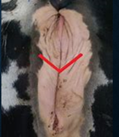
Caslick’s Procedure
Vestibule
Common pathway for reproductive and urinary systems
Hymen - thin piece of mucosal tissue that surrounds or partially covers the external vaginal opening. Physiological function? No nerve endings
Contains the opening to the urethra
Contains the clitoris - a small organ at base of vulva/vestibular junction
Comprised of a small amount of erectile tissue
High percentage of sensory nerves
Signaling the female for correct position for mating
The Vaginal Vault:
Past Vestibule
Highly muscular tissue, is capable of both constriction and expansion
Three distinct tissue layers:
The Tunica serosa - the tissue which separates the vagina from other tissues in the peritoneal cavity (The peritoneal cavity is a potential space between the parietal peritoneum and visceral peritoneum, that is, the two membranes that separate the organs in the abdominal cavity from the abdominal wall.)
The Tunica muscularis - the muscle layer
The Tunica mucosa - a glandular epithelial cell layer that lines the lumen of the vaginal canal
Epithelial layer (thin outer portion) and muscle layer respond to hormones
Ex: estrus, the epithelial layer, water mucus to facility sperm movement and act as a lubricant during the time of mating
Muscle cells of the Tunica muscularis, rhythmic contractions move sperm up the track
Vagina - Organ of Copulation
In some species, site of sperm deposition, very acidic pH - urine pooling, serves as barrier to bacteria and sperm
The vagina has a high concentration of antibodies
Prevent or limits infections in the reproductive and urinary tracks
Sometimes vagina recognizes sperm as a foreign material leading to decreased fertility and inflammation
“pH levels of semen are higher than the pH levels of vaginas, which can mess with the growth of healthy bacteria or “vaginal flora”
Parturition, vagina and cervix must open to form the birth canal
Due to presence of estrogens and relaxin in the track at once
Vaginal fornix - where vagina and the cervix join together
“blind pouch” inside the vagina
“often mistaken as the opening to the cervix”
Vagina
Functions
Copulatory organ
Site of semen deposition
Absorbs seminal plasma
Acidic pH-
Ig G and Ig A
Antibodies to sperm sometimes found in infertility
Birth canal
Urinary canal
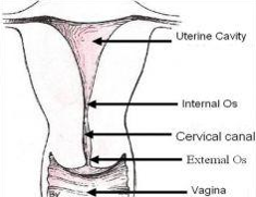
The Cervix
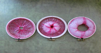
The Cervix is round in shape and has a hole in the middle
The can running through the cervix lined by a glandular epithelium, secrets mucous which is responsive to progesterone and estrogen
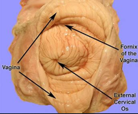
Os - mouth or opening
Os Cervix and Os Uterus
Interior of the canal is irregularly shaped
Horse, mucosal folds
Sheep and Cows, annual rings
Pig, corkscrew shaped
These shapes play an important role in our ability to pass an AI rod through the cervix when we attempt artificial insemination
So what is the function of the cervix???
Serves as “plug” for the uterus: pregnancy, non estrus, mucus will be thickened and block sperm movement (a barrier)
Serves as “transport tunnel”: estrus, the mucus is thinned, contractions and cilia on the epithelial line help move sperm through the canal
The environment in the canal is much more supportive of sperm survival than the low pH of the vaginal vault
Some species, most notably the boar with his corkscrew-shaped penis and sometimes the stallion, site of sperm deposition
Cervical canal will become part of the birth canal at parturition: two-millimeter hole must be able to dilate to allow passage of the fetus
Number of hormones which allow the muscles to pull back to the peritoneal wall, forming a much larger opening
Also called cervix uteri
Thick walled, inelastic tube with small lumen
Functions
Site of semen deposition
Sperm reservoir
Prevent microbial contamination of uterus
Produces cervical mucous
The Uterus: An Overview of Uterine Function
Unique organ of the mammalian female reproductive system
Two Functions:
Incubator where the developing fetus will grow and develop. During gestation, the placenta attaches to uterus which will supply the nutrients the embryo/fetus need
Makes the hormones
Relaxin, allows the uterus to large as the fetus grows
Makes PGF2a which helps with gamete movement and parturition
Uterus: Parts of the Uterus
Three Layers:
Serosal layer: which separates the organ from other organs in the peritoneal cavity
Myometrium: the muscle layer of the uterus which can relax to allow for space or contract to empty the uterus at parturition
Endometrium: Separating the myometrium from the lumen of the uterus
Secretes mucus, nutrients, and hormones supports the growth of the fetus and reservoir for sperm movement.
Irregular shape: Crypts - lining crevices, in response to hormones
Uterus can be both a barrier and reservoir movement
Uterus: Uterine Shapes
Species Specific! Unlike other parts of the reproductive tract
Adapted evolutionarily to accommodate each species
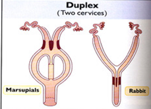
Duplex
Bicornate
Bipartite
Simplex
1) Duplex - no common area (uterine body) and a separate cervical opening
Rat, mouse, rabbit, guinea pig
Two cervices
No uterine body
Uterine horns completely separate
2) Bicornate - a single cervical opening, a small uterine body, and long uterine horns
The body may have a tissue separation across most the body called a septum
Pig
One cervix
Uterine body very small
Uterine horns long and convoluted