Valves of the heart
Ensure blood flows in one direction
Composed of connective tissue and endocardium
4 valves divided into 2 categories:
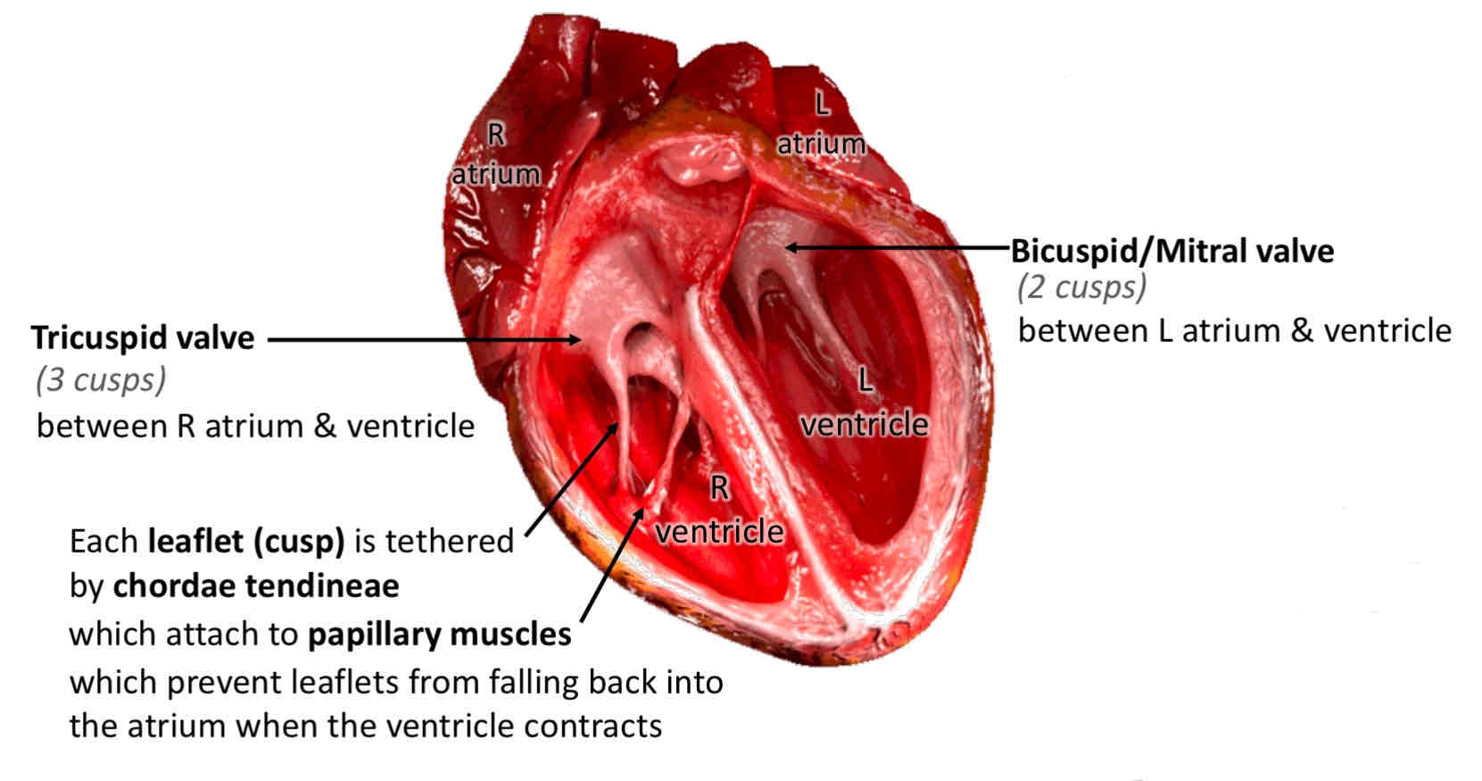
Atrioventricular - tricuspid and mitral (bicuspid). Located between the atria and corresponding ventricle
Semilunar - pulmonary and aortic valve. Located between the ventricles and their corresponding artery and regulate flow of blood leaving the heart
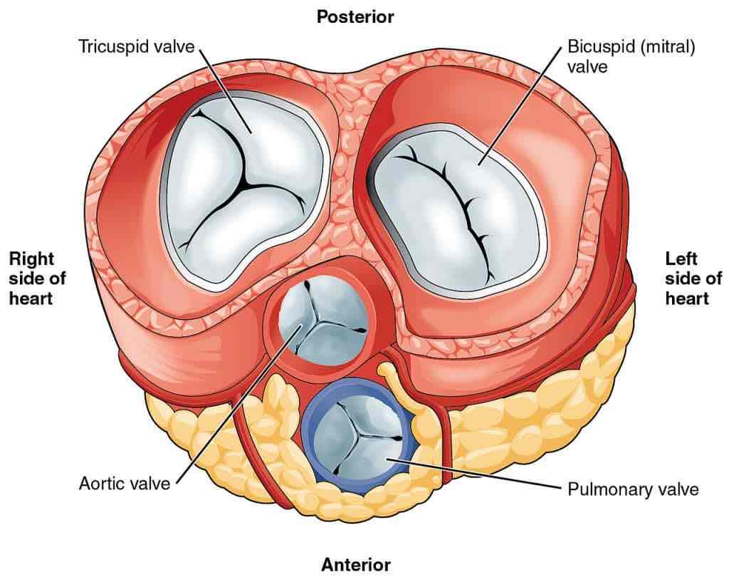
Atrioventricular valves:
located between the atria and ventricles
Close during the start of ventricular contraction (systole), producing the first heart sound
Tricuspid valve - between RA and RV (right atrioventricular orifice)
3 cusps (anterior, septal and posterior)
Base of each cusp anchored to a fibrous ring that surrounds the orifice
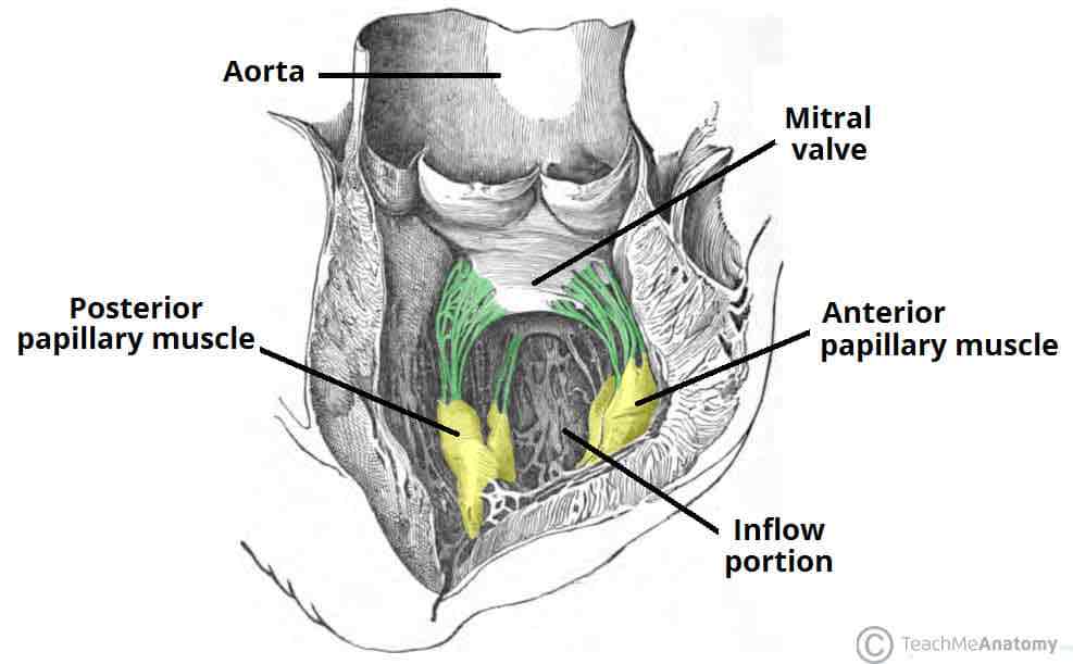
Mitral/bicuspid valve - located between LA and LV (left atrioventricular orifice)
2 cusps
Supported by attachment of fibrous cords (chordae tendineae) to the free edges of the valve cusps
Chordae tendineae are attached to papillary muscles on interior surface of ventricles - these muscles contract during ventricular systole to prevent prolapse of the valve leaflets into the atria
5 papillary muscles in total - 3 in RV supporting the tricuspid valve, 2 in LV acting on the mitral
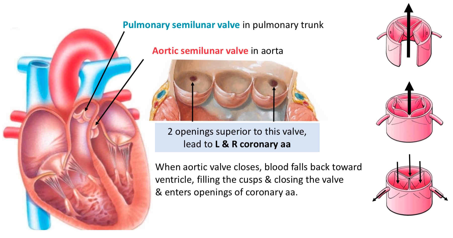
Semilunar valves:
Located between ventricles & outflow vessels
Close at the beginning of ventricular relaxation (diastole), producing the 2nd heart sounds
Pulmonary valve - between the RV and pulmonary trunk (pulmonary orifice)
3 cusps (left, right and anterior - named by position in foetus before heart undergoes rotation)
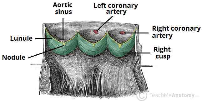
Aortic valve - between LV and ascending aorta (aortic orifice)
3 cusps (left, right, posterior)
Left and right aortic sinuses mark origin of left and right coronary arteries. As blood recoils during ventricular diastole, it fills the aortic sinuses and enters the coronary arteries to supply the myocardium
Have similar structure
Sides of each valve leaflet are attached to the walls of the outflow vessel, which is slightly dilated to form a sinus
Free superior edge of each leaflet is thickened (the lunule), and is widest in the midline (the nodule)
At beginning of ventricular diastole, blood flows back towards heart, filling the sinuses and pushing the valve cusps together - closes the valve