The Axial Skeleton and Odontology
The Axial Skeleton and Odontology
Instructor: Dr. Kori Filipek
Introduction to Forensic Anthropology
Forensic anthropology is the identification and analysis of human skeletal remains for medicolegal purposes. This field requires the ethical treatment of human remains with dignity and respect. As a reminder, practitioners in this domain utilize clinical terminology to accurately communicate their findings and practices concerning skeletal analysis. Bone is classified as a living organ characterized by a high mineral content, and the human body typically has an average of 206 bones.
Laboratory Guidelines
During practical laboratory sessions, students are encouraged to follow essential rules to ensure a conducive learning environment:
Always wear a lab coat to participate in activities.
Remain within designated areas; avoid spreading into other bench spaces or off allocated mats.
Consult your bone manual or class materials during exercises.
Treat the equipment and remains with respect; failure to do so may result in expulsion from the lab.
Attending readings and hands-on practicals is crucial for passing the course successfully.
Weekly Objectives
This week's focus is on the Axial Skeleton and Odontology.
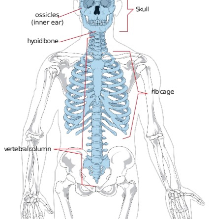
The Axial Skeleton
The axial skeleton, derived from the term "axis," comprises the central core of the human skeleton including:
Skull
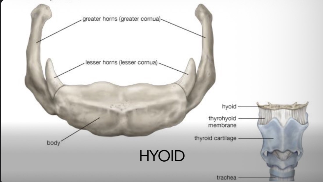
Hyoid bone
Rib cage
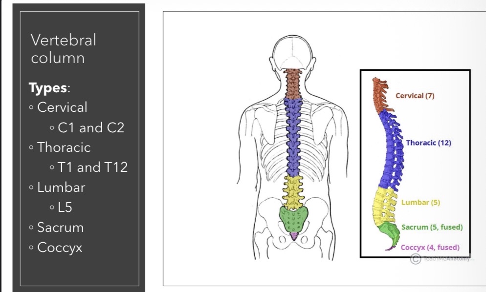
Vertebral column
Breakdown of the Axial Skeleton Components
Skull
Comprised of:
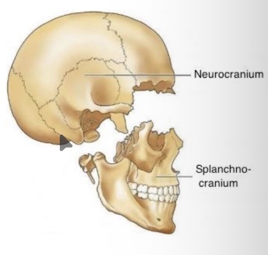
Neurocranium (8 bones)
Splanchnocranium (Facial skeleton, 14 bones)
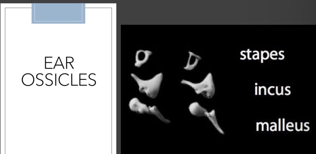
Additional components: Hyoid and ear ossicles (total of 7)
Rib Cage
Consists of:
12 pairs of ribs
Manubrium
Sternum (including the xyphoid process)
Vertebral Column
Constituted of:
7 cervical vertebrae
12 thoracic vertebrae
5 lumbar vertebrae
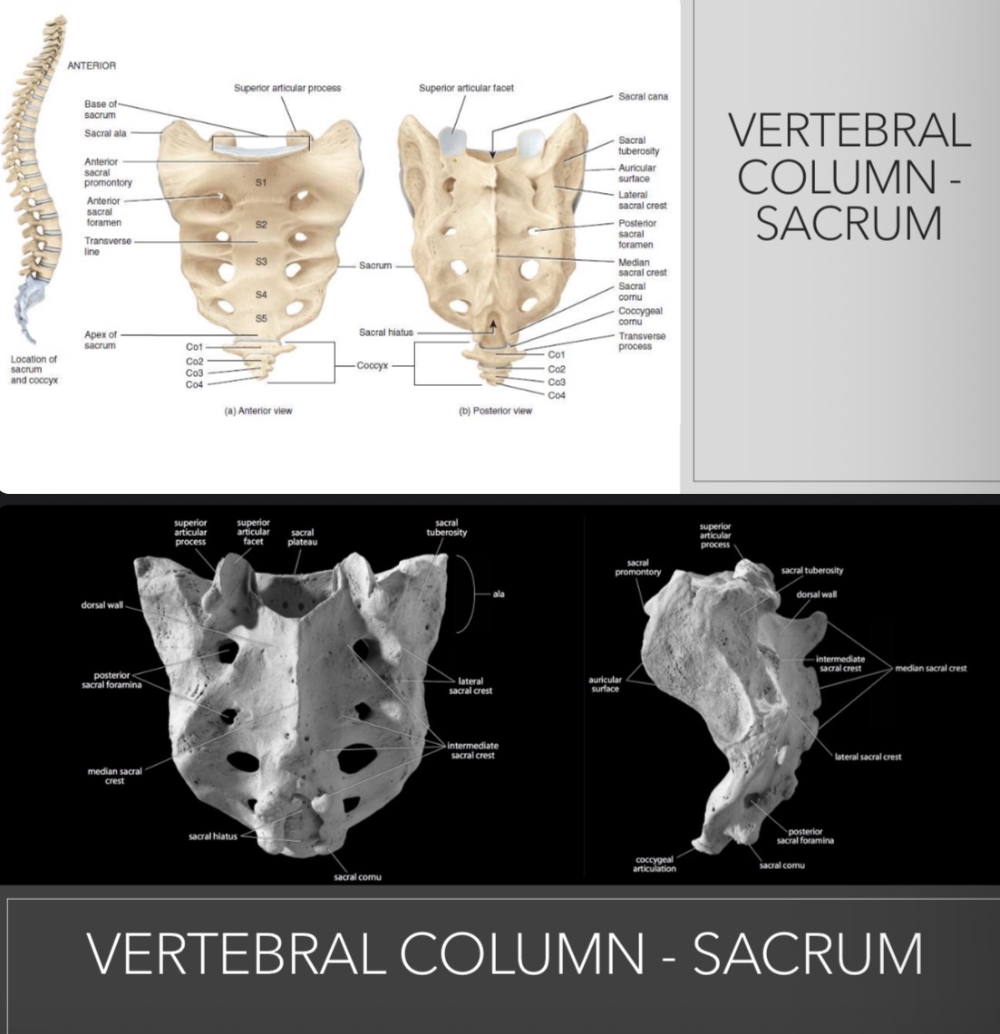
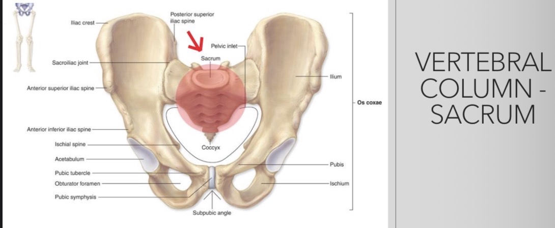
Sacrum
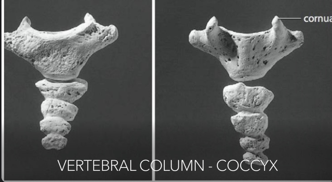
Coccyx
Detailed Anatomy of the Skull
Major Components
The skull consists of the following components:
Neurocranium (8 Bones):
Frontal
Parietals (2)
Temporals (2)
Occipital
Ethmoid
Sphenoid
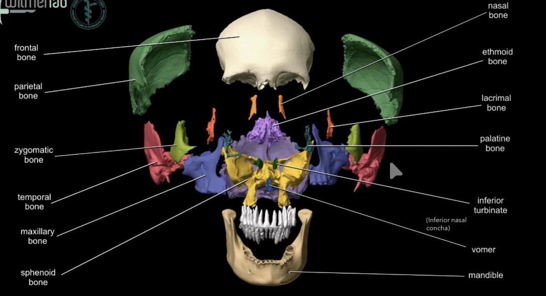
Splanchnocranium (Facial Skeleton, 14 Bones):
Maxillae (2)
Mandible
Nasal bones (2)
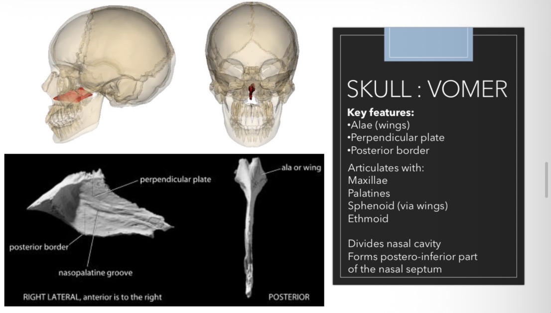
Vomer - line down middle of nose
Inferior nasal conchae (2)
Palatines (2)
Lacrimal bones (2)
Zygomatic bones (2)
Key Definitions
Cranium: The cranium refers to the skull excluding the mandible.
Skull Definition: The skull includes both cranium and mandible.
Definitions and Key Features of Skull Bones
Cranial Bone Terms:
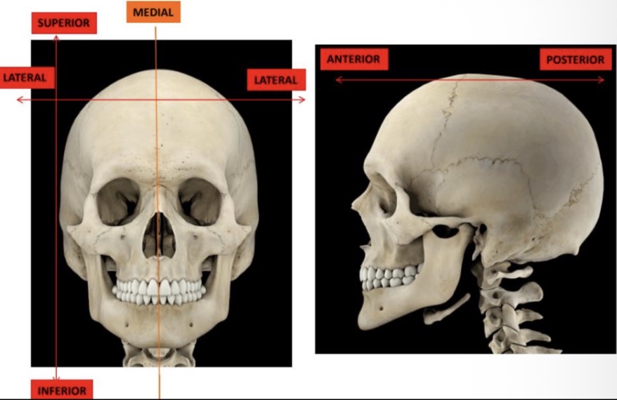
Endocranial Surface: The inner surface of the skull that houses the brain.
Ectocranial Surface: The outer surface of the skull that is visible externally.
Specific Bones with Anatomical Features
Frontal Bone
Key features: Frontal squama, temporal line, frontal eminence, supraorbital margin, lacrimal fossa, etc.
Articulates with: Parietals, Sphenoid, Zygomatic, Nasals, Maxilla, Lacrimals, Ethmoid.
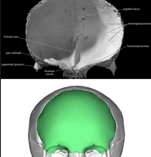
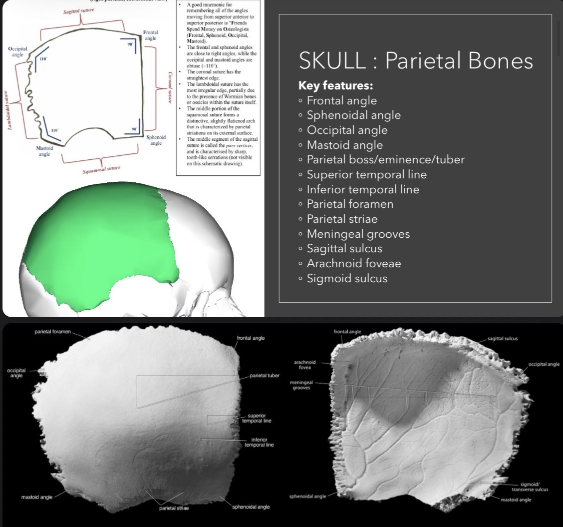
Parietal Bones
Key features: Frontal angle, occipital angle, superior temporal line, and parietal foramen.
Articulates with: Opposite parietal, frontal, occipital and temporal bones.
Temporal Bones
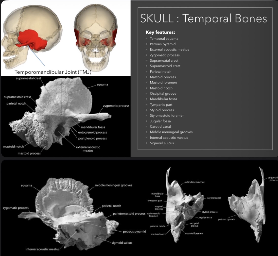
Key features: Temporal squama, external acoustic meatus, zygomatic process.
Articulates with: Parietals, Occipital, Sphenoid, Zygomatics, Mandible.
Occipital Bone
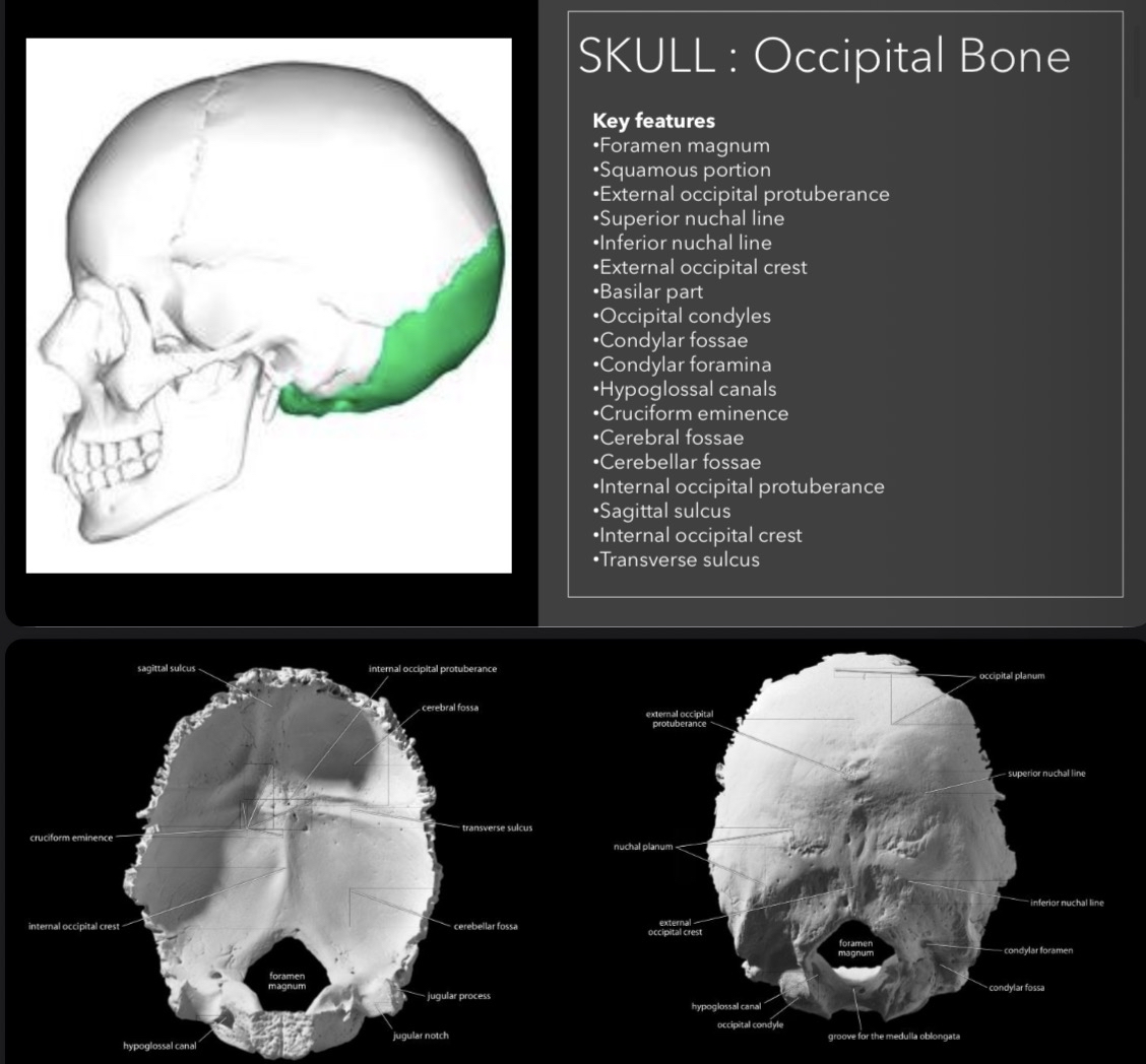
Key features: Foramen magnum, external occipital protuberance, condylar fossae.
Articulates with: Temporal bones, Sphenoid, Parietal bones, C1 vertebra (atlas).
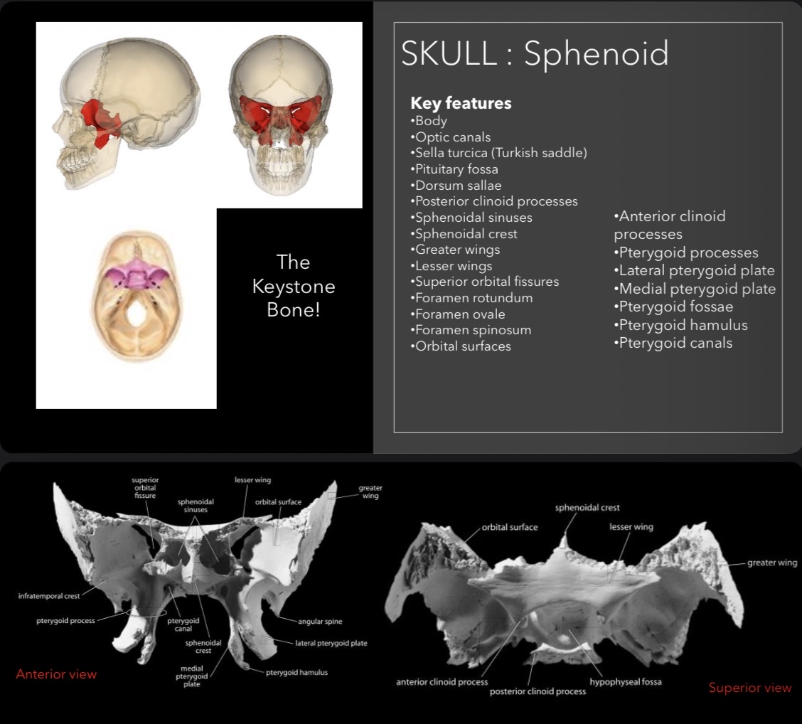
Sphenoid Bone
Key features: Body, optic canals, sella turcica (Turkish saddle), and pterygoid processes.
Noted for articulating with all neurocranium bones, plus vomer, zygomatics, and palatines.
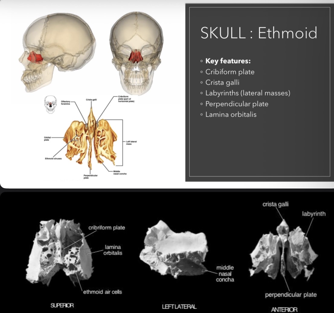
Ethmoid Bone
Key features: Cribiform plate, crista galli, labyrinths.
Articulates with: Frontal, Sphenoid, Nasals, Maxillae, Lacrimals, Palatines, and Vomer.
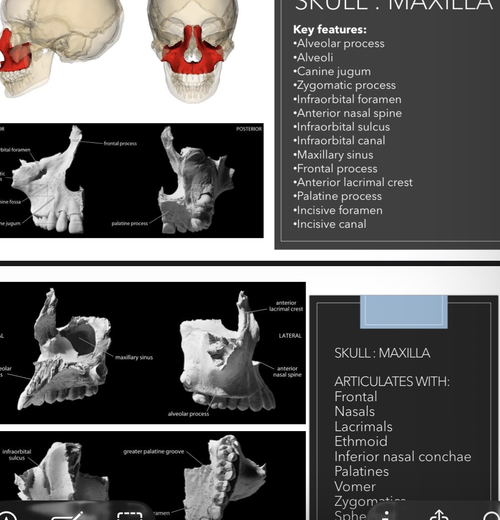
Maxilla
Key features: Alveolar process, infraorbital foramen, maxillary sinus.
Articulates with: Frontal, Nasals, Lacrimals, Ethmoid, and others.
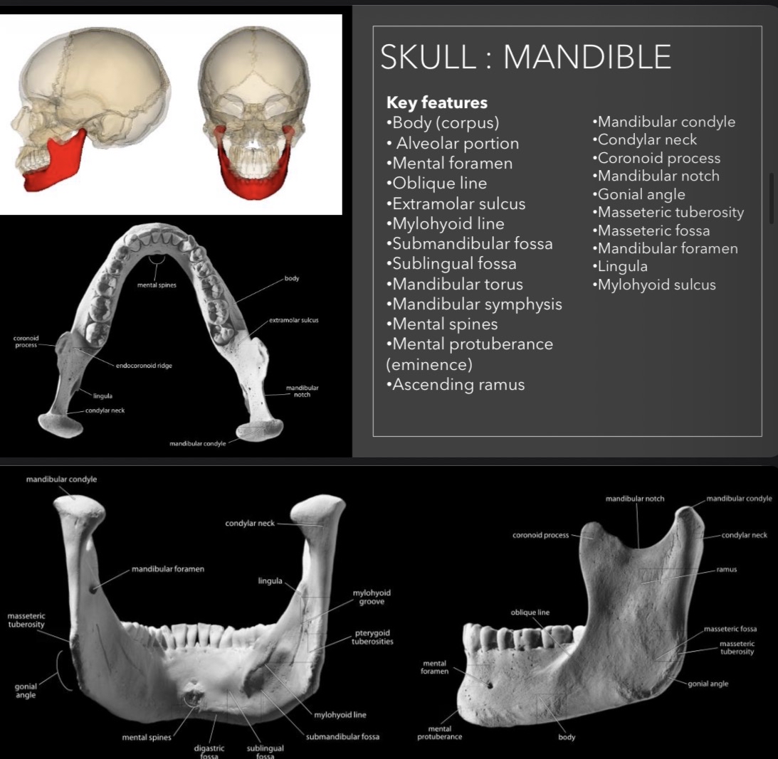
Mandible
Key features: Body, mental foramen, mandibular condyle, and ramus.
Articulates with: Frontal, Sphenoid, Nasals, Maxillae, etc.
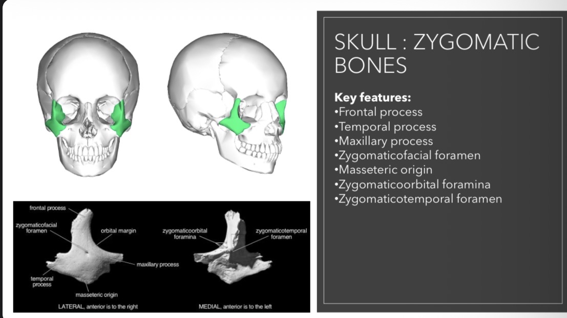
Zygomatic Bones
Key features: Frontal process, temporal process, maxillary process.
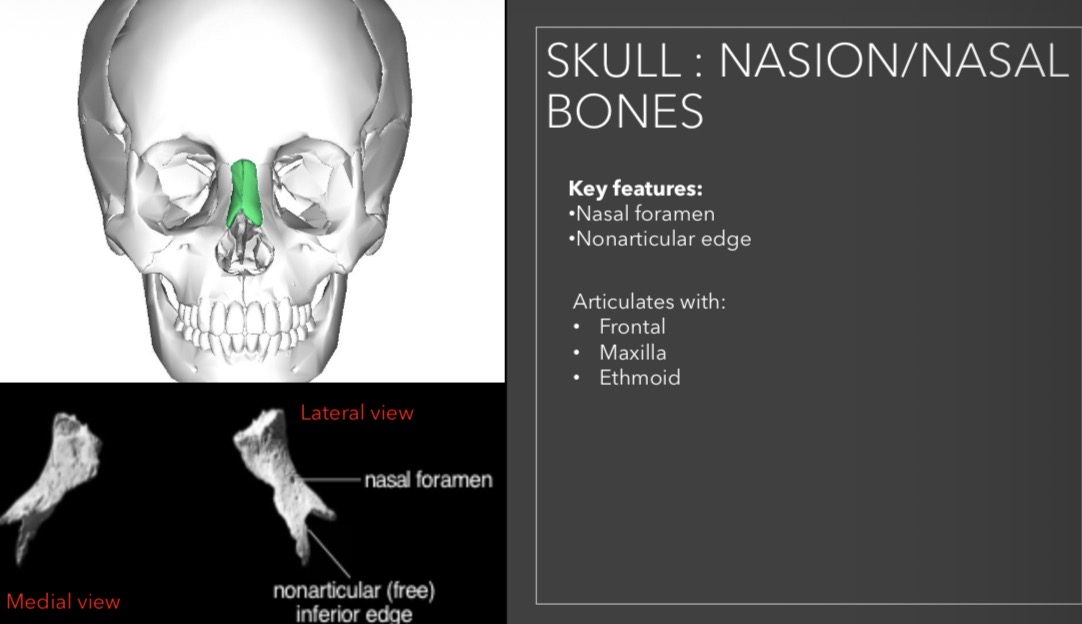
Nasal Bones
Key features: Medial view and lateral view anatomy.
Articulates with: Frontal, Maxilla, Ethmoid.
Vomer
Key features: Alae (wings) and perpendicular plate.
Functions: Divides nasal cavity and forms the posterior-inferior part of the nasal septum.
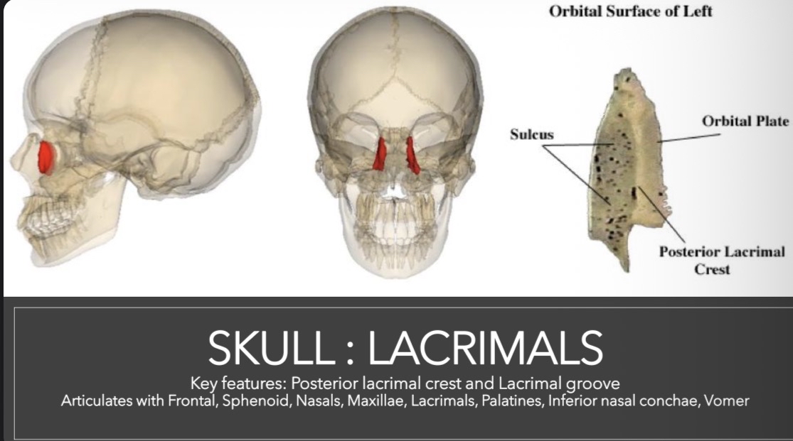
Lacrimal Bones
Key features: Posterior lacrimal crest and lacrimal groove.
Articulates with: Frontal, Sphenoid, Nasals, Maxillae.
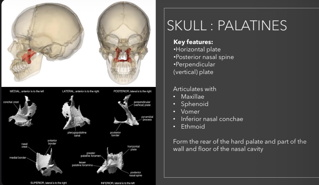
Palatine Bones
Key features: Horizontal plate and vertical plate.
Articulates with: Maxillae, Sphenoid, Vomer, Ethmoid.
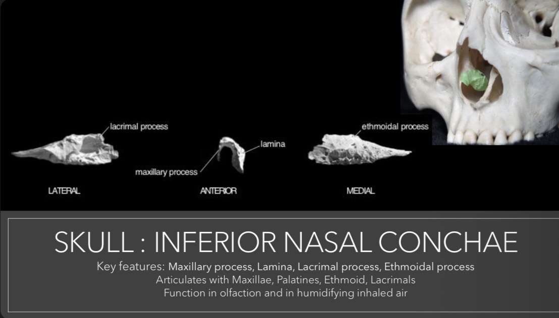
Inferior Nasal Conchae
Key features: Maxillary and lacrimal processes.
Functions: Aid in the humidification of inhaled air.
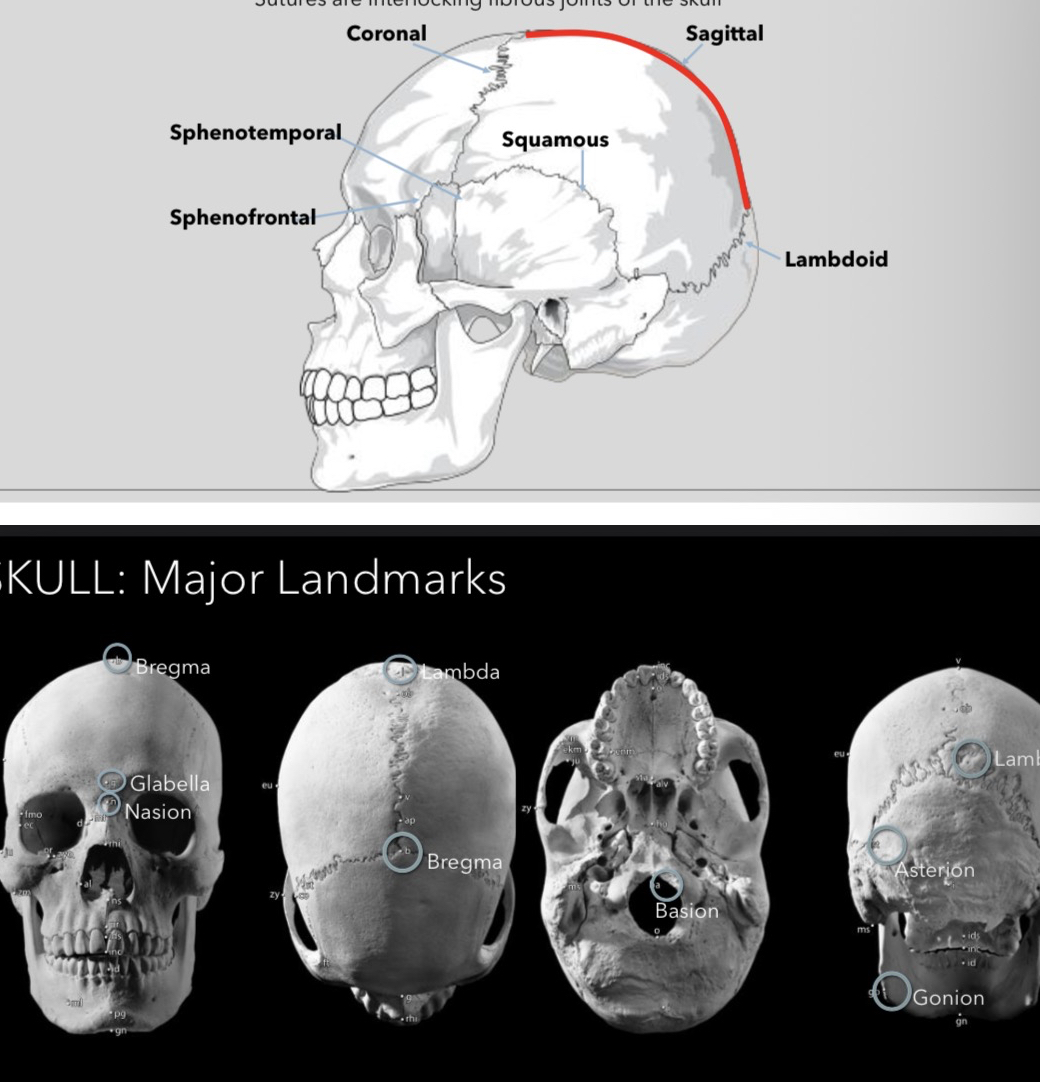
Identifying Major Landmarks of the Skull
Craniometric landmarks are used for measurements: Bregma, Glabella, Nasion, Lambda, Gonion, Asterion. These features allow anthropologists to perform comparisons in craniometric analyses.
Vertebral Column
Structure of the Vertebral Column
The vertebral column consists of various sections with distinct types:
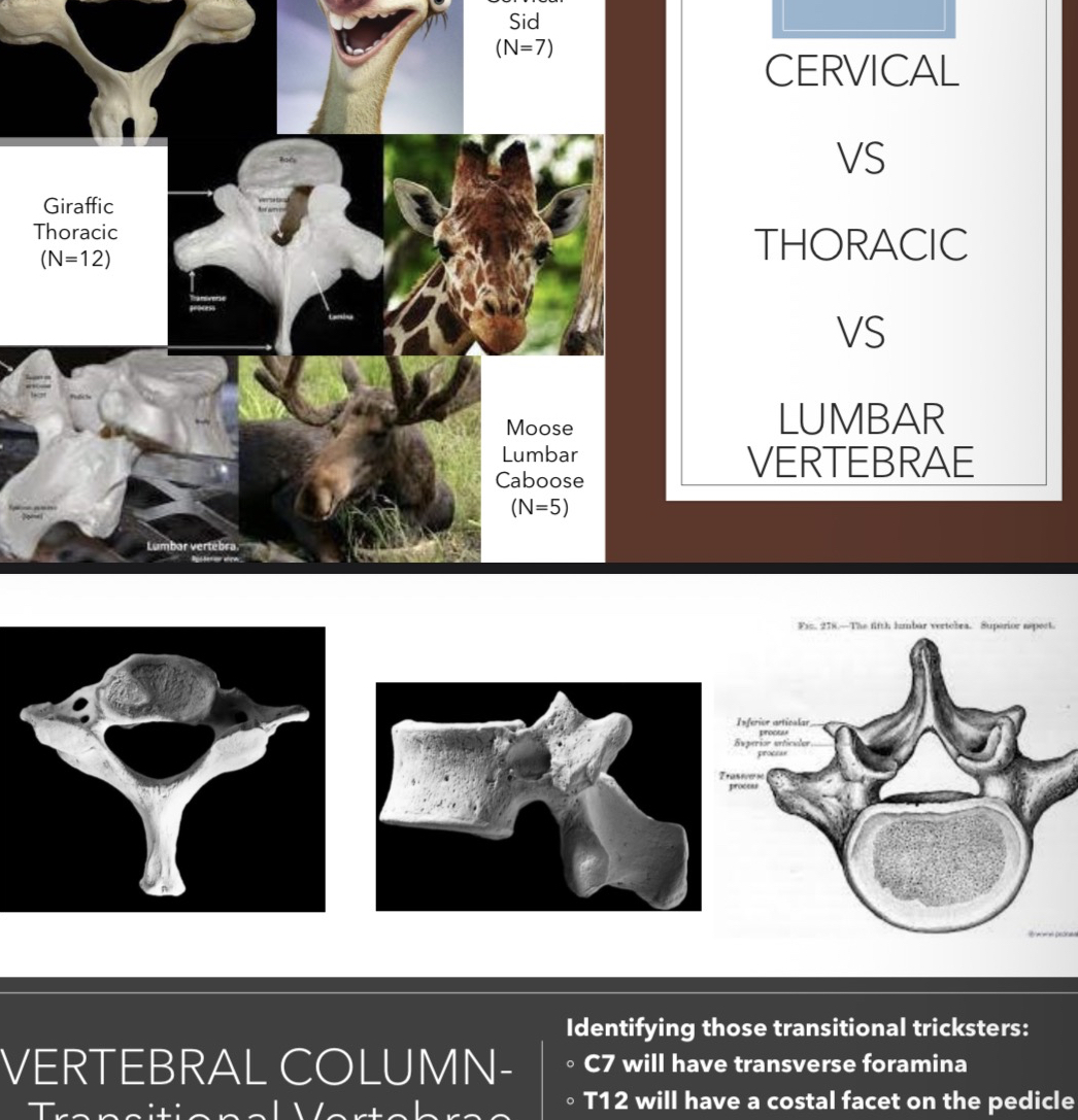
Cervical (C1-C7)
Thoracic (T1-T12)
Lumbar (L1-L5)
Sacrum (5 fused vertebrae)
Coccyx (typically 4 fused vertebrae)
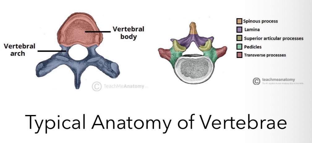
Anatomy of Typical Vertebrae
For a typical vertebra, the main components include:
Vertebral body
Vertebral arch
Spinous process
Transverse processes
Superior and inferior articular facets
Pedicles
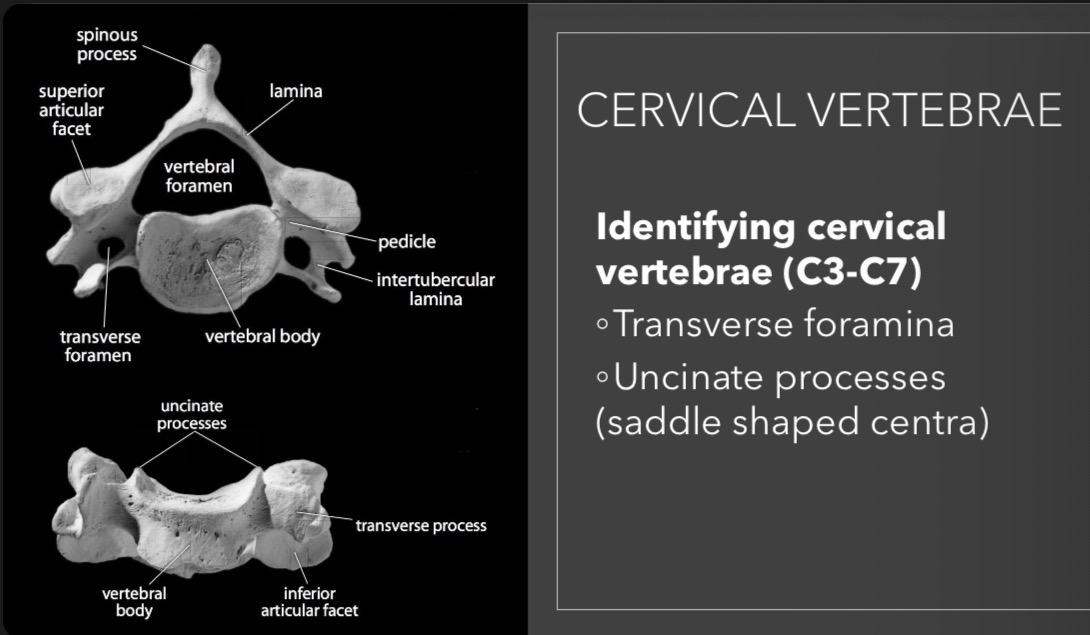
Specific Vertebra Types
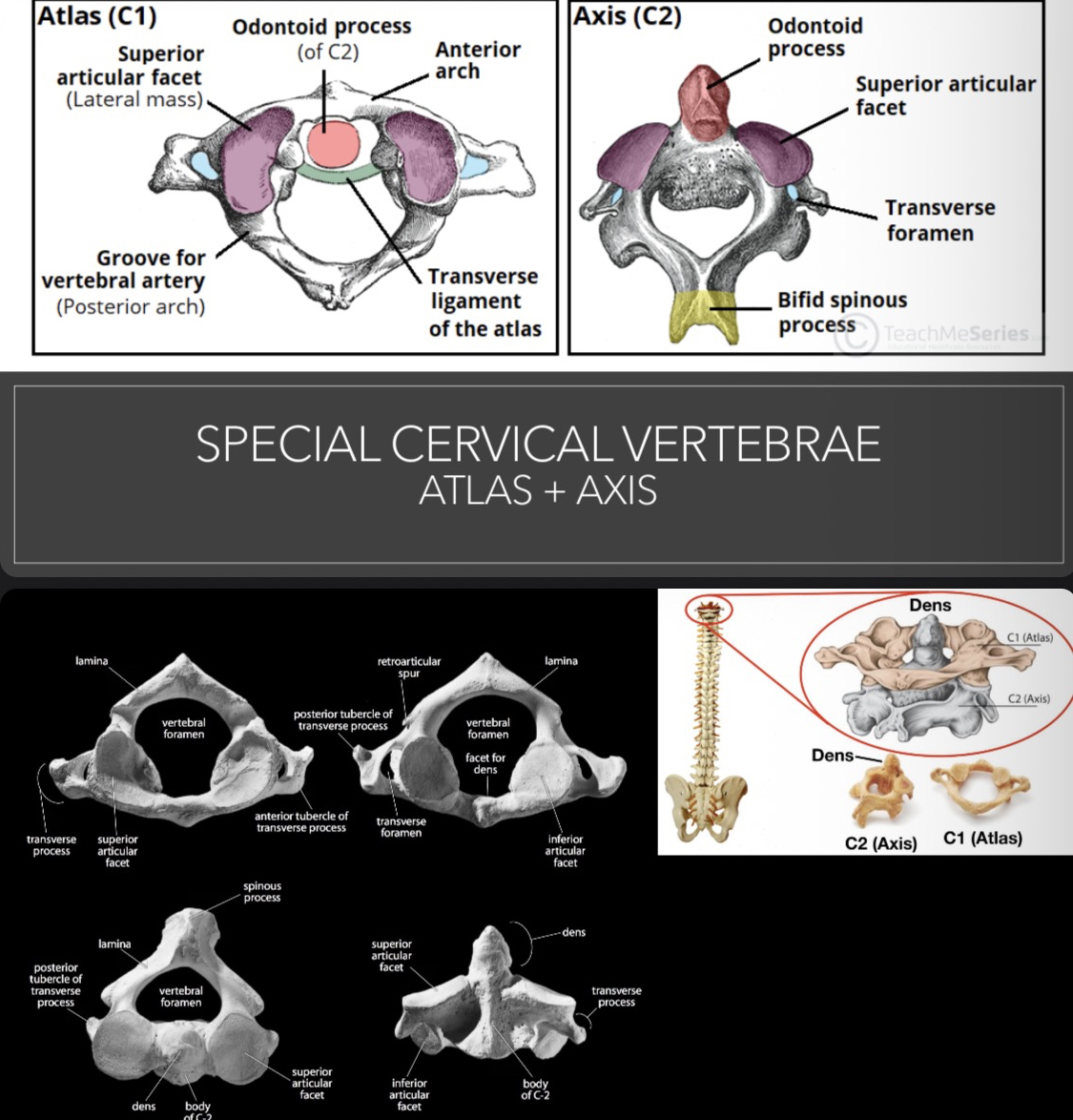
Atlas (C1): Holds the skull (complex structure); Features include anterior and posterior arches, superior facets for occipital condyles, etc.
Axis (C2): Has the odontoid process (dens) that acts as a pivot for rotation of the skull.
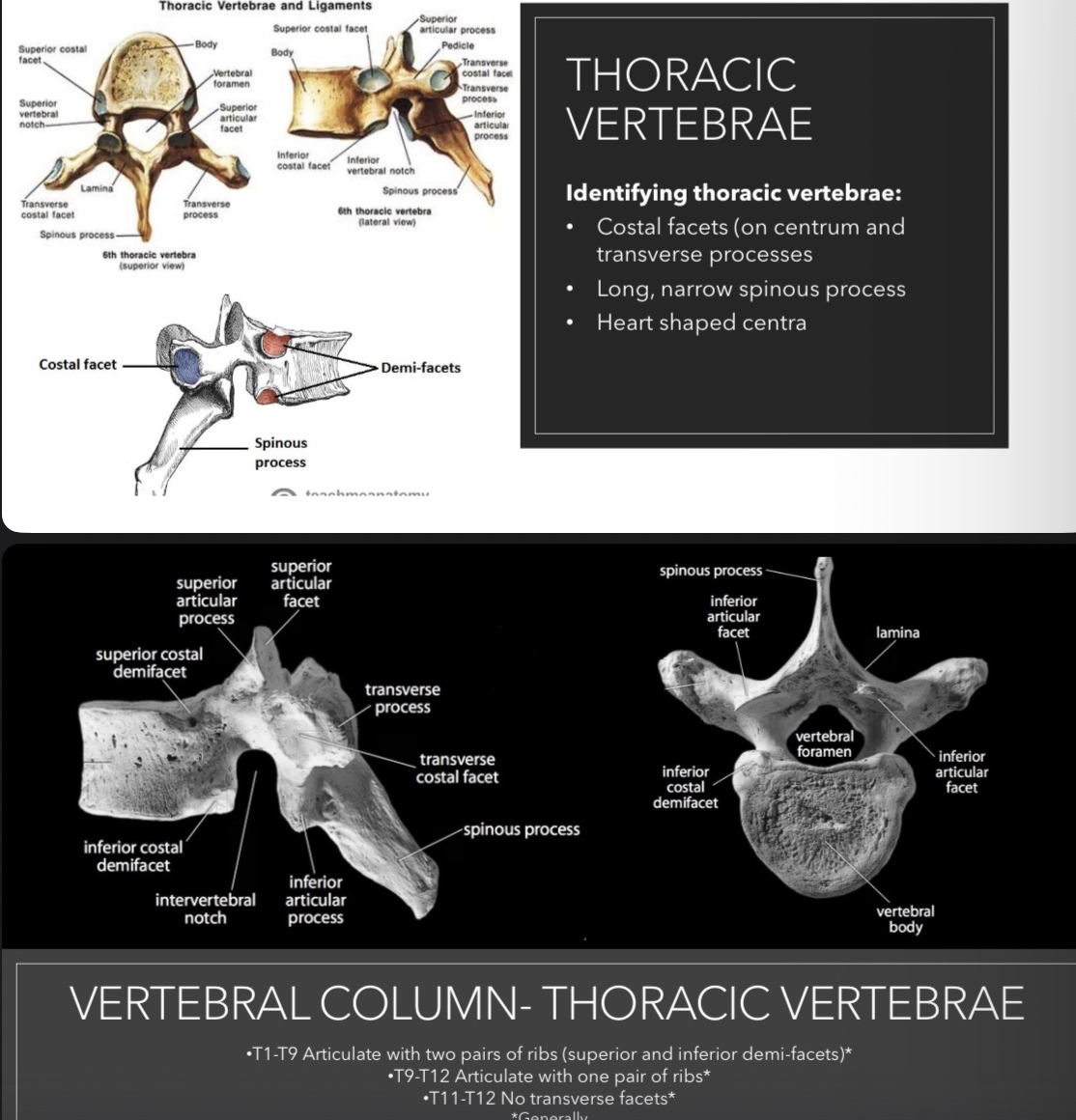
Thoracic Vertebrae: Characterised by long, narrow spinous processes and costal facets.
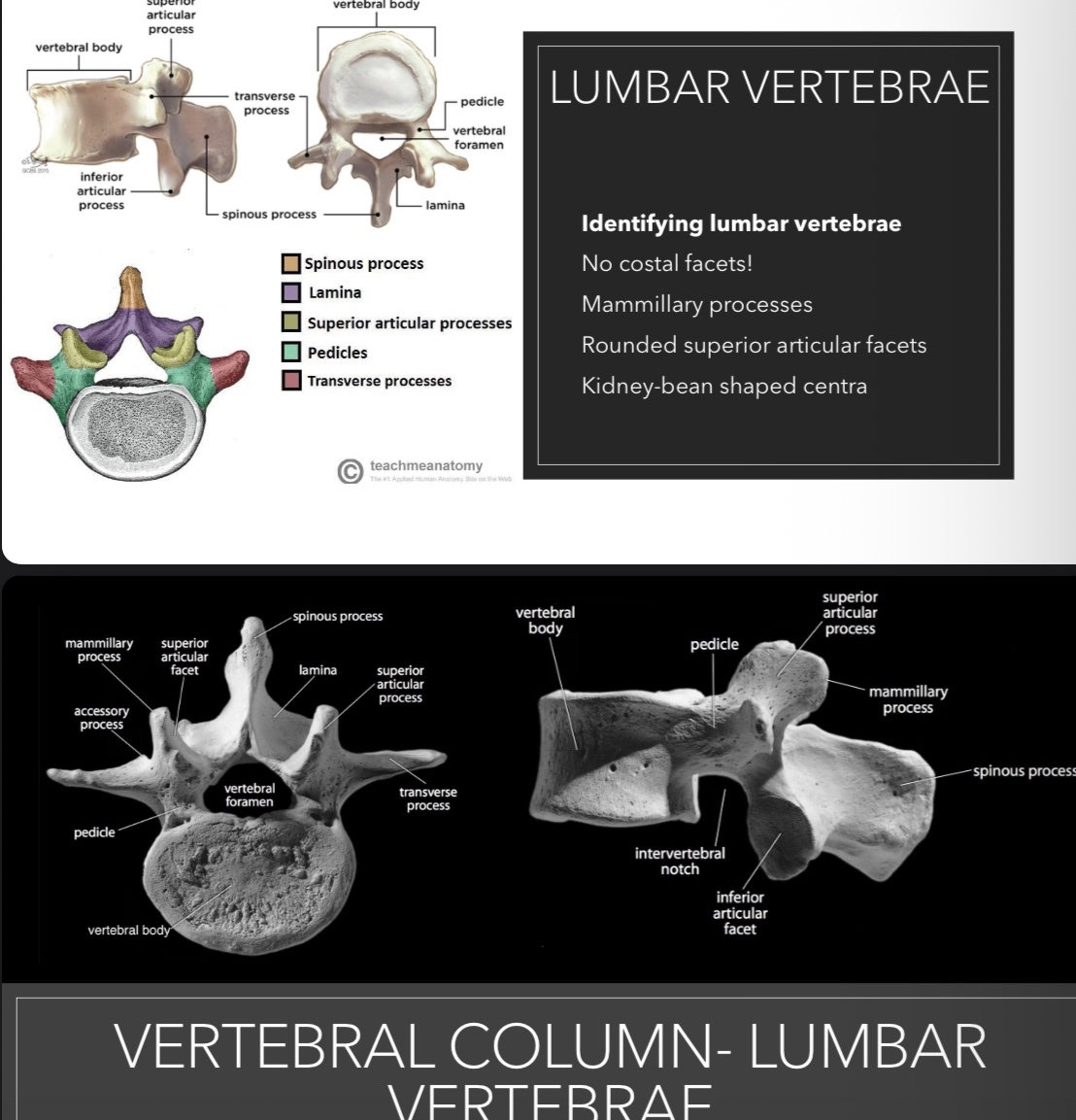
Lumbar Vertebrae: Designated by no costal facets and a larger body for bearing weight.
Sacrum: Comprised of fused vertebrae articulating with the pelvic girdle.
Coccyx: The rudimentary tailbone structure.
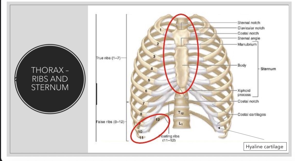
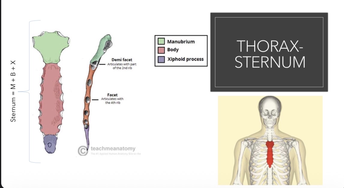
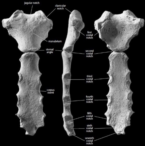
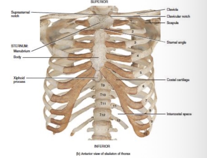
Thorax - Ribs and Sternum
Thorax - Sternum: 3 sections and key features
Manubrium
Jugular notch
First costal notch
Clavicular notch
Sternal angle
Sternal body
Costal notches
Xiphoid process
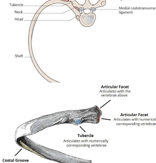
Thorax - Ribs:
2 sets of 12
1-7 true ribs
8-10 false ribs
11-12 floating ribs
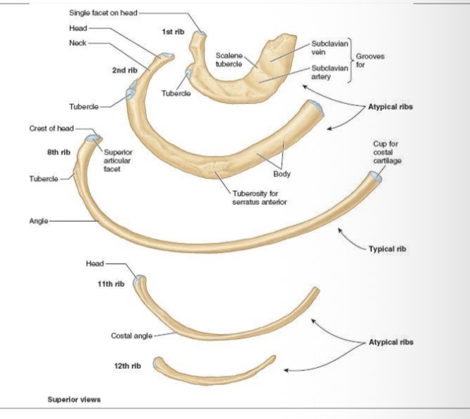
Key features- ribs:
Head
Tubercle
Crest of neck
Angle
Costal groove
Cranial edge
Caudal edge
Sternal. End
Used in ageing - Iscan 1984
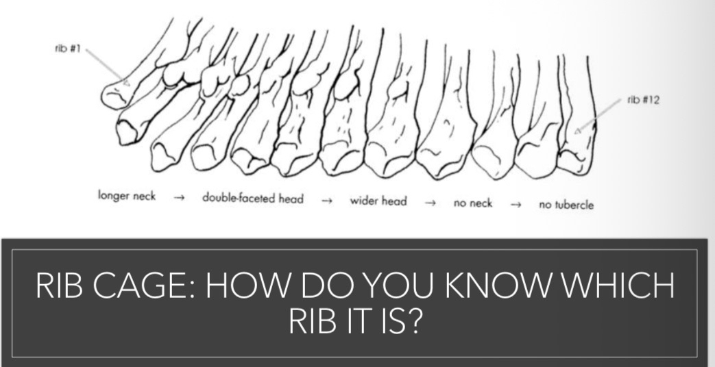
Atypical Ribs:
1st rib
2nd rib
11th and 12th rib
Joints of the Axial Skeleton
Sutures of the skull are fibrous joints, creating interlocking structures with little to no movement.
Synarthrosis allows little movement between elements, structures of the skull, coccygeal elements, teeth sockets.
Amphiarthrosis refers to slightly movable joints formed by fibrocartilage (e.g., vertebral discs).
Diarthrosis (synovial) joints allow greater movement and articulation, like between the articular facets of vertebrae.
Odontology
Importance of Dental Knowledge
Understanding teeth provides insights into:
Human evolution
Dating/DNA analysis
Ethnic origin
Age at death estimates
Dietary habits, migration, and diseases
Stress and famine indicators
Hygiene practices and occupational indicators
Personal adornments and identity in forensic contexts
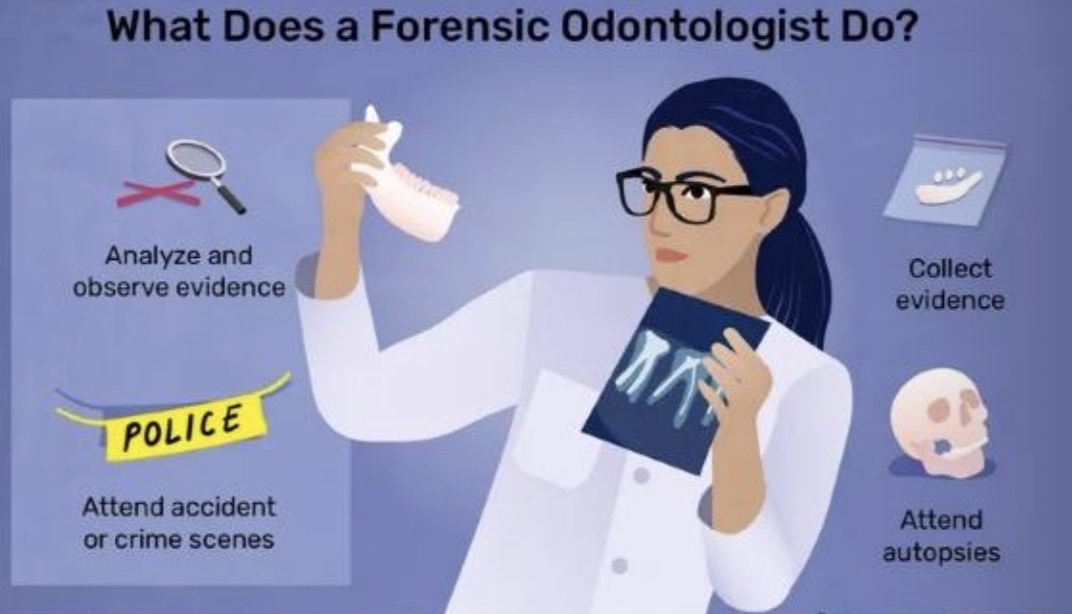
Roles of Forensic Odontologists
Forensic odontologists engage in various tasks including:
Analyzing and observing dental evidence
Collecting evidence from police or crime scenes
Attending autopsies to provide consultation
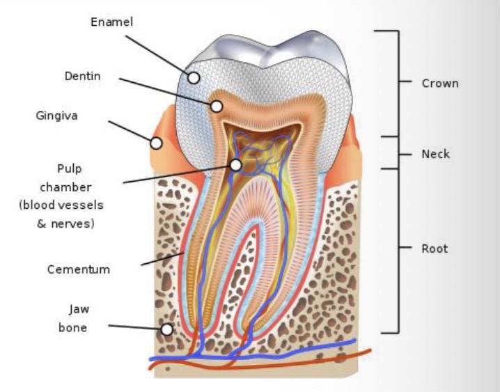
Composition of Teeth
Teeth consist of:
Organic material (25%)
Inorganic material (75%)
This inorganic nature is critical for preservation, making teeth some of the most durable remains.
Types of Teeth
Humans are diphyodonts, having two sets of teeth:
Deciduous teeth: 20 total, starts developing in utero by the 6-8th week, usually shed by around 12 years of age.
Permanent teeth: 32 total, begin forming around 4 months of age, with the first permanent tooth (1st molar) erupting around age 6, and the last (3rd molar) emerging between ages 15-25.
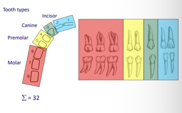
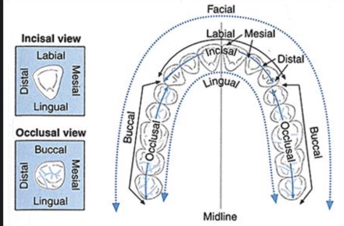
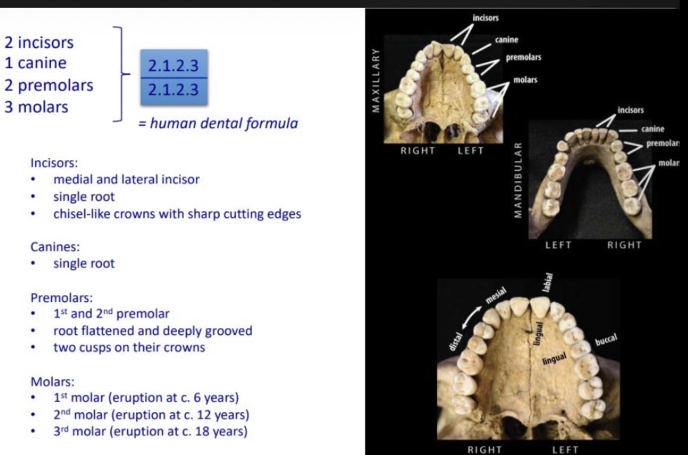
Dental Terminology and Structure
Permanent teeth
Dental formula: 2.1.2.3 x 2 (upper and lower jaws)
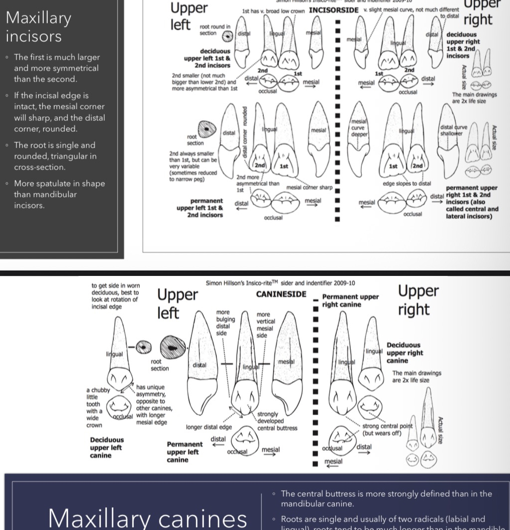
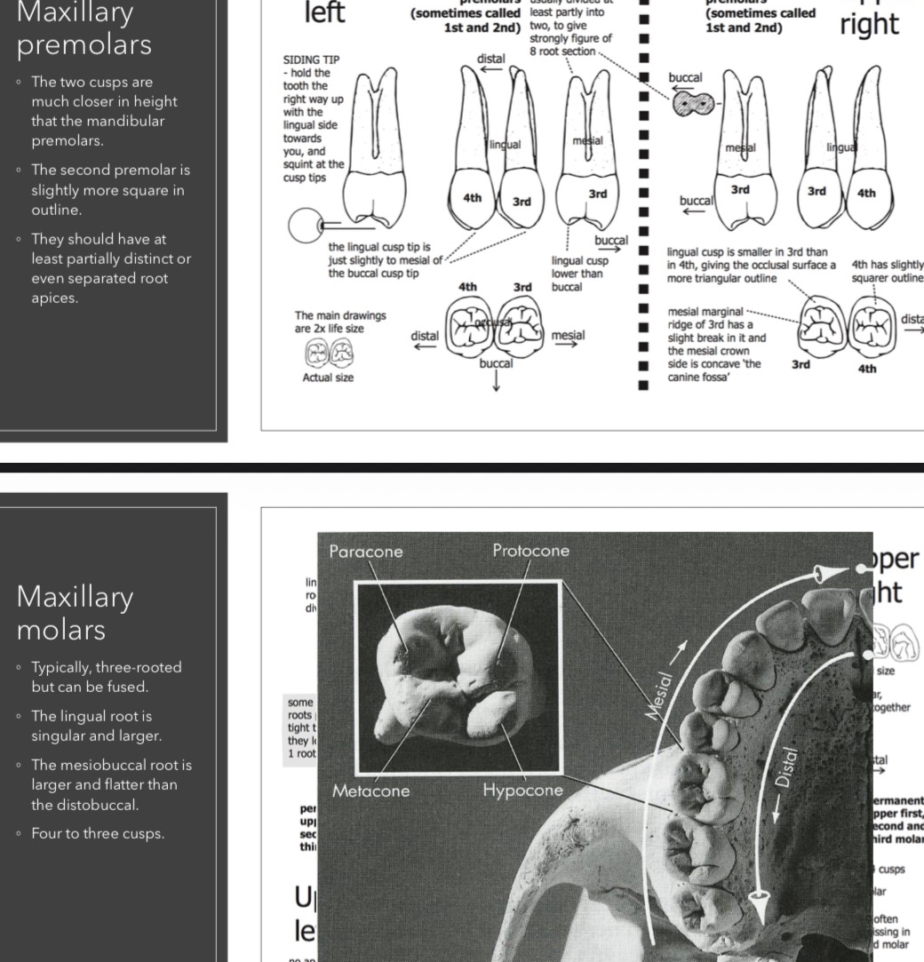
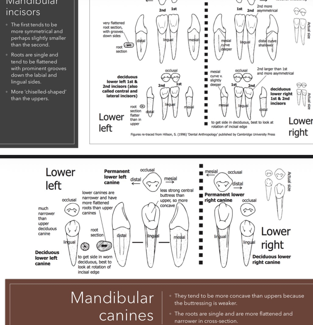
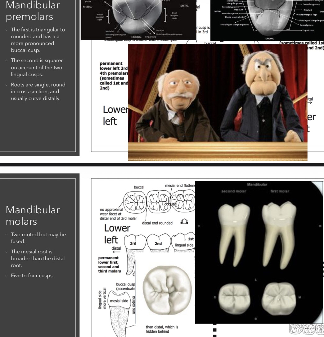
The arrangement includes incisors, canines, premolars, and molars with distinct root and crown features.
Deciduous teeth structure differs from permanent teeth in root size and crown proportion.
Summary of Eruption Sequence for Teeth
Upper teeth erupt as follows:
Cental Incisor: 8-12 months
Lateral Incisor: 9-13 months
Canine (Cuspid): 16-22 months
First Molar: 13-19 months
Second Molar: 25-33 months
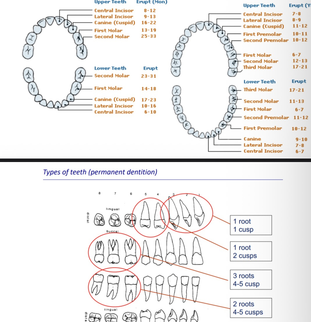
Differences Between Types of Permanent and Deciduous Teeth
Permanent vs. Primary teeth have eruption timelines and structural differences (e.g., permanent molars have multiple cusps compared to single-rooted primary molars).
Specific structural comparisons (e.g., maxillary vs. mandibular) illustrate notable variations needed for identification.
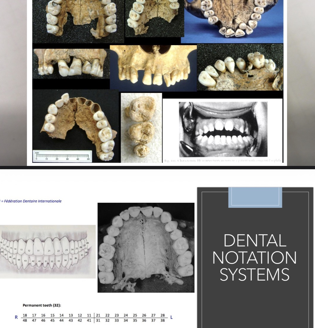
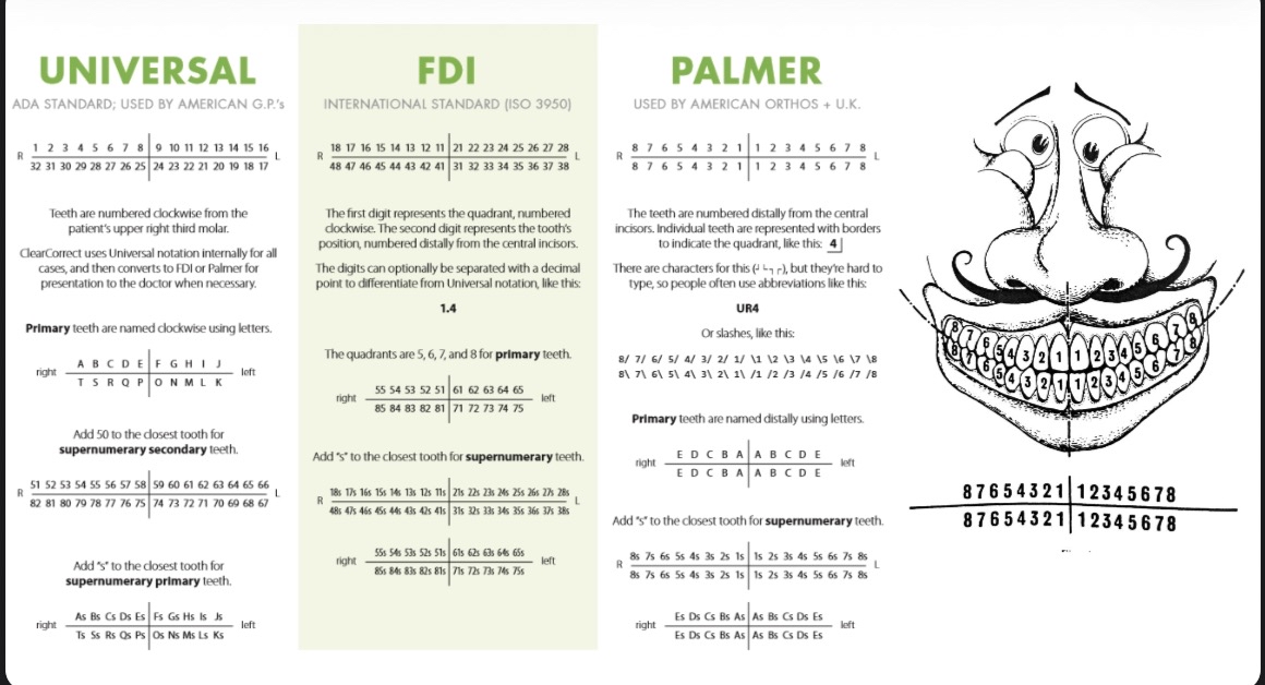
Dental Notation Systems
FDI (Fédération Dentaire Internationale):
Quadrant-tooth numbering for easy identification (e.g., permanent teeth noted with two-digit systems).
Universal Notation:
Widely used in clinical and educational settings, represents teeth in a clockwise manner starting from the upper right third molar.
Palmer Notation:
Used primarily by orthodontists, indicates teeth with specific symbols for quadrants.
Laboratory Activities and Objectives
During lab sessions, you should focus on the following objectives:
Tooth positioning
Articulating Beauchene skulls
Layout of the axial skeleton
Identifying and labeling features of the axial skeleton
Remember to bring your lab coat, worksheets, and bone manual.
Time management is crucial for success in lab exercises.