Unit 8
Gas exchange - exchange of carbon dioxide and oxygen gases at cells/tissues via diffusion
cells doing aerobic respiration require
oxygen → into cell
carbon dioxide → outside of cell
cells doing photosynthesis require
carbon dioxide → into cell
oxygen → outside of cell
specialized gas exchange surfaces
unicellular organisms → large surface area:volume ratio
exchange gases directly through plasma membrane
animals get bigger → SA:V decreases → less SA for gas exchange relative to size
cells cannot get sufficient oxygen
gases exchanged by diffusion
slow process
bigger animals → too much time for oxygen to diffuse to all cells
large animals → specialized gas exchange/transport systems → sufficient oxygen for cells
Adaptations of gas exchange surfaces
large SA
increases quantity of gas exchanged
thin tissue layers
reduces distance gases travel
exchange tissues → 1 cell thick
permeable membranes
gases diffuse through them
concentration gradient for diffusing gases
gases diffuse from high → low concentration
exchange surfaces covered in moisture
gases dissolve/diffuse rapidly
alveoli - where gas exchange occurs in the lungs

composed of 2 types of cells facilitating rapid exchange of gases
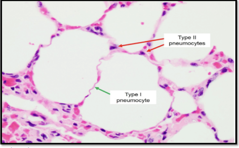
Type I Pneumocytes
long/flat
Type II Pneumocytes
cuboid shaped
secrete surfactant → reduces surface tension + provides liquid for rapid diffusion
secreted by secretory vesicles (lamellar bodies)
Maintaining concentration gradients
gas exchange via diffusion
diffusion - passive transport of particles from high → low concentration
high concentration gradient needed for gases to diffuse rapidly
adaptations
dense network of capillaries around tissues in gas exchange
constant blood flow through capillaries around tissues in gas exchange
Lungs for gas exchange ventilate lungs w air → high concentration of oxygen to alveoli → remove carbon dioxide from alveoli
Gills move water through gills → high concentration of oxygen → move carbon dioxide away from gills
gas exchange through gills
gills adapted for rapid exchange of gases
large SA
constant supply of blood flow
water continuously moves through gills
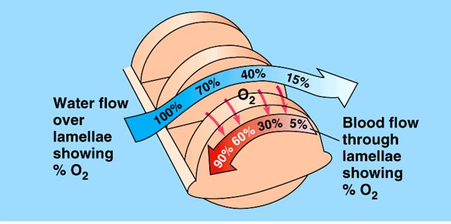
“counter-current exchange”
water flows over gill filaments in 1 direction → blood flows through capillaries in gill filaments in opposite direction
ensures blood is exposed to water w high oxygen concentration → continuous oxygen diffusion
very efficient
gas exchange, ventilation, and respiration
ventilation - movement of air in/out of alveoli in lungs → facilitate gas exchange
breathing
maintains concentration gradients of oxygen and carbon dioxide
respiration - release of ATP from organic compounds within cells
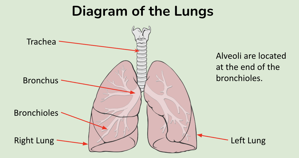
Lungs - allow for exchange of oxygen from air → bloodstream and carbon dioxide from bloodstream → air
adaptations
branching bronchioles connect to alveoli
all alveoli have large SA
gas exchange
alveoli secret surfactant
prevents alveoli walls adhering + moist surface for gas exchange
alveoli surrounded by extensive capillary bed
maintains high concentration gradients for oxygen and carbon dioxide btwn blood and alveoli
capillaries give constant supply of blood w low oxygen + high carbon dioxide concentration → alveoli
Ventilation of the lungs
Inspiration (breathing in)
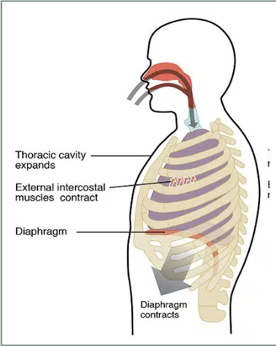
diaphragm contracts + moves down
external intercostal muscles contract → ribcage moves up/out
volume in thorax increases → decreases pressure in lungs
air moves from air (high pressure) → lungs (low pressure)
Expiration (breathing out)
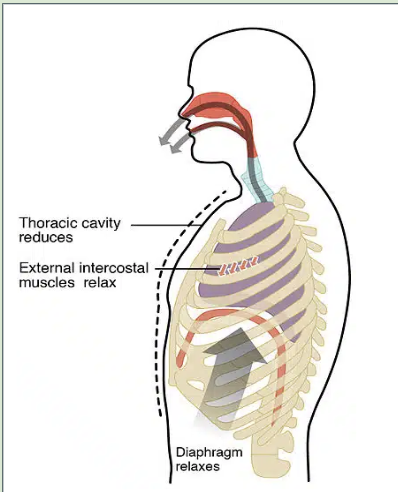
abdominal muscles contract → push diaphragm up
external intercostal muscles relax + internal intercostal muscles contract → ribcage moves down/in
volume in thorax decreases → increases pressure in lungs
high pressure lungs moves air out of lungs → surrounding air (lower pressure)
Lung volumes
tidal volume - volume of air moving in/out normally
inspiratory reserve - more volume of air inhaled w max effort
expiratory reserve - more volume of air exhaled w max effort
vital capacity - greatest volume of air expelled from lungs after deepest possible breath
vital capacity = tidal + inspiratory + expiratory
measured by spirometers
Partial Pressures of Gases
partial pressure - pressure exerted by a single gas in a mixture of gases
dependent on
total pressure exerted by all gases in a mixture
concentration of the gas in the mixture of gases
correlated w concentration of gas in solution
used when looking at oxygen + carbon dioxide in blood
Oxygen dissociation curve - shows affinity for hemoglobin for oxygen
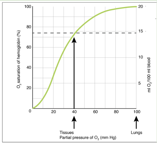
low partial pressures of oxygen → hemoglobin low affinity for oxygen → slow increase in saturation of hemoglobin
partial pressure of oxygen increases → hemoglobin affinity for oxygen increases
1 oxygen binds to hemoglobin → conformational change in hemoglobin shape → greater affinity of hemoglobin for oxygen
results in rapid increase in oxygen saturation
example of cooperative binding
high partial pressures of oxygen → curve flattens
most hemoglobin molecules have 4 oxygen molecules already
“saturated”
sigmoid shape
4 molecules of oxygen can bind to 4 haem groups in hemoglobin
in lungs → high partial pressure of oxygen that diffuses into capillaries around alveoli
respiring tissues use oxygen → low partial pressure of oxygen around respiring tissues
hemoglobin releases oxygen at low partial pressures → diffuses into respiring tissues for aerobic respiration
Hemoglobin
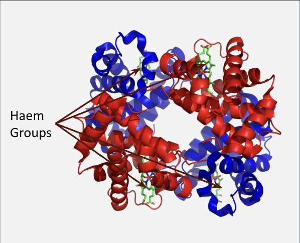
haem groups bind to oxygen molecules
every hemoglobin molecule has 4 haem groups → can bind to 4 oxygen molecules
cooperative binding of oxygen - binding of 1 oxygen to hemoglobin facilitates binding of other oxygen molecules
no oxygen bound to hemoglobin → low oxygen affinity
partial pressure of oxygen must be high for oxygen to bind
a single oxygen molecule binds to a haem group → shape of the hemoglobin molecule changes → hemoglobin affinity for oxygen increases
adult vs fetal hemoglobin
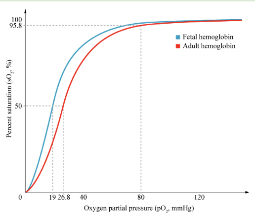
Adult hemoglobin
2 alpha + 2 beta chains of polypeptides
high partial pressure of oxygen in maternal blood
results in release of oxygen
fetal hemoglobin
2 alpha + 2 gamma chains of polypeptides
low partial pressure
results in binding of oxygen
greater affinity for oxygen than adult hemoglobin
oxygen more likely to be transferred from adult → fetal hemoglobin
Bohr Shift - the shift of the oxygen dissociation curve due to carbon dioxide partial pressures
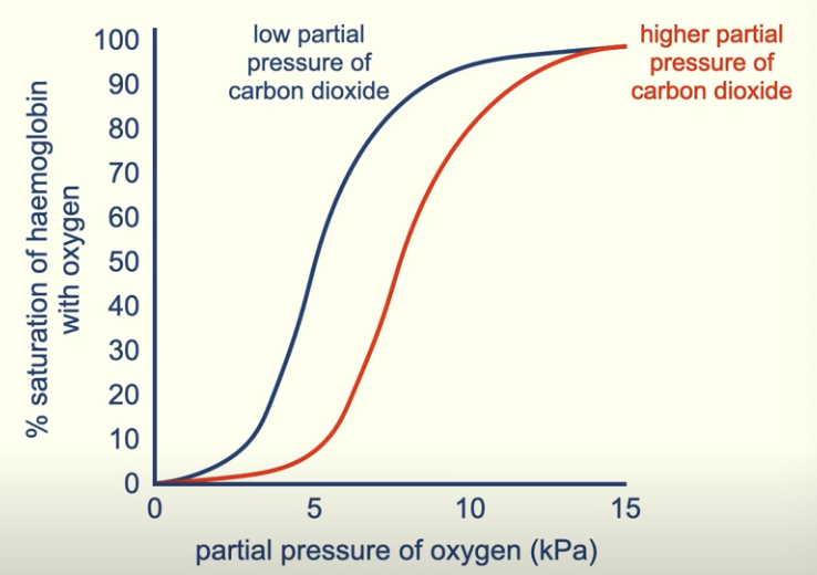
high partial pressure of carbon dioxide → reduces affinity of hemoglobin for oxygen → shifts oxygen dissociation curve right
In respiring tissues
tissues use oxygen + produce carbon dioxide during aerobic respiration → very low partial pressure of oxygen → reduces affinity of hemoglobin for oxygen
high partial pressure of carbon dioxide → bohr shift → reduces affinity of hemoglobin for oxygen
hemoglobin releases oxygen at respiring tissues
In lungs
high concentration of oxygen + low concentration of carbon dioxide in alveoli
oxygen diffuses from alveoli to blood → high partial pressure of oxygen → high affinity of hemoglobin for oxygen
carbon dioxide diffuses from blood to alveoli → low partial pressure in blood → bohr shift → stronger affinity of hemoglobin for oxygen
high oxygen + low carbon dioxide partial pressures → oxygen binds to hemoglobin at high saturation levels
Carbon Dioxide
most CO2 in blood diffuses into RBCs
CO2 + water → carbonic acid
CO2 + H2O → H2CO3
carbonic acid dissociates → hydrogen carbonate ions + hydrogen ions
reaction catalyzed by carbonic anhydrase
chloride shift
hydrogen carbonate leaves cell + chloride ions enter cell
reaction reversible, releasing CO2 when partial pressure of CO2 is low in blood plasma
transported 3 ways in blood
CO2 dissolved in blood plasma
CO2 bound to hemoglobin
CO2 reversibly converted → hydrogen carbonate ions + hydrogen ions (H+) in RBCs
hydrogen ions bind to hemoglobin → conformational change of protein → affinity for oxygen
most CO2 transported this way
Muscle Tissue
muscles - contract and are involved in movement
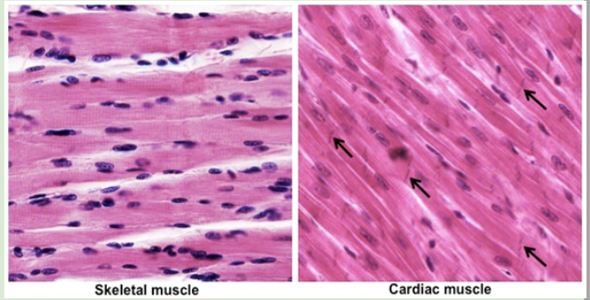
cardiac muscle
located in heart
responsible for heart beat
myofibrils
branched + connected by intercalated discs
allows rapid transmission of electrical impulses through heart tissue
electrical impulses trigger contraction of cardiac muscle cells
intercalated discs - gap junctions allowing ions to flow between cells
striated/skeletal muscle
attached to skeleton
involved in bone movement
myofibrils
long, multinucleated fibers formed via cell fusion
debated if they should be considered cells
smooth muscle
Capillaries
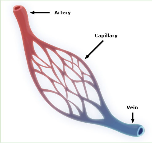
small blood vessels connected arteries → veins
exchange materials btwn blood and cells
adaptations
large SA
highly branched w narrow diameter
narrow lumen
wide enough for 1 RBC at a time
thin walls
allow rapid diffusion
capillaries 1 cell thick
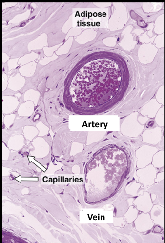
Micrograph of arteries vs veins
arteries
thick wall
narrow lumen
veins
thin wall
wide lumen
arteries
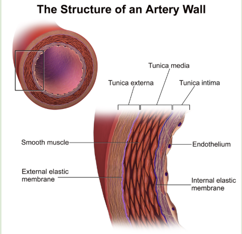
transport blood away from heart
adapted to withstand/maintain high blood pressure
thick wall
withstand high blood pressure
collagen in outer wall (tunica extrema)
strengthens artery to withstand high blood pressure
smooth muscle in artery
contracts to maintain blood pressure btwn heart beats
elastic fibers in artery wall
allow stretch/recoil as pressure increases/decreases from heart beats
recoil keeps blood moving
narrow lumen
maintains high blood pressure
lumen lined w smooth endothelial cells
reduces friction during blood flow
measuring pulse rate
felt using fingertips
radial artery in wrist
carotid artery in neck
Veins
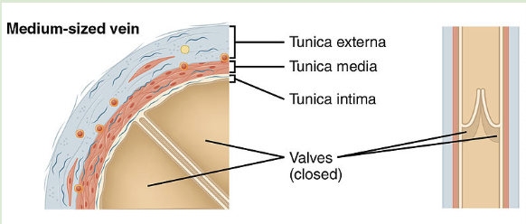
return blood to heart
blood returning moves slowly + not under high pressure
adaptations
thin wall
allows vein to be compressed by skeletal muscles
compression moves blood back to heart
wide lumen
allows vein to carry high volume of blood
valves
prevent backflow
Atherosclerosis
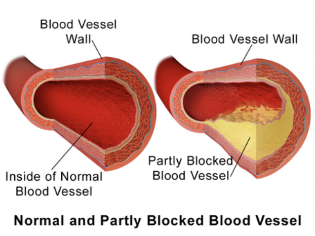
hardening/narrowing of arteries
coronary arteries branch off from main artery (aorta) → supply heart w oxygen/nutrients
can be occluded (blocked) by atherosclerosis
can lead to death of heart tissue + heart attack
inner lining of artery is damaged bc high blood pressure, leading to:
macrophages (type of WBC) attracted to sites of damage → release growth factors stimulating fibrous tissue growth
macrophages consume cholesterol → plaque
plaque grows and blocks artery
can break away and cause blood clot
risk factors
genetics
age
older ppl arteries more likely damaged
gender
men more likely
obesity
higher blood pressure
physical inactivity
can lead to obesity
smoking
increases blood pressure
diet of fats/cholesterol
Correlation coefficients (r)
quantifies correlations btwn variables
allows strength of relationship to be assessed
close to 0 → no relationship btwn variables → disproves hypothesis
correlation is not causation
The lymphatic system
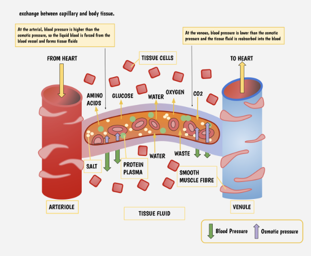
tissue fluid surrounds cells → enables exchange of materials btwn blood and cells
fluid formed by liquid part of blood (plasma) leaking from capillaries
blood leaves artery (arteriole) at high pressure → enters capillary → high hydrostatic pressure of blood filters plasma through capillary gaps → tissue fluid
blood pressure decreases as blood moves along capillary → plasma proteins decrease blood osmotic potential
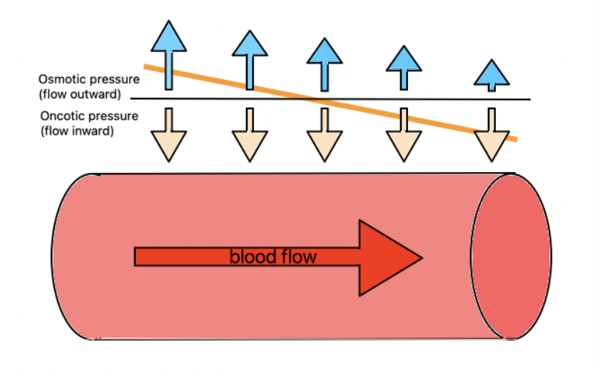
most tissue fluid returns to blood by osmosis via oncotic pressure
higher than hydrostatic pressure
exerted by protein molecules in a fluid that pulls fluid back into blood vessels
hydrostatic pressure pushes fluid out of capillaries
oncotic pressure pulls it back in
blood plasma and tissue fluid
high hydrostatic pressure of blood → filters blood through capillary wall gaps
large particles (ex. BCs, proteins) too large to pass through gaps
small particles/dissolved solutes leave blood → tissue fluid
composition
blood
plasma
cell components
tissue fluid
plasma w/o plasma proteins
erythrocytes
platelets
lymph
plasma w/o plasma proteins
erythrocytes
platelets
more lipid droplets
larger number of lymphocytes
by lymph nodes
Blood plasma and tissue fluid shared components
dissolved nutrients
glucose
amino acids
fatty acids
dissolved oxygen
metabolic wastes
carbon dioxide
WBCs
can move through gaps
blood plasma components not in tissue fluid
RBCs
platelets
large plasma proteins
tissue fluid and cells
tissue fluid surrounds cells
exchange of materials
fluid has high concentration of nutrients/oxygen + low concentration of carbon dioxide/metabolic wastes
metabolism in cells use nutrients/oxygen → produce metabolic waste
cells have low concentration of oxygen/nutrients + high concentration of metabolic wastes
oxygen/nutrients diffuse tissue fluid → cells
metabolic wastes (carbon dioxide) diffuse cells → tissue fluid
Draining tissue fluid
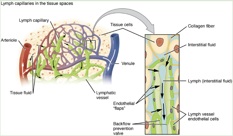
most tissue fluid returns to blood plasma
tissue fluid not re-entering blood → taken up by lymph ducts
known as lymph
lymph ducts collect excess tissue fluid and return to blood
lymph travels through lymphatic system → fluid returned to blood via lymph nodes
adaptations of lymph vessels/ducts
gaps in wall of lymph ducts
allows fluid to enter
thin walls
compressed by skeletal muscles → move lymph fluid
valves
prevent backflow of lymph fluid
thoracic duct - returns lymph to blood
drains into subclavian vein
circulation systems in fish vs mammals
bony fish
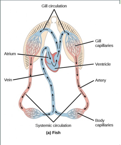
single circulatory system
due to 2 chambered heart
heart ventricle pumps blood → gills
oxygen/carbon dioxide exchanged as blood passes through capillaries in gills
oxygenated blood leaves gills → transported to tissues
gas exchange occurs as blood passes through body tissues
deoxygenated blood returns to heart
mammals
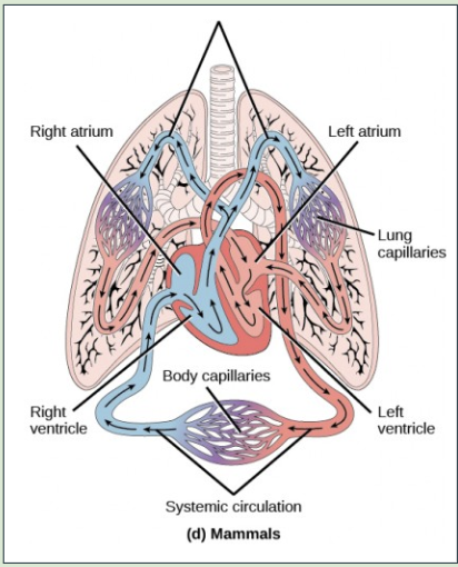
double circulatory system
due to 4 chambered heart
right side pumps blood to lungs
oxygen/carbon dioxide exchanged as blood passes through lung capillaries
oxygenated blood returns to left side of heart + is pumped to body
gas exchange occurs as blood passes through body tissues
deoxygenated blood returns to right side of heart
The heart
pericardium
protective membrane
secretes a fluid → reduces friction as heart beats
has 4 chambers
right + left atria
collecting reservoirs for blood returning to heart
right + left ventricles
pumps ejecting blood to body
separated by valves
prevent backflow
4 heart valves
right atrioventricular valve (tricuspid valve)
left atrioventricular valve (mitral valve)
right semilunar valve (pulmonary valve)
left semilunar valve (aortic valve)
path of blood
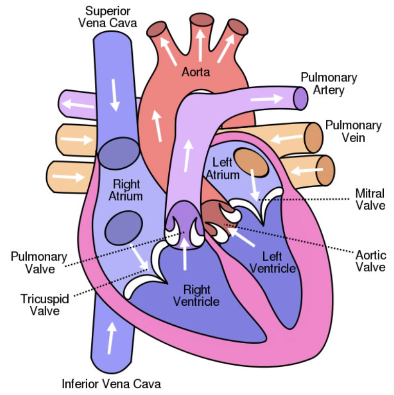
deoxygenated blood
superior/inferior vena vaca → right atrium → right atrioventricular valve (tricuspid) → right ventricle → right ventricle → right semilunar valve (pulmonary) → lungs (becomes oxygenated)
oxygenated blood
lungs → pulmonary vein → left atrium → left atrioventricular valve (mitral) → left ventricle → pumps blood to whole body → left semilunar valve (aortic) → aorta (brings blood to tissues → deoxygenated)
adaptations
atria
gets blood from body and lungs
ventricles
lots of cardiac muscle pumps blood
cardiac muscle
allows heart to contract to create high pressure
thicker on the left ventricle than right
left ventricle needs high pressure to move blood to body
pacemaker (sinoatrial node)
initiates/controls rate of heart beat
atrioventricular valves
prevents backflow of blood from ventricles to atria
semilunar valves
prevent backflow of blood from arteries to ventricles
septum
prevents oxygenated/deoxygenated blood from mixing
arteries
move blood away from heart at high pressure
veins
return blood to heart
control of the cardiac cycle
medulla oblongata
has 2 nerves connected to sinoatrial node (pacekamer)
nerves control the rate the heart beats
cardiac muscle = myogenic
contracts w/o stimulation
sinoatrial node controls rate of heart beat
initiates action potential (electrical signal) → rapidly spreads across atria → atrial systole (contraction)
fibrous tissue prevents action potential from traveling to ventricle
pause before signal reaches ventricles → 4 chambers do not contract at the same time
action potential travels to ventricles via atrioventricular node → then Purkinje fibers at apex of heart
action potential travels up walls of ventricle → initiates ventricular systole from apex → pumps blood out of ventricles
systole - contraction
diastole - relaxation
left atrium + ventricle in diastole
most blood flow directly through atrium → ventricle
action potential from sinoatrial node → left atrium enters systole
pressure increases in atrium as it contracts → blood forced to ventricle
action potential from atrioventricular node → ventricle enters systole
ventricle contracts → pressure in ventricle increases → atrioventricular valve closes
pressure higher in ventricle than atrium
high blood pressure in ventricle increases until semilunar valve opens + blood moves to aorta
ventricle enters diastole → pressure in ventricle decreases
pressure greater in aorta than ventricle → semilunar valve closes
pressure lower in ventricle than atrium → atrioventricular valves open
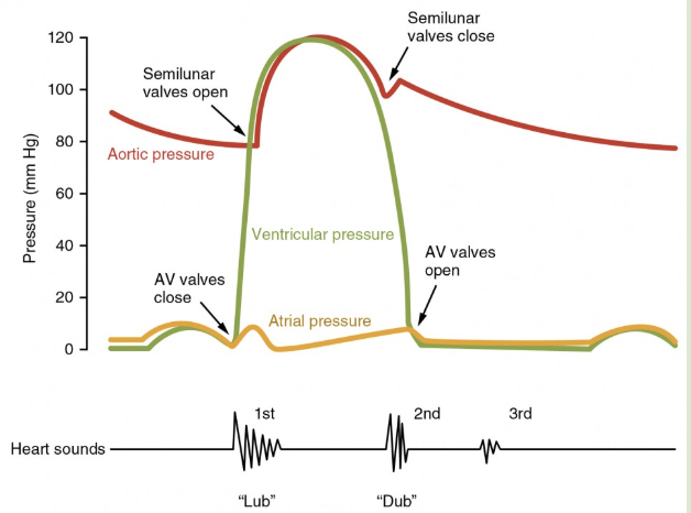
closing atrioventricular/semilunar valves → sound of heartbeat
measuring blood pressure
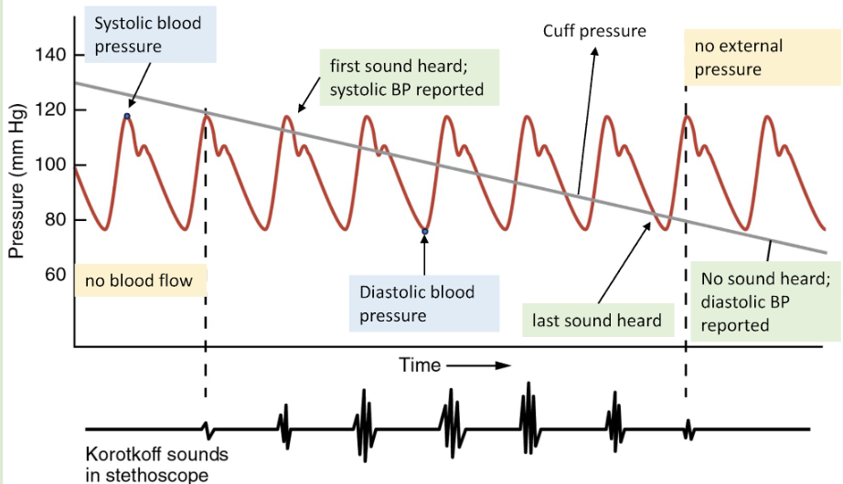
systolic pressure
caused by ventricular systole
diastolic pressure
btwn ventricular contractions