A+P: 4.6-Auditory Processes and Occipital Processes
Auditory Processes:
Structure of the Ear:
- ^^Tympanic Membrane (Ear Drum)^^
- ^^Auditory Ossicle ( Small Bones)^^
- ^^Malleus (Hammer)^^
- ^^Incus (Anvil)^^
- ^^Stapes (Stirrup)^^
- ^^Eustachian Tube^^
- %%Oval Window%%
- %%Cochlea%%
- %%Basilar membrane%%
- %%Organ of Corti (Hairs within Cochlea)%%
- %%Bone Labryinth of Cochlea%%
- %%Cochlear Nerve%%
How Sound Travels Through Your Ears to the Brain
- The of the outer ear and using the
Auricles → Auditory Canal
- The ^^tympanic membrane^^ or the eardrum captures the sound in the middle ear and sends the vibrations created by the sound to the ^^hammer, anvil, and stirrup.^^ They pass the vibrations to the oval window of the ^^cochlea.^^
Eardrum→ auditory aussicles → Cochlea.
Sound Waves → Mechanical Vibrations
- The vibrations in the %%cochlea%% cause fluid to move in the basilar membrane. This fluid moves the hair in the cochlea or the %%organ of corti.%% The organ of corti contain receptors that are sent the info on the movement to the %%cochlear nerve.%% The nerve sends the info to the brain.
Oval Window → cochlea → basilar membrane → organ of corti → cochlear nerve
Mechanical Vibrations → Fluid Vibrations.
Additional Information
- The eustachian tube and the space around the auditory aussicles (tympanic membrane space) controls pressure. Hurting ears on airplane + Quesiness → Changes in pressure in the ear.
- The auditory aussicles work like a chain reaction. The vibrations cause the hammer to move the anvil which causes the anvil to “flip” the stirrup like a lever. The stirrup moves to touch the oval window.
- The ==semicircular canals== of the cochlea helps with balance.
- Frequency Theory
- Different sound waves of different frequencies → triggers different hairs on the organ of corti → percieved as different sounds.
- Ex) High frequency sounds will reach hairs on the organ of corti that are higher up the cochlea while low frquency sounds will reach hairs that are closer to the oval window.
Occipital Processes
Eye Anatomy
Tunics:
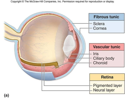
- Vascular Tunic:
- Function: Nourishment
- %%Ciliary Body-%% Provides aqueous solution for eye; It is around the iris and lens and it is disc-shaped.
- %%Iris%%
- Choroid
- Neural Tunic:
- Function: Processing and Communication to the brain
- %%Retina%%
- Fibrous Tunic:
- Function: Protection
- %%Sclera-%% White Tough Layer around the Eye
- %%Cornea%%- Clear layer in which light passes through the eye. It protects the eye from dust etc.
Major Parts:
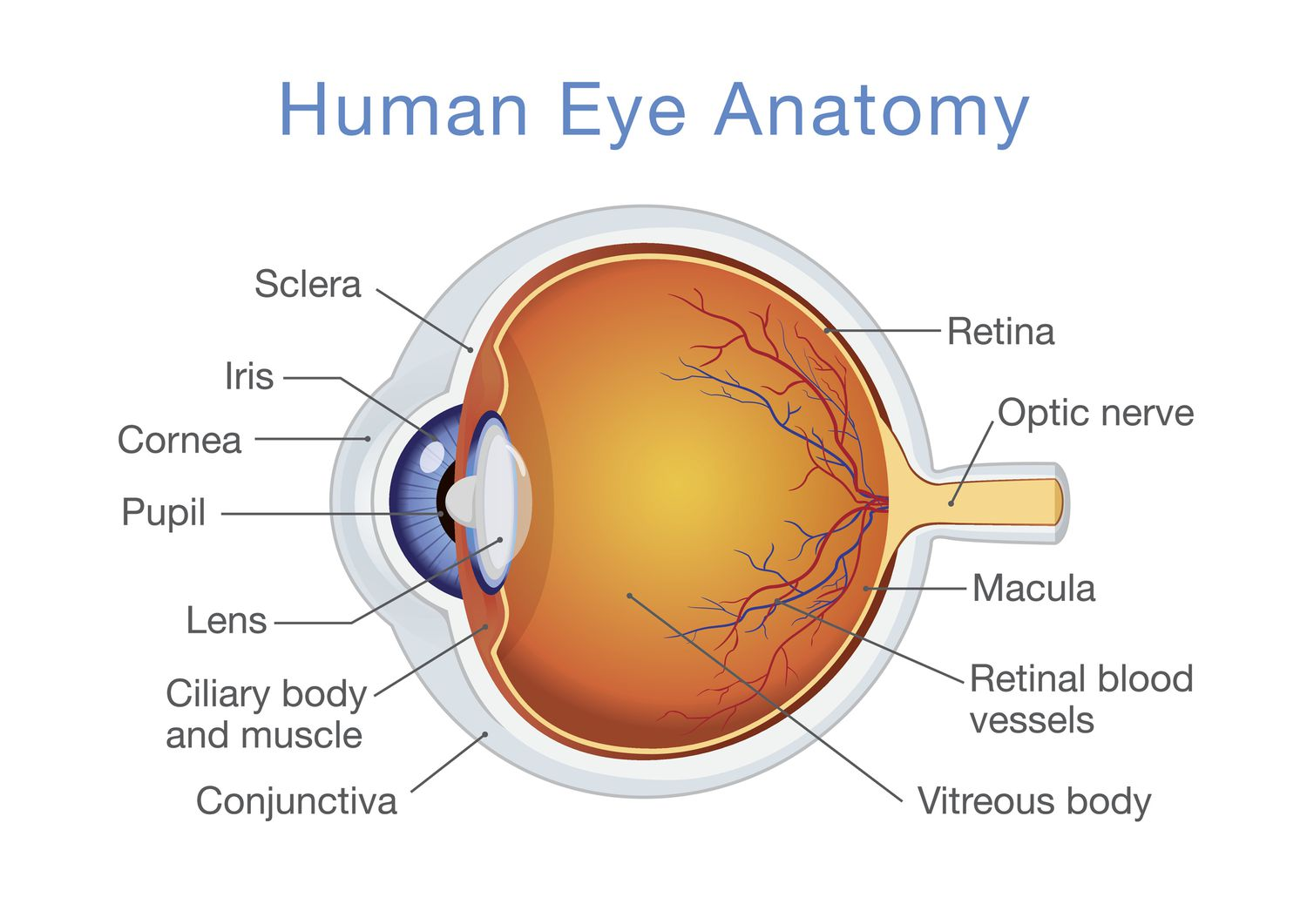
- Cornea: outer covering of eye.
- Pupil: Place where light is directed to
- Iris: Controls how much light is directed to pupil.
- Choroid: Contain blood vessels that bring oxygen and nutrients to eye. In-between retina and schlera.
- Lens: A lens after the pupil that redirects the lights path to the retina.
- Retina: The place where light is directed to. It has photoreceptors that sends info to the brain.
- Conjuctiva: Mucuos membrane that covers the front of the eyelids.
Moisturization of the Eye:
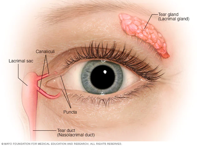
- Mucus, Tears, and Secretion moisturize the eye and protects the eye from irritants and pathogens.
- The Lacrimal Gland which is located at the above your eye produces tears. These tears are distributed around the eye. When we cry, the tears go down the lacrimal canal and down to the lacrimal duct. These places are in-between the nose and the eye. The duct is connected to the nose.
- The lacrimal punctas are two holes in the eye where the tears drain into the canal.
How do we see using our eyes:
- Light enters the cornea → Pupil → Lens.
- The Lens focuses the light to the retina.
- The photoreceptors (rods & cones) of the retina processes the images and sends the info to the brain using the optic nerve.