Vertebral column
Curvatures
Primary - present at birth - anteriorly concave
Thoracic
Kyphosis
Pelvic/ pelvic
Kyphosis
Secondary - posterior concave
Cervical
Lordosis
Develops when you start supporting your head
Lumbar
Lordosis
When infants begin to crawl and walk
Typical vertebral sections
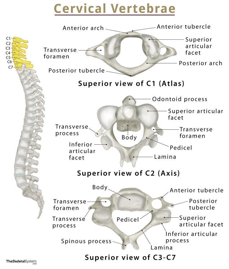
Cervical
C3-C7
Transverse foramen
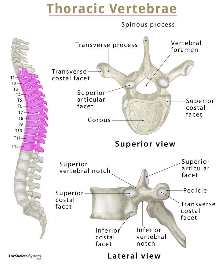
Thoracic
T5-T8
costal facets
demifacets
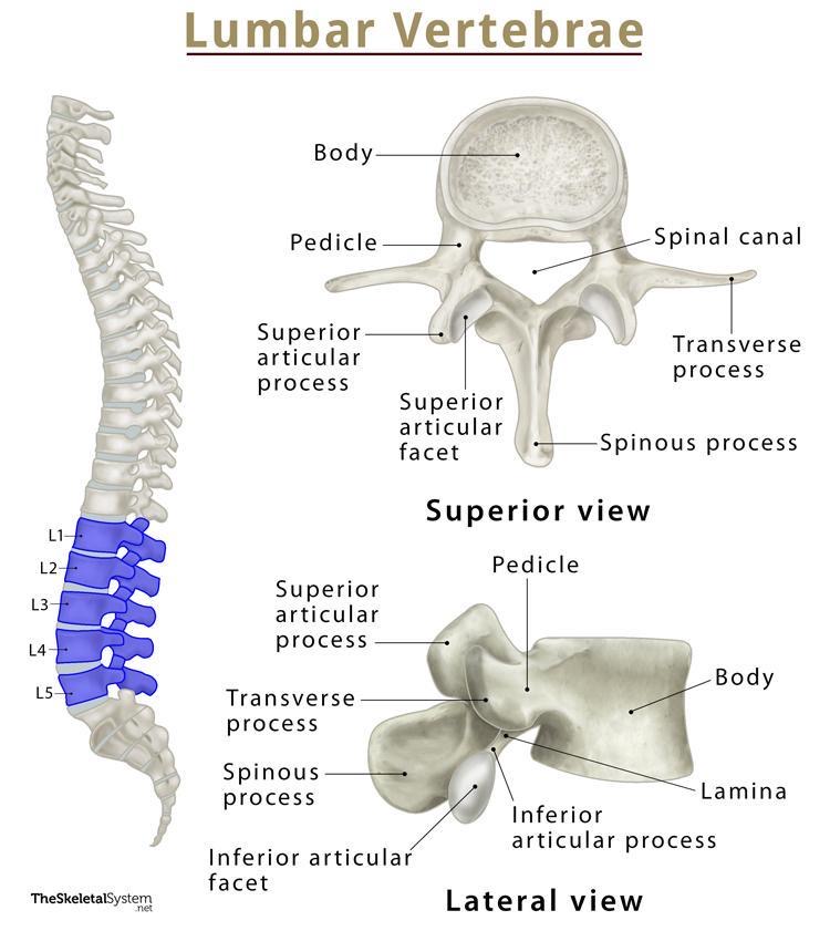
Lumbar
L5
Weight bearing one
Creates the lumbo-sacral angle
Largest movable vertebrae
Has stout transverse processes
All are typical L1-L5
Sacral S1-S5
Sacral canal for nerve roots
Base is superior - L5-S1
Sacral promontory - anterior edge of S1
Sacral hiatus, canal and cornua (horns)
Hiatus = space (absent SP and lamina of S5)
Canal = nerve roots, terminal filament
Ventral
Smooth and concave
Dorsal - 5 ridges
Median sacral crest - reminant of SP
2 Intermediate crest - reminant of articular processes
2 Lateral crests - reminant of articular processes
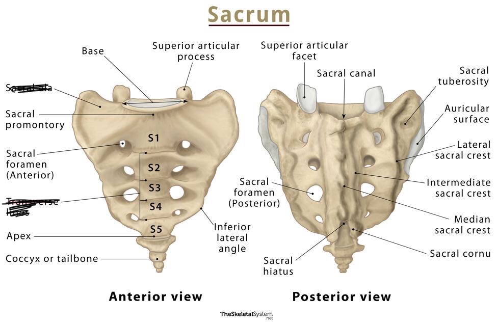
Coccyx
3-5 vertebrae fused together
4 is the most common that people have
Atypical vertebrae
C1 (Atlas)
Wide and ring shaped
No SP or body
Anterior/posterior arches with tubercle and lateral masses (short and chubby TP)
Superior articular facets are concave and receive occipital condyles
C2 (Axis)
strongest vertebra in the body
Dens (ondontoid process)
Transverse ligaments of the atlas hold odontoid in place
Bifid spinous process → 1st palpable SP
T1-T4
Features like cervical vertebrae (smaller and more delicate)
T1 has a complete costal facet for the 1st rib
Located on superior edge of body
Demifacets for 2nd rib
Located on inferior edge
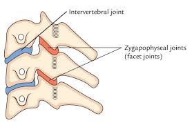
Arthrology of vertebral column
Intervertebral Joints
Between bodies of C2-S1
Symphyses → amphiarthroidal (slightly moveable) → strength and weight bearing
Articulating surfaces (hyaline cartilage) & connected by fibrocartilage
Vertebral bodies
United by longitudingal ligaments
Anterior longitudinal ligament (ALL)
Connects anterior aspects of vertebral bodies
Extends from sacrum to occipital bone
Fibers are fixed to the discs and periosteum
Their function is to maintain the stability of the vertebral column and prevent hyperextension of the vertebrae
Posterior longitudinal ligament (PLL)
(Weaker and narrower) connects posterior aspects of vertebral body
Extends from within vertebra from sacrum to C2
Well innervated with pain fibers
Functions to prevent protrusion of the discs and prevent hyperflexion
Continuous with tectorial membrane (superior continuation of PLL)
Intervertebral discs
Plates of fibrocartilage
Weight bearing
Superior disc between C2 and C3
Inferior disc between L5 and S1
Thickest in cervical and lumbar regions
Not thick in thoracic regions because you want the thoracic closer together for rib articulation
Disc is composed of 2 regions
Anulus fibrosus → outer fibrous portion
Nucleu pulposus → center shock absorbing portion
Avascular and contains a lot of water
Clinical application
Discs get weaker as we age but are SUPER strong in childhood, therefore children are more likely to break a bone before a disc
As we age, water content is lost in discs and become more compressed and lose height
Joints of vertebral arches
Called zygapophyseal facet (z-facet) joints
Synovial
Located between inferior articular processes of superior vertebral + superior articular processes of the inferior vertebrae
Permits gliding movement
Each joint supplied by 2 nerves
each arises from the dorsal rami of the spinal nerves
Accessory ligaments of intervertebral joints
Ligamenta flava → broad (possibly yellow)
Broad elastic bands → thickest in the lumbar region
Extend from lamina above to lamina below
Function to preserve normal curvature and prevent abrupt flexion
Interspinous ligaments
weakest
join adjacent spinous processes
Supraspinous ligament
Strong, attach adjacent SP
Runs from sacrum to C7 (superior to interspinous ligaments)
Intertransverse ligaments
Connects adjacent ligaments
Craniovertebral joints
Known as the subocciptial joints
Occiput (CO-C1) & C1-C2
Alanto-occipital → C0 and C1
Alanto-axial → C1 and C2
Differ in 2 aspects from others in vertebral column:
Only synovial
No vertebral discs
Atlanto-occipital joint
Location → occipital condyles that articulate with superior facets of C1 (lateral masses)
Movement → flexion and extension
Permits nodding and slight sideways tilt
Synovial condyloid joint
Associated ligaments and membranes
Anterior/posterior atlanto-occiptial membranes
Span from C1 to foramen magnum
Posterior is weaker than anterior
Function is to stabilize joint
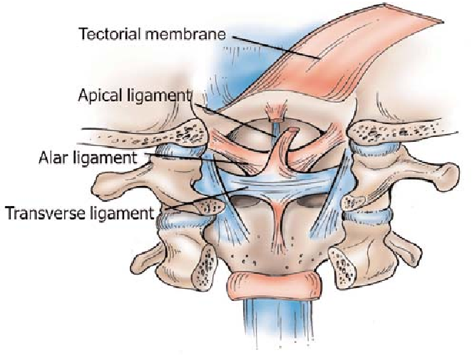
Transverse ligament of Atlas
Strong band that extends between lateral masses of C1
Holds dens of C2 against anterior art of C1
Forms posterior wall of socket for dens
Synovial joint
Cruciform ligament (crux/cross)
Superior/inferior longitudinal bands that run from C1-C2
There + transverse ligaments form a cross
Alar Ligaments
Extend from dens to foramen magnum
Function to check rotation and prevent excessive motion
Tectorial membrane
Extend from arch of C1 to occipital bone
Superior continuation of PLL
Covers alar and transverse ligaments
Atlanto-axial joint
Articulations between C1 and C2
2 lateral joints + 1 median joint
Central/medial alantoaxial joint (C1 and dens of C2)
Pivot synovial joint
Lateral alantoaxial joint
Zygopophyseal joint between C1 & C2)
Plane synovial joint
Excessive rotation of this joint is prevented by alar ligaments
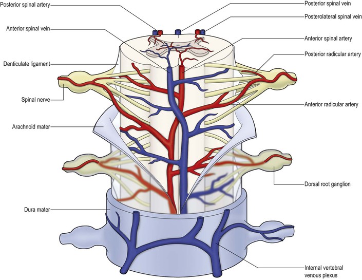
Vasculature
Spinal arteries receive blood supply in a regional distribution
Spinal artery enters through the intervertebral foramen
Then divide to form the radicula arteries
Spinal veins
Form internal/external vertebrae venous plexuses
Basivertebral veins drain into body
Larger vessels serve spinal cord and vertebral column bone