Basic Clinical Biochemistry
Clinical Biochemistry is the division of medical lab that deals with the measurement of biochemicals in blood, urine, and other bodily fluids.
Test results are useful for:
Detecting health problems and diagnosing disease.
Screening for latent infection (i.e. infection that is dormant, hidden etc.)
Determining prognosis for a patient.
Monitor progress and guiding treatement.
Examples of Clinical Samples:
Blood (whole, plasma, serum, cells, etc.)
Urine
Cerebrospinal Fluid (CSF)
Synovial Fluid
Gastric Fluid
Duodenal Fluid
Tears
Saliva
Biopsy Material
Example of a Clinical Biochemistry Sample:

Precision vs Accuracy:
Precision:
Repeated measurements of an analyte in the sample should give similar results.
*An analyte is a substance whose chemical constituents are being identified and measured.
Accuracy
Test result should reflect the true value.
Coefficient of Variation

Sensitivity vs Specificity:
Sensitivity:
Limit of detection of an analyte must be appropriate.
Specificity:
Must be able to distinguish the analyte from other molecules.
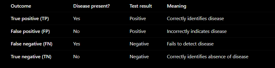
Sensitivity Equation:

Specificity Equation:

Physiological Factors and Reference Ranges:
Physiological Factors:
Genetic background
Gender
Age
Ethnicity
Inter-individual
Intra-individual
Diet
Fitness
Diurnal variation
Reference Ranges:
To be clinically useful, a test needs to be determined as abnormal or normal.
So test results are compared with reference ranges.
The range is constructed such that 95% of normal values lie within it.
Positive Predictive Value (PPV)
Positive predictive value (PPV) is the proportion of patients who test positive who actually have the disease.


Enzymes in Diagnosis:
Normal plasma contains very low levels of enzymes.
When tissue damage occurs, enzymes leak into the bloodstream:
Cytosolic enzymes are released quickly when cell membranes are damaged.
Membrane-bound enzymes are released when damage is more severe or cells die.
Therefore, elevated plasma enzyme levels are indicators of tissue injury.
Common causes of tissue damage and enzyme release:
Cause | Example |
|---|---|
Hypoxia | Myocardial infarction (heart attack) |
Toxic substances | Drugs, alcohol abuse, heavy metals |
Infections | Viral hepatitis |
Trauma | Crush injuries |
Immune destruction | Autoimmune diseases |
Isoenzymes (Isozymes):
Isoenzymes are different forms of an enzyme that:
Are encoded by different genes.
Catalyse the same reaction/substrate.
Differ in amino acid sequence and sometimes kinetic properties.
Have different distributions in different tissues.
Measuring which isoenzymes are present in blood can help identify the location of tissue damage.
Clinically important diagnostic enzymes
AST and ALT (aminotransferases) → commonly used to assess liver damage.
CK (CPK) (creatine kinase) → muscle and cardiac damage.
LDH (lactate dehydrogenase) → general tissue damage.
ALP (alkaline phosphatase) → liver and bone disorders.
ACP (acid phosphatase).
GGT (γ-glutamyltransferase) → liver and biliary disease.
Amylase → pancreatic disorders.
LDH isoforms in clinical testing:
LDH (lactate dehydrogenase) is a useful example of how isoenzymes can help identify the possible source of tissue damage.
LDH catalyses the same reaction in different tissues, but exists in different molecular forms. Measuring the pattern of these forms in blood can provide clues about which tissues are releasing LDH.
The five LDH isoenzymes:
LDH is a tetramer, meaning each enzyme molecule contains four subunits. There are two main subunit types:
H subunits: historically called heart-type subunits.
M subunits: historically called muscle-type subunits.
Different combinations of these subunits produce five main isoenzymes.

Measurement of LDH Isoforms:
Electrophoresis can separate the isoforms.

The different subunit combinations give the isoenzymes different electrical properties. When an electric field is applied, they migrate at different rates through a supporting medium, forming separate bands.
The laboratory can then assess the relative amounts of each isoenzyme.
There are two main approaches:
Total LDH measurement: Measures the overall LDH activity in the sample.
LDH isoenzyme analysis: Separates and measures the different forms to help investigate the likely tissue source.
Clinical Significance of LDH Isozymes:

Creatine kinase (CK):
CK is an enzyme involved in rapid energy regeneration in muscle.
It catalyses the reversible transfer of a phosphate group from phosphocreatine to ADP to produce ATP.
CK is abundant in skeletal muscle, cardiac muscle and brain.
CK isoenzymes:
Isoenzyme | Main tissue association |
|---|---|
CK-MM | Skeletal muscle |
CK-MB | Cardiac muscle; also present in skeletal muscle |
CK-BB | Brain and other tissues |
Isoenzyme measurements can help identify the possible source of tissue injury.
Detecting myocardial infarction (MI):
Myocardial infarction occurs when inadequate blood supply causes damage to heart muscle.
Damaged cells release intracellular proteins and enzymes into the blood. Measuring these biomarkers can help detect myocardial injury.
Biomarkers and their time courses
Biomarker | Approximate pattern after MI | Clinical significance |
|---|---|---|
CK / CK-MB | Rises within hours, peaks around 24 hours, returns towards normal in 2–3 days | Historical marker of muscle and cardiac injury |
AST | Rises within hours, peaks around 24–36 hours | Nonspecific; also found in liver and skeletal muscle |
LDH | Rises later, peaks around 2–3 days, may remain elevated for 7–10 days | Historical marker of tissue injury |
Cardiac troponin I/T | Rises within hours and remains elevated for days | Preferred biomarker for diagnosing MI |
Time courses are approximate and depend on the assay and clinical circumstances.
LDH flip
Normally, LDH-2 is more abundant than LDH-1 in serum.
Historically, following MI, LDH-1 could become greater than LDH-2.
This is known as the LDH flip.
It is not specific to MI and can also occur in haemolysis.
Liver and skeletal muscle enzymes
Enzyme | Clinical association |
|---|---|
ALT | Liver cell injury; relatively liver-specific |
AST | Liver and skeletal muscle injury, among other causes |
ALP | Cholestasis and bone disorders |
GGT | Liver and biliary disease; may increase with chronic alcohol use |
CK | Skeletal muscle injury |
LDH | General tissue injury; nonspecific |
Important distinction: ALT and AST are markers of cellular injury rather than direct measurements of liver function. Albumin and prothrombin time/INR provide information about aspects of liver synthetic function.
Laboratory enzyme assays
Enzyme assays measure enzyme activity in a patient sample.
General procedure
Add the patient sample to reagents containing appropriate substrates and cofactors.
The enzyme catalyses a reaction.
Monitor a measurable signal, such as absorbance, fluorescence or luminescence.
Calculate the reaction rate to estimate enzyme activity.
Initial reaction rate
Enzyme activity is commonly measured using the initial rate of the reaction.
The initial rate is measured while substrate is abundant and the reaction rate is approximately linear.
Under suitable conditions, the initial rate reflects the amount of active enzyme present.
Spectrophotometry at 340 nm
NADH and NADPH absorb UV light at approximately 340 nm.
NAD⁺ and NADP⁺ absorb very little at this wavelength.
Changes in absorbance at 340 nm can therefore be used to monitor reactions involving these cofactors.
Coupled reactions allow an enzyme that cannot be measured directly to be assessed through a second, measurable reaction.
Example: CK assay
CK activity is coupled to reactions involving hexokinase and glucose-6-phosphate dehydrogenase, ultimately producing NADPH.
The change in absorbance at 340 nm is used to calculate CK activity.
Enzyme electrophoresis
Electrophoresis separates enzyme isoenzymes according to their migration properties in an electric field.
Different isoenzymes form separate bands.
Enzyme activity can be measured in the separated bands.
Band intensity can provide information about the relative activity of each isoenzyme.
Clinical use: Helps investigate the possible tissue source of elevated enzyme activity.
Serum protein electrophoresis (SPEP):
Definition: An inexpensive laboratory method that separates serum proteins according to their electrophoretic properties, particularly their net charge.
The separated protein fractions are quantified using densitometry.
The five main serum protein fractions
Fraction | Main components / significance |
|---|---|
Albumin | Major serum protein; synthesised by the liver |
Alpha-1 globulins | Includes alpha-1 antitrypsin |
Alpha-2 globulins | Includes haptoglobin and alpha-2 macroglobulin |
Beta globulins | Includes transferrin and some complement proteins |
Gamma globulins | Mainly immunoglobulins (antibodies) |
Clinical patterns
Condition | Typical SPEP findings |
|---|---|
Liver cirrhosis | Reduced albumin, increased gamma globulins; possible beta–gamma bridging |
Nephrotic syndrome | Reduced albumin, increased alpha-2 globulin fraction |
Monoclonal gammopathy | Narrow, prominent monoclonal protein peak, often in the gamma region |
Chronic inflammation | Broad increase in the gamma globulin region may occur |
SPEP patterns support diagnosis but are not definitive on their own.