Nervous Tissue
Nervous System
The master controlling and communicating system of the body
The most powerful system in the body and can override anything
Functions
Sensory input—monitoring stimuli occurring inside and outside the body
Sensory = afferent; toward CNS
CNS: brain & spinal cord; our computer
Integration—interpretation of sensory input; computing
Motor output—response to stimuli by activating effector organs
Motor = efferent; away from CNS
Effector organs: muscles & glands
Carries out orders of brain and spinal cord


Organization of the Nervous System
Central nervous system (CNS)
Brain & spinal cord
Integration (interprets sensory input) & command center
Peripheral nervous system (PNS)
Paired spinal and cranial nerves
Ganglia—clusters of cell bodies of neurons
Neurons—individual nerve cells
Carries messages to (sensory/afferent) and from (motor/efferent) the spinal cord and brain
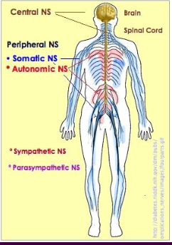
Peripheral Nervous System (PNS): Two Functional Divisions
All of the branches that are coming off of the brain and spinal cord; does not actually include brain and spinal cord
Sensory (afferent) division
Sensory afferent fibers—carry impulses from skin, skeletal muscles, joints, and special senses organs to the brain
Gonna be carrying info from the outside of the body (skeletal muscles, joints, and special sense organs are considered external)
Visceral afferent fibers—transmit impulses from visceral organs and blood vessels to the brain
Asks what’s going on internally (are you hungry/is your stomach empty, is your appendix hurting, are you about to have a baby, etc.)
Motor (efferent) division
Transmits impulses from CNS to effector organs
Motor Division: Two Main Parts
Somatic nervous system
Conscious control of skeletal muscles (somatic & skeletal both begin w/ an S)
Autonomic nervous system (ANS)
Regulates smooth muscle, cardiac muscle, and glands
Divisions: sympathetic & parasympathetic

Receptor Class by Location
Sensory receptors are going to begin afferent pathway, which sends info toward CNS
Exteroceptors
Respond to stimuli arising outside the body
Sensitive to touch, pressure, pain, and temperature
Include the special sense organs (vision, hearing, taste, smelling)
Interoceptors
Respond to stimuli arising within the body
Found in internal viscera and blood vessels
Sensitive to chemical changes, stretch, and temp changes
Proprioceptors
Gets us to where we need to be and helps maintain balance
Detects body’s position by detecting stretch in all of these things down below (skeletal muscles, tendons, etc.)
Respond to stretch in skeletal muscles, tendons, joints, ligaments, and CT coverings of bones and muscles
Constantly “advise” the brain of one’s movements
Histology of Nerve Tissue
The two principal cell types of the nervous system are
Neurons—excitable cells that initiate and transmit electrical signals
Supporting cells—cells that surround and wrap neurons
Helps neurons grow, live, prop them up, and sometimes even feed them and take away waste
Neurons (Nerve Cells)
Structural units of the nervous system
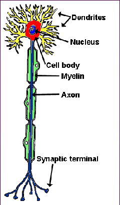
Composed of a body, axon, and dendrites
Long-lived, amitotic, and have a high metabolic rate
Their plasma membrane functions in
Electrical signaling
Cell-to-cell signaling during development

Neuron Cell Body (Perikaryon or Soma)
Contains the nucleus and nucleolus (inside the nucleus that produces parts of ribosomes, which lead to the production of proteins)
Is the major biosynthetic center
Is the focal point for the outgrowth of neuronal processes
Has no centrioles (hence its amitotic nature)
They can not get cancer bc the only way to grow a tumor is to have actively dividing cells
Has well-developed Nissl bodies (rough ER) that cause gray color in gray matter
Makes stuff for ribosomes, which produce proteins; proteins are big building blocks of the cell body
Contains an axon hillock (aka trigger center)—cone-shaped area from which axons and action potentials (sparks) arise

Neuron Processes
Armlike extensions from the cell body
Bundles of these are called tracts in the CNS and nerves in the PNS
There are no nerves in the brain and spinal cord
Two types
Dendrites—convey electrical signals (varying in amount) toward the cell body
Axons—longer and also called nerve fibers
Convey electrical signals away from the cell body toward another neuron, muscle, or gland
Release NT (Neurotransmitter)
Neuron Structural Classification
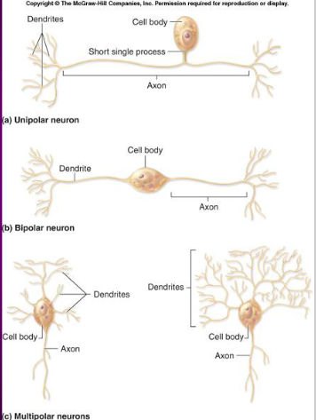
Neurons vary in the number of cell body processes
Unipolar—one process; sensory neurons
Bipolar—two processes; typically only found in special sense organs
Multipolar—at least 3 processes; motor neurons, interneurons, etc.
Interneurons (aka Association Neurons)

A) Sensory (afferent)—transmit impulses toward CNS
B) Motor (efferent)—carry impulses away from the CNS
C) Interneurons—multipolar neurons entirely within the CNS that shuttle signals through CNS pathways
Communicate w/ sensory and motor neurons
Supporting Cells: Neuroglia
The supporting cells (neuroglia or glial cells)
Not going to send electrical signals
Provide a supportive scaffolding for neurons in CNS and PNS
Segregate and insulate neurons
Makes sure that some cells or parts of cells are not inappropriately affected by electrical signals
Guide young neurons to the proper connections
Promote health and growth
Very numerous, can divide, do not transmit impulses/action potential/electrical signals/sparks
Can form tumors due to dividing nature
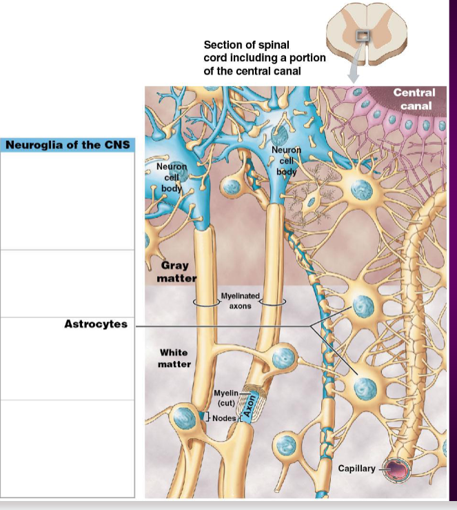
Glial Cells: Astrocytes
Most abundant, versatile, and highly branched glial cells
Clings to neurons and their synaptic endings, and covers capillaries (forms the blood-brain barrier)
Functionally, they:
Support and brace neurons
Anchor neurons to their nutrient supplies
Guide migration of young neurons
Control the chem environ
Microglia and Ependymal Cells
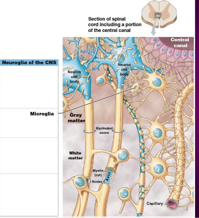
Microglia—small, ovoid (oval-shaped) cells w/ spiny processes
Phagocytes that destroy wastes and pathogens
Kinda like spiders waiting on a web for bad things to come along
Ependymal cells—range in shape from squamous to columnar
Line central cavities of the brain and spinal column to circulate cerebrospinal fluid (CSF)
Satellite cells—squamous cells surrounding ganglia
Ganglia (singular: ganglion)—Clusters of cell bodies in PNS
Clusters of cell bodies are like mini brains scattered around the body
Regulate chem of nutrients, gases, and NT (neurotransmitter) for ganglia

Oligodendrocytes and Schwann Cells Perform Myelination
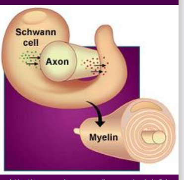
Myelin sheath—whitish, fatty (protein-lipoid), segmented sheath that protects the axon, electrically insulates fibers from one another, and incs the speed of nerve impulse transmission
Forms a protective outer structure
Fats insulate against electricity
Schwann cells (neurolemmocytes)—myelinate nerve fibers (axon) of PNS
Oligodendrocytes—branched cells that myelinate CNS nerve fibers (axons)
These guys basically do the same functions, but in different parts of the body (CNS v. PNS)


Saltatory Conduction
Current passes through a myelinated axon only at the nodes of Ranvier (neurofibral nodes)
Jumps over myelin sheath and lands on neurofibral nodes

Action potentials are triggered only at the nodes and jump from one node to the next
Much faster (and uses less ATP) than conduction along entire length of unmyelinated axons
Conduction Velocities of Axons
Varies widely among neurons
Rate of impulse propagation is determined by
Axon diameter—the larger the diameter (thicker), the faster the impulse
Presence of a myelin sheath—myelination dramatically incs impulse speed
Axon Regeneration
CNS axons generally can’t regenerate
Damaged PNS axons may regenerate if some of the neurolemma is still present
Depends on distance btwn severed ends
Depends on secretion of nerve growth factors
Nerve/Tract CT
Axons are bundled by 3 CT
Epineurium—surrounds entire nerve or tract; dense irregular CT
Perineurium—surrounds fascicles of axons; dense irregular CT
Endoneurium—surrounds the individual axon; loose (areolar) CT

Synpases
Axons end w/ axonal terminals at synapses
Presynaptic neurons—release NT into synapse for comms
Postsynaptic neurons—receive NT from presynaptic neuron
Axons can have synaptic contacts at any unmyelinated surface on another neuron

Neurotransmitters
Nerve impulses reach the axonal terminal of the presynaptic neuron and open Ca2+ channels
Neurotransmitter is released into the synaptic cleft via exocytosis (when vesicle bubble merges w/ cell membrane to release NT)
Neurotransmitter crosses the synaptic cleft and binds to receptors on the postsynaptic neuron
Postsynaptic membrane experiences Na+ influx, causing an excitatory or inhibitory effect
Synaptic delay is the time elapsed
Nerve impulse travels down presynaptic neuron’s axon → opens the gateways for Ca2+ → Ca2+ triggers synaptic vesicles which carry NT (ACh) → vesicles move to end and merge w/ cell membrane → exocytosis (release NT) → now we’re in the synapse! → once NT hits postsynaptic receptors, allows Na+ to come in (can be either excitatory or inhibitory)

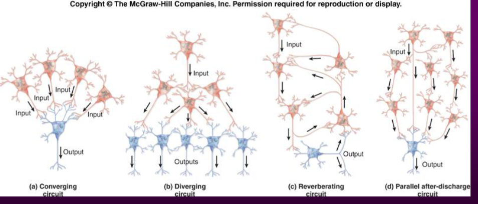
Neuronal Pools/Circuits
Interneurons form patterns (pools) in the CNS, based on function
A pool may be localized, or may be distributed regionally throughout the CNS