CV - Clinical Assessment 2
Three tests discussed
Nagel Anomaloscope
City University Colour Vision Test
Medmont C-100
Nagel Anomaloscope
Gold standard instrument for red green (RG) colour vision deficiency (CVD) classification.
only test able to reliably classify dichromats from anomalous trichromats (AT) and protan types (exact wording in transcript).
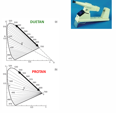
Test setup:
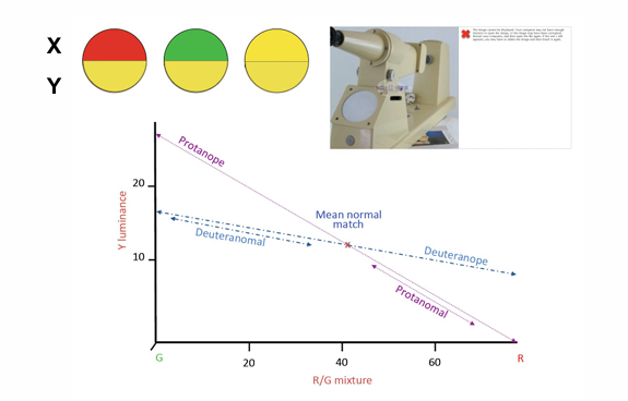
2-degree circular field, split horizontally.
Upper hemifields: mixture of spectral red (670 nm) and green (546 nm) at constant luminance.
Lower hemifields: spectral yellow (589 nm) with variable luminance.
Based on the “Rayleigh Equation”:
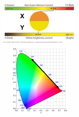
Scales:
Red/Green scale (x-axis): from 0 (pure green) to 73 (pure red).
Yellow luminance scale (y-axis): from 0 to 87 (bright yellow maximum).
Subject’s matching mid-point, range, and profile define their congenital RG CV status.
Key concepts:
Mid-point (SFM): the subject’s exact colour-match point on RG vs Y fields.
Range: how far the RG field can be shifted while still achieving an exact match via adjustments of the Y field.
Profile: the overall shape of the RG vs Y matching across the x-y plane.
Explain to subject how to respond
An exact match is required in both colour and brightness; otherwise the subject must report “no match.”
Testing procedure (two-step)
Step 1: Subject makes several exact colour matches by simultaneous adjustment of the RG and Y fields.
First match is the Subject’s First Match (SFM).
If an exact match cannot be achieved, the subject reports “no match.”
Repeat this process twice; average the SFM coordinates (x, y) from 3 attempts.
Plot this averaged SFM point on the graph.
Step 2: Examiner sets the RG field (x-axis) at 5 or 10 units on either side of the SFM and the subject adjusts the Y field (brightness; y-axis) to attempt exact matches.
Stop when no match is possible on both sides of the SFM, or if the x-axis limit is reached (0 or 73).
Record the range achieved on the graph.
NA - Example 1
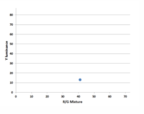
Step 1 matches: 42/12, 40/13, 41/13 → Average SFM = 41/13.
Step 2: Examiner adjusts x-axis to ±5 units around 41 (i.e., x = 36 and x = 46).
x = 36: No Match → record 36/ no match
x = 46: No Match → record 46/ no match
only graph the matched points
NA - Example 2
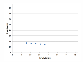
Step 1 matches: 33/15, 25/16, 35/14 → Average SFM = 31/15.
Step 2: Examiner sets x-axis at several points and subject’s matches recorded:
x=26, subject matches y=16: record 26/16
x=21, matches y=16: record 21/16
x=16, matches y=17: record 16/17
x=11, no match: record 11/No Match
x=36, matches y = 14: record 36/14
x=41, no match: record 41/No Match
Graph shows x-range from 16 to 36 with mid-point at 26.
Nagel Anomaloscope interpretation
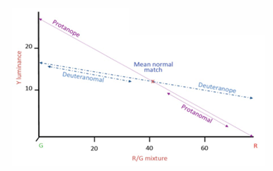
CV normals: match a single point (typical around ~42/13, with ±2 in x and small y-range).
CV can have small x - axis range (1-5 units)
Dichromats: will match for all x-axis settings (0–73)
cannot differentiate hue settings on RG scale
Y-axis matches reflect brightness only.
Deuteranopes (deuteranopes)
Y-axis value relatively constant (flat) across x-range.
Protanopes (protanopes)
Sloping function
Lower red sensitivity → less bright and Y brightness reduced to match
Anomalous Trichromats (AT) characteristics
Mid-points lie on one or the other side of the normal match (42/13):
Deuteranomals: x-midpoints < 40 → need to add more green to match.
Protanomals: x-midpoints > 50 → need to add more red to match.
Range reflects AT severity: a larger range indicates more severe CVD.
Severe AT often matches to one end of the x-axis
AT usually reject the normal 42/13 match but may accept if the AT is very severe.
Graph interpretation notes (visual patterns from the data)
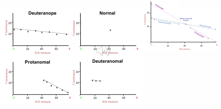
Normal: single, consistent match near 42/13 with small x-range.
Dichromats: broad ability to match across x-range; Y-brightness alignment dominates.
AT (deutanomal, protanom): midpoints shift left or right; broader ranges indicate greater severity.
Severe AT: may cluster near an end of the x-range (0 or 73) and can be difficult to obtain a normal-match.
Severity ranges for AT
PA range (severity measure):
mild < 10
mod 10–15
severe > 15
Double for DA (likely referring to a dichromat anomaly or a dual-type assessment)
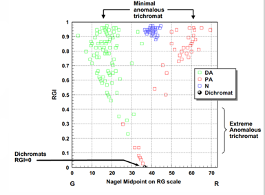
Red-green discrimination index (RGI):
Formula: RGI = 1 - (patients age / 74)
CV normal: RGI ≈ 1 (graphically blue region)
Dichromats: RGI = 0 (will accept any RG mixture)
AT: RGI falls between 0 and 1 depending on range
City University Test (CUT)
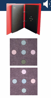
Matching test using the D-15 Munsell colours.
Format: 4 alternate forced choice, with 10 plates (2nd edition).
Page layout: 5 colours per page; 1 central colour and 4 peripheral colours.
Task: Select the peripheral dot that most closely matches the central dot.
Confusion lines: One peripheral colour is adjacent to the D-15 sequence; the other 3 lie on the D, P, and T confusion lines.
Scoring: 2 errors or more on CUT predict a pass/fail on D-15 well.
Reasonable test with severity classification similar to D-15.
Less sensitive than Ishihara for RG-CVD detection.
P vs D sensitivity ~60% (similar to Ishihara).
D-15 is better at distinguishing protans from deutan
Protans make fewer errors, due to luminous contrast variations
Medmont C-100
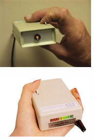
Handheld electronic device with a small circular flickering disk.
Mechanism: Flickering red and green lights in counter-phase.
Operational setup: 40 cm working distance; normal room lighting; switch off fluorescent lights if flicker is detected.
Test: patient adjusts RG ratio to eliminate or minimize flicker perception (the “null point”).
Null point ranges:
CV normals: between −1 and +1.
Protans: between −2 and −5 (reduced red sensitivity).
Deutans: between +2 and +5 (reduced green sensitivity).
Experimental protocol: average of 3 settings recorded.
Classification role:
Colour classification test for RG defects only.
Excellent sensitivity/specificity for protan vs deutans.
Protans may be less occupationally safe (in both dichromats and AT).
Protans show reduced red signal detection by up to ~40%.
Cannot screen for CVD; classifies RG defect once RG defect is confirmed.
Useful in conjunction with Ishihara.
Cannot assess CVD severity.
Dichromats cannot be distinguished from AT by this test.
Colour Naming & Occupational Lantern Tests
Lantern tests are occupational tools that test colour naming of signals relevant to specific industries (aviation, maritime, armed forces, police, railway, etc.).
Determines if signal lights would be identified accurately in the field
Generally RG based using:
Small stimuli to simulate distance
Dimming filters to simulate unfavourable weather conditions
Key lanterns:
Farnsworth Lantern (FALANT)
Holmes Wright Lantern A and B (HW A/B)
Eldridge-Green and other lanterns
Farnsworth Lantern (FALANT)
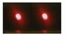
Setup: paired red, green, and white stimuli arranged vertically
white may appear yellowish and lie on RG confusion lines for CVD.
Filters used to remove brightness cues.
Procedure:
Distance: 2.4 m; moderate room lighting.
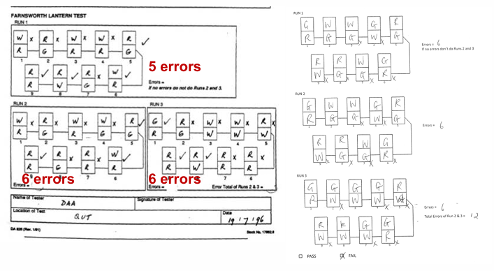
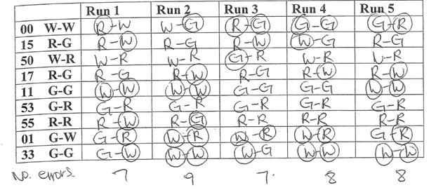
1 run= presents 9 colour pairs (all possible combinations) for 2 seconds each.
The patient names the top and bottom colour for each pair; responses must be red, green, or white only.
Any miscalls (one or both lights) count as 1 error.
Interpretation:
Errors accumulate across runs; a failure is declared after predefined error totals (as per example runs shown in the transcript).
Holmes Wright B Lantern (HW B Lantern)
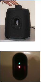
Used in maritime occupations to simulate ship navigation lights
horizontally arranges pairs of red, green, white colours
Distance: 1.5 nautical miles; 9 pairs shown after 10 minutes of dark adaptation.
Two forms:
HW-B (full lantern, brighter/larger lights) and HW-A (vertical arrangement, lights are brighter and larger).
Test procedure specifics (illustrated in sample runs):
3 runs are typically used; a single error can lead to failure.
Diagnostic tendency:
Approximately 98% of CV abnormals will probably fail.
About 8% of CV normals may also fail.
Who should be assessed? (General considerations)
All school children:
Colour often used as an aid to learning; abnormal CV can impact learning.
Males more likely to have CV defects, but implications similar for females.
Severe CV defects observed early (incorrect colour naming or choosing colours) may be noted from an early age.
If CV defect is mild, errors may be subtle or unnoticed; screening helps identify risk.
Identifying CV deficiencies in adulthood is often late for career planning.
If a CV defect is detected, discuss implications for career paths early.
Assessment of CV in children
Age considerations:
Testing can start around age 4.
By age 6+, tests become more reliable.
Screening is more likely until around age 8.
D-15 and Nagel tests become more reliable from around age 10+; Nagel may not be reliable under 10.
always confirm findings as child ages (tentative dx for young children)
treat with urgency if evidence of acquired CVD in a child → Possible neuro-ophthalmic disease
Screening tools for children:
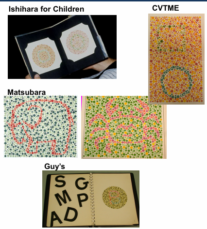
Ishihara PIC plates: generally adequate for RG defects by age 6; Ishihara for children from age 4; shapes better than trails.
Matching approaches: Colour Vision Testing Made Easy (CVTME) – pictures/shapes but RG defects only.
Matsubara/Ishihara for Children (Matsubara Ishihara for Children).
Guy’s tests; CVTME; Matsubara
others exist but validation varies.
early role is identifying risk and managing parental anxiety by providing information and reassurance
Who should be assessed:
All px with the folloiwng?
reporting colour disturbances or difference between eyes
taking medications known to affect CV
with confirm or suspected ocular, neurological or systemic disease (e.g., reduced VA, VF defects, RAPD)
All px considering specific occupations:
With known CV occupational requirements
Aviation, maritime, armed forces, police, railways, fire brigade
Where normal CV would be an advantage, but not a requirement
Textiles, fashion, paint, electronics
Test battery in general practice
Recommended minimum:
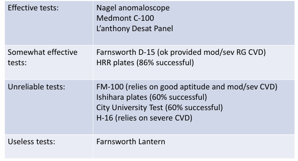
PIC plates (Ishihara) or Richmond HRR;
D-15 panel test for severity indication (P, D, T dichromats and AT severity);
Medmont C-100 to differentiate Protan vs Deutan defects (not CVD severity).
Practical note: In practice, clinics may have fewer tests; the goal is to identify RG defects and provide basic vocational/lifestyle guidance.
Test battery in a CV clinic
Ishihara PIC (and Richmond HRR PIC) used for initial screening.
Pass on Ishihara: further testing not routinely indicated.
Fail on Ishihara: proceed to classification of type and severity.
Medmont C-100 differentiates Protan/Deutan defects but does not provide severity.
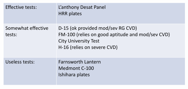
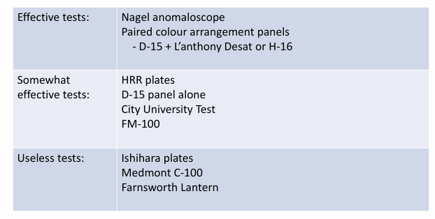
D-15: Identifies and classifies Protan, Deutan, and Tritan dichromats and moderate/severe AT
cannot separate mild AT from very mild AT.
L’anthony Desat panel provides separation of mild vs very mild AT.
Anomaloscope: definitive RG CVD classification (gold standard).
Farnsworth Lantern: vocational aptitude assessment for aviation and other industries.
Classifications: Protans vs Deutans
Classifications: Tritans
Determining CVD severity
Agreement between tests
Performance on CV tests does not always align across tests.
Ishihara is a poor predictor of CV severity and does not reliably predict outcomes on D-15, Nagel Anomaloscope, or Farnsworth Lantern.
Can clinical CV tests predict if people with CVD pass or fail the Farnsworth Lantern?
D-15: 67% of those who fail D-15 (2+ diam crossing) fail Farnsworth Lantern; 94% who pass D-15 pass FL.
Nagel: 87% who fail Nagel (range > 10 units) fail FL; 57% who pass Nagel pass FL.
Neither Nagel nor D-15 alone is a perfect predictor of FL performance.
Approximately 35% of CVD cases pass Farnsworth Lantern
mostly mild deuteranomals
some may still fail simulated flight tasks (e.g., PAPI).