chpter 14 Nervous system
Chapter 14: Nervous System Disorders
Overview of the Nervous System
Components:
Central Nervous System (CNS):
Includes the brain and spinal cord.
Peripheral Nervous System (PNS):
Includes cranial and spinal nerves.
Contains sensory neurons and neuromuscular junctions.
Brain
Function:
Acts as the communication and control center of the body.
Receives, processes, and evaluates inputs.
Decides on actions and initiates responses.
Manages both involuntary actions (to maintain homeostasis via the autonomic nervous system) and voluntary actions.
Responsible for reflex activities.
Protection of the Brain
Meninges:
Dura Mater: The outermost layer adjacent to the skull.
Arachnoid Mater: The middle layer.
Pia Mater: The innermost layer adhering to the brain's surface.
Subarachnoid Space: Contains cerebrospinal fluid (CSF).
Cerebrospinal Fluid (CSF):
Provides cushioning for the brain and spinal cord.
Appears similar to plasma but with different concentrations of electrolytes, glucose, and proteins.
Changes in CSF characteristics can be diagnostic.
Formed by choroid plexuses in the ventricles and maintains intracranial pressure (ICP).
Normal CSF Characteristics (TABLE 14-1)
Appearance: Clear and colorless
Pressure: or
Red Blood Cells: None
White Blood Cells: Occasional
Protein:
Glucose:
Electrolytes: Sodium , Potassium
Specific Gravity:
pH:
Volume in the system at one time:
Volume formed in 24 hours:
Blood-Brain Barrier and Blood-CSF Barrier
Blood-Brain Barrier:
Located at brain capillaries, limits material passage, and controls CNS balance of electrolytes.
Lipid-soluble substances can easily pass through.
Blood-CSF Barrier:
Found at choroid plexus, regulating CSF constituents.
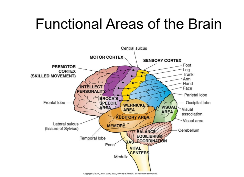
Functional Areas of the Brain
Major Areas:
Cerebral Hemispheres:
Divided into: Prefrontal, Frontal, Parietal, Temporal, and Occipital lobes.
Diencephalon:
Functions as relay station (Thalamus) and homeostatic control (Hypothalamus).
Cerebellum:
Responsible for coordination and balance.
Brain Stem:
Connects the brain to the spinal cord; includes the midbrain, pons, and medulla oblongata.
Key Functions (from TABLE 14-2):
Frontal Lobe: Intellectual function, personality, voluntary movements, speech (Broca's area).
Parietal Lobe: Sensation (e.g., touch, pain).
Occipital Lobe: Vision (visual cortex).
Temporal Lobe: Hearing, smell, and speech comprehension (Wernicke's area).
Cerebral Hemispheres
Structure:
Largest and most visible part of the brain, separated by the longitudinal fissure.
Comprised of grey matter (cortex) and white matter (corpus callosum).
Each hemisphere is functionally specialized: the left tends to manage language and analytical tasks, while the right manages creativity and spatial ability.
Diencephalon
Thalamus: Relay station for sensory impulses.
Hypothalamus: Governs autonomic nervous system functions like temperature, appetite, and sleep cycles.
Brain Stem
Connective area between the brain and spinal cord consisting of:
Midbrain: Most superior part, important for reticular activating system that regulates alertness.
Pons: Acts as a bridge between areas of the brain, contains cranial nerve nuclei.
Medulla Oblongata: Controls vital functions such as respiration and heart rate.
Cerebellum
Position: Dorsal to pons and medulla.
Functions: Coordinates movements, maintains posture and equilibrium by integrating information from various sensory pathways.
Blood Supply to the Brain
Internal Carotid and Vertebral Arteries:
Major arteries supplying the brain; branches into anterior and middle cerebral arteries.
Circle of Willis: An anastomotic arrangement providing collateral circulation.
Cranial Nerves
There are 12 pairs, each associated with specific functions (TABLE 14-3).
Olfactory (I): Sensory for smell.
Optic (II): Sensory for vision.
Oculomotor (III): Motor; responsible for eye movements and pupil constriction.
Trigeminal (V): Mixed; sensory from face and motor for mastication.
Abducens (VI): Motor; responsible for lateral eye movement.
Facial (VII): Mixed; involved in facial expression and taste sensations from the anterior two-thirds of the tongue.
Vestibulocochlear (VIII): Sensory; responsible for hearing and balance. Glossopharyngeal (IX): Mixed; involved in taste sensations from the posterior one-third of the tongue and contributes to swallowing.
Vagus (X): Mixed; plays a crucial role in parasympathetic control of the heart, lungs, and digestive tract.
Accessory (XI): Motor; controls the muscles of the neck and shoulders to facilitate head movement.
Hypoglossal (XII): Motor; responsible for regulating tongue movements essential for speech and swallowing.
Abducens (VI): Motor; responsible for controlling lateral eye movement by innervating the lateral rectus muscle.
Trochlear (IV): Motor; responsible for innervating the superior oblique muscle, allowing for downward and lateral eye movement.
Spinal Cord and Spinal Nerves
Structure: Protected by the vertebral column, it ends at the lower border of the first lumbar (L1) as a collection of nerve roots (cauda equina).
White Matter: Comprises ascending and descending tracts for sensory and motor pathways respectively.
Gray Matter: Contains anterior horns (motor neurons), posterior horns (interneurons), and lateral horns (visceral motor neurons).
There are 31 pairs of spinal nerves named according to their emergence from the vertebral column.
Reflexes
Automatic Responses: Rapid, involuntary responses to stimuli, conducted through sensory and motor fibers, synapses occurring in the spinal cord or brain.
Neurons and Conduction of Impulses
Neurons: Specialized cells that conduct impulses; dependent on glucose and oxygen for metabolism.
Structure:
Axons: Conducts impulses away from the cell body.
Dendrites: Receives impulses.
Myelin Sheath: Insulates fibers to speed up conduction, formed by Schwann cells (PNS) or oligodendrocytes (CNS).
Synapses
Structure: Comprises the presynaptic terminal, synaptic cleft, and postsynaptic receptors.
Neurotransmitters: Chemical messengers (e.g., Acetylcholine, Dopamine) that transmit signals across synapses.
Autonomic Nervous System (ANS) internal regulation involves synapses that facilitate communication between the central nervous system and peripheral organs, ensuring homeostasis.
Comprised of sympathetic and parasympathetic systems, responsible for involuntary functions.
Sympathetic Nervous System: Prepares the body for 'fight or flight' responses; increases heart rate and blood pressure.
Parasympathetic Nervous System: Predominantly controls 'rest and digest' functions.
Neurologic Dysfunction Consequences
Local Effects: Symptoms based on specific brain or spinal cord lesions, such as paresis or paralysis.
Global Effects: Represented by increased ICP, potential for seizures, and changes in consciousness.
Levels of Consciousness
Evaluated by assessing responsiveness, from confusion to coma. Utilizes the Glasgow Coma Scale for assessments.
Types of Neurologic Disorders
Brain Tumors: Space-occupying lesions affecting intracranial pressure.
Vascular Disorders: Include transient ischemic attacks and strokes (cerebrovascular accidents).
Infections: Meningitis, encephalitis, brain abscesses, and their complications.
Spinal Cord Injuries: Results from trauma leading to varying degrees of impairment and complications.
Chronic Degenerative Disorders: Such as Multiple Sclerosis, Parkinson's Disease, and Amyotrophic Lateral Sclerosis.
Mental Disorders: Including schizophrenia, depression, and panic disorders.
This transcript highlights extensive details pertaining to the structure and function of the nervous system along with various disorders that can affect it. Each section elaborates on critical anatomical and physiological concepts and clinical implications, making it useful for comprehensive understanding and study of nervous system disorders.
Chapter 14: Nervous System Disorders
Overview of the Nervous System
Components:
Central Nervous System (CNS):
Includes the brain and spinal cord, acting as the primary control center, integrating sensory information and coordinating motor responses.
Peripheral Nervous System (PNS):
Comprises cranial and spinal nerves that extend from the CNS to the rest of the body.
Contains sensory (afferent) neurons that transmit signals to the CNS and motor (efferent) neurons that transmit signals from the CNS to effector organs (muscles and glands) via neuromuscular junctions.
Brain
Function:
Acts as the complex communication and control center of the body, orchestrating all conscious and unconscious activities.
Receives diverse sensory inputs, processes this information, and evaluates it against stored memories and current needs.
Decides on appropriate actions and initiates coordinated responses, ranging from simple muscle contractions to complex behaviors.
Manages both involuntary actions (e.g., heart rate, breathing, digestion) to maintain homeostasis via the autonomic nervous system, and voluntary actions stemming from higher cognitive functions.
Responsible for rapid reflex activities, ensuring immediate protective responses.
Protection of the Brain
Meninges: Three protective layers of connective tissue surrounding the brain and spinal cord, providing structural support and protection.
Dura Mater: The outermost, thick, fibrous layer adjacent to the skull, forming dural folds (e.g., falx cerebri, tentorium cerebelli) that partition the cranial cavity and contain dural venous sinuses for blood drainage.
Arachnoid Mater: The middle, web-like layer, separated from the dura mater by the subdural space.
Pia Mater: The innermost, delicate layer that closely adheres to the surface of the brain and spinal cord, following all contours.
Subarachnoid Space: Located between the arachnoid mater and pia mater; it contains cerebrospinal fluid (CSF) and major blood vessels supplying the brain.
Cerebrospinal Fluid (CSF):
A clear, colorless fluid that circulates within the ventricles, subarachnoid space, and central canal of the spinal cord.
Provides vital cushioning for the brain and spinal cord, protecting them from mechanical shock and trauma.
Appears similar to plasma but with distinctly different concentrations of electrolytes, glucose, and proteins (much lower protein content).
Changes in CSF characteristics (e.g., presence of blood, increased white blood cells, altered protein/glucose levels) can be highly diagnostic for conditions like infections (meningitis), hemorrhage, or tumors.
Formed continuously by choroid plexuses within the brain's ventricles and reabsorbed primarily through arachnoid villi into the dural venous sinuses, which helps maintain stable intracranial pressure (ICP) and facilitates waste product removal.
Normal CSF Characteristics (TABLE 14-1)
Appearance: Clear and colorless
Pressure: or
Red Blood Cells: None. Presence indicates hemorrhage.
White Blood Cells: Occasional (usually ). Elevated levels suggest infection or inflammation.
Protein: . Increased in infections or tumors.
Glucose: (approx. two-thirds of blood glucose). Decreased in bacterial meningitis due to bacterial consumption.
Electrolytes: Sodium , Potassium . Similar to plasma.
Specific Gravity:
pH:
Volume in the system at one time:
Volume formed in 24 hours: (meaning CSF is completely replaced several times a day).
Blood-Brain Barrier and Blood-CSF Barrier
Blood-Brain Barrier: A highly selective semipermeable border formed by endothelial cells of the capillaries in the brain.
Located at brain capillaries, it strictly limits the passage of substances from the blood into the brain interstitial fluid, thereby controlling the chemical balance of electrolytes and neurotransmitters in the CNS.
Formed by tight junctions between endothelial cells, a thick basement membrane, and astrocyte foot processes.
Lipid-soluble substances (e.g., oxygen, carbon dioxide, alcohol, anesthetics) can easily pass through, while water-soluble molecules and larger substances require specific transporters or are actively excluded.
Blood-CSF Barrier: A similar protective barrier, less stringent than the blood-brain barrier.
Found at the choroid plexus, it regulates the constituents of cerebrospinal fluid, allowing for selective transport of substances from the blood into the CSF.
Functional Areas of the Brain
Major Areas: The brain is broadly divided into major regions, each with specialized functions.
Cerebral Hemispheres: The largest part of the brain, responsible for higher cognitive functions.
Divided into: Prefrontal, Frontal, Parietal, Temporal, and Occipital lobes, each with distinct functional roles.
Diencephalon: Located between the cerebral hemispheres and the brainstem.
Functions as a crucial relay station (Thalamus) for sensory impulses and a primary center for homeostatic control (Hypothalamus) over basic bodily functions.
Cerebellum: Positioned posterior to the brainstem.
Primarily responsible for coordination of voluntary movements, balance, and maintaining posture and muscle tone.
Brain Stem: Connects the cerebrum and cerebellum to the spinal cord.
Its components (midbrain, pons, and medulla oblongata) regulate vital life functions and serve as a pathway for major ascending and descending nerve tracts.
Key Functions (from TABLE 14-2): Localization of specific functions within cerebral lobes.
Frontal Lobe: Governs intellectual function, personality, abstract thought, planning, voluntary motor movements (primary motor cortex), and speech production (Broca's area, typically in the left hemisphere). Damage can lead to personality changes or motor deficits.
Parietal Lobe: Processes and interprets sensory information such as touch, pain, temperature, and pressure (somatosensory cortex). Involved in spatial awareness and navigation.
Occipital Lobe: Integrates and interprets visual information (visual cortex).
Temporal Lobe: Responsible for hearing (auditory cortex), olfaction (smell), memory formation, and speech comprehension (Wernicke's area, typically in the left hemisphere).
Cerebral Hemispheres
Structure:
The largest and most visible part of the human brain, characterized by gyri (ridges) and sulci (grooves), and separated into left and right halves by the longitudinal fissure.
Comprised of an outer layer of grey matter (the cerebral cortex, containing neuronal cell bodies, dendrites, and unmyelinated axons for processing) and an inner core of white matter (myelinated axons forming tracts for communication).
The two hemispheres are connected by the corpus callosum, a large bundle of white matter that allows for interhemispheric communication.
Each hemisphere is functionally specialized to some degree: the left tends to manage language, logic, and analytical tasks, while the right manages spatial ability, artistic and musical appreciation, and creativity.
Diencephalon
Thalamus: A large, bilobed mass of grey matter that serves as the primary relay station for all sensory impulses (except smell) ascending to the cerebral cortex. It filters and processes sensory information.
Hypothalamus: A small but immensely vital region inferior to the thalamus, governing crucial autonomic nervous system functions such as regulating body temperature, hunger and satiety (appetite), thirst, sleep-wake cycles (circadian rhythm), emotional responses, and endocrine function through its connection to the pituitary gland.
Brain Stem
Connective area between the brain and spinal cord, essential for transmitting motor and sensory pathways and controlling vital autonomic functions, consisting of:
Midbrain: The most superior part of the brainstem, containing structures important for auditory and visual reflexes, and the reticular activating system (RAS) that regulates alertness, wakefulness, and sleep.
Pons: Located superior to the medulla oblongata, it acts as a bridge, relaying signals between the cerebrum and cerebellum, and contains nuclei for several cranial nerves involved in facial sensation, mastication, and eye movements, as well as respiratory control centers.
Medulla Oblongata: The most inferior part of the brainstem, continuous with the spinal cord. It is critical for controlling vital autonomic functions, including the respiratory rhythmicity center, cardiac center (heart rate and contractility), and vasomotor center (blood pressure regulation), along with reflexes like vomiting, coughing, and sneezing.
Cerebellum
Position: Located dorsal to the pons and medulla oblongata, beneath the occipital and temporal lobes of the cerebrum.
Functions: Often called the