Foot and Ankle Joints
Ankle and Foot Joints
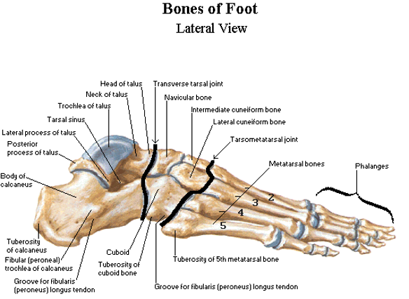
The foot can be divided into 3 regions:
- Hindfoot (rear foot) : Talus and Calcaneus
- Midfoot: Navicular, Cuboid, and Three Cuneiforms
- Forefoot: Metatarsals, Phalanges
Functions of the foot:
- Stability to support the weight of the body
- Shock absorption
- Propels body through space
- Flexibility to adapt to uneven terrains
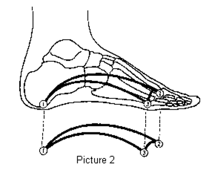
Arches of the foot:
- Medial longitudinal arch: Largest arch of the foot; runs on the MEDIAL side (“ the arch of the foot”)
- Lateral longitudinal arch: Runs on the lateral side of the foot
- Transverse arch: Runs transversely (L to R) across the foot
Pes Cavus: HIGH arched foot (Supinated foot)
Pes Planus: LOW arched foot (Pronated foot)
Plantar fascia:
- Dense fibrous tissue on the plantar aspect of the foot
- Runs from Calcaneal Tuberosity Posteriorly to the MTP joint proximally
- Purpose= to maintain and stabilize arches of the foot
many intrinsic muscles of the foot attach into the plantar fascia so if they are weak, the plantar fascia loses its support and the arches may fall. this can result in PES PLANUS 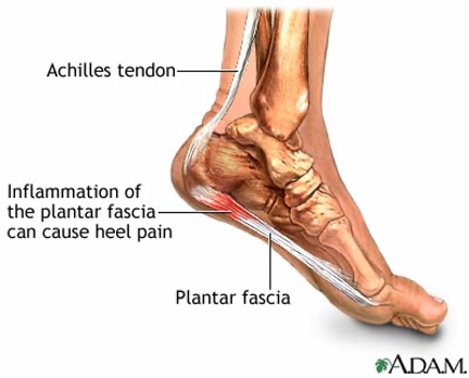
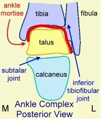
Ankle Joint: Talocrural Joint
- Articulating surfaces:
Proximal: Distal end of the tibia and malleoli of tibia and fibula
Distal: Trochlear surface of the talus
(often referred to as the mortise joint : wooden dowel fitting in to hole)
 2. Motions: Dorsiflexion and plantarflexion
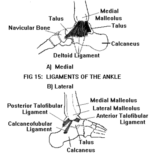
3. Ligaments:
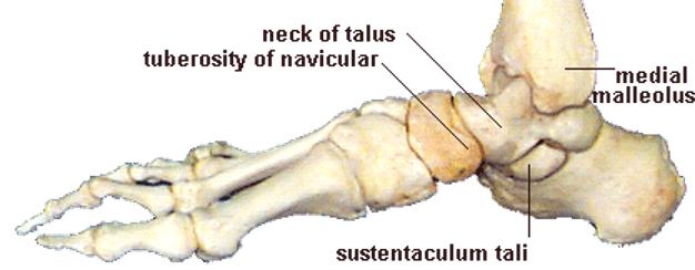
- Medial collateral ligament (MCL or deltoid ligament): runs from medial malleolus of tibia and fans out on the talus to attach to the medial aspect of the talus, sustentaculum tali of the calcaneus and the navicular tuberosity.
@@Function: Prevents eversion sprains@@; VERY STRONG and this in combination with the longer fibula laterally makes eversion sprains very Uncommon (avulsion fracture before sprain of deltoid ligament)
Lateral collateral ligament (LCL): composed of 3 ligaments- all begin proximally at the lateral malleolus of the fibula with their distal attachments=
a. Anterior talofibular (ATF): attaches to anterior talus
c. Calcaneofibular (CF): attaches into lateral aspect of calcaneus
p. Posterior talofibular (PTF): attaches to posterior talus
@@FUNCTION of LCL: Prevents inversion sprains@@- ATF (anterior talofibular) most commonly injured, then CF (calcaneofibular) and PTF (posterior talofibular)
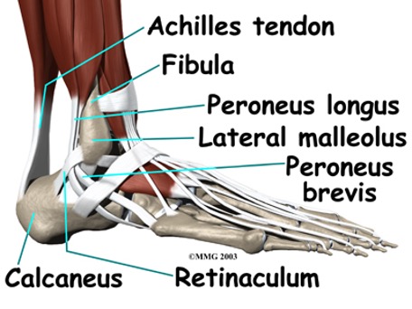
- Retinacula: Fibrous sheaths that function to hold down the tendons that cross the ankle joint
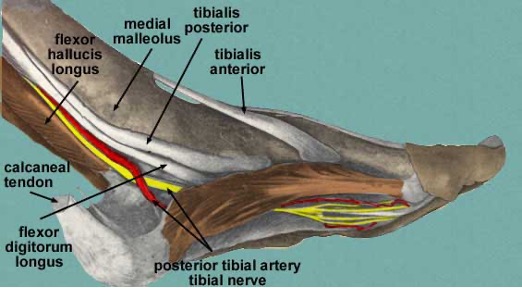
==Muscles that support the ankle joint==:
==Laterally==: muscles located in the lateral compartment (PL and PB)
==Medially==: 3 or 4 of the muscles located in the deep posterior compartment (TP, FDL, FHL - Tom, Dick and Harry)
Subtalar Joint: Talocalcaneal
- Articulation surfaces: 3 separate articulations between the posterior, middle, and anterior facets of the talus and calcaneus; ; a space exists laterally know as the
- Motions:
^^Pronation^^ (eversion, DF, and abduction)- refers to ^^eversion in OKC and pronation in CKC^^
%%Supination%% (inversion, PF, and adduction)- refers to %%inversion in OKC and supination in CKC%%
%%Closed-packed position of subtler joint: Supination%%
Transverse Tarsal (Chopart’s) Joint: talonavicular and calcaneocuboid
- Articulating Surfaces: talus and navicular (talonavicular) and calcaneus and cuboid (calcaneocuboid)
- Motions:
• @@Pronation@@ (eversion, DF and abduction) – refers to as @@eversion in OKC and pronation in CKC@@ • (inversion, PF and adduction) –refers to as
%%Closed-Packed Position of subtalar joint: supination%%
Other Joints:
Tarsometarsal Joints: between the distal row of tarsals (1st, 2nd and 3rd cuneiforms, cuboid) and the bases of MTs
Intermetatarsal Joints: between the MTs
Metatarsophalangeal (MTP) Joints: between the MT heads and the bases of the proximal phalanges
==Motions: flexion/extension, abduction/adduction==
%%Closed-packed position of MTP joints: extension%%
- Interphalangeal (IP) Joints: between the head of the more proximal phalanx and the base of the more distal phalanx
==Motions: flexion/extension==
%%Closed-packed position of IP joints: extension%%