Lesson 2: EKG
Lesson 2: EKG
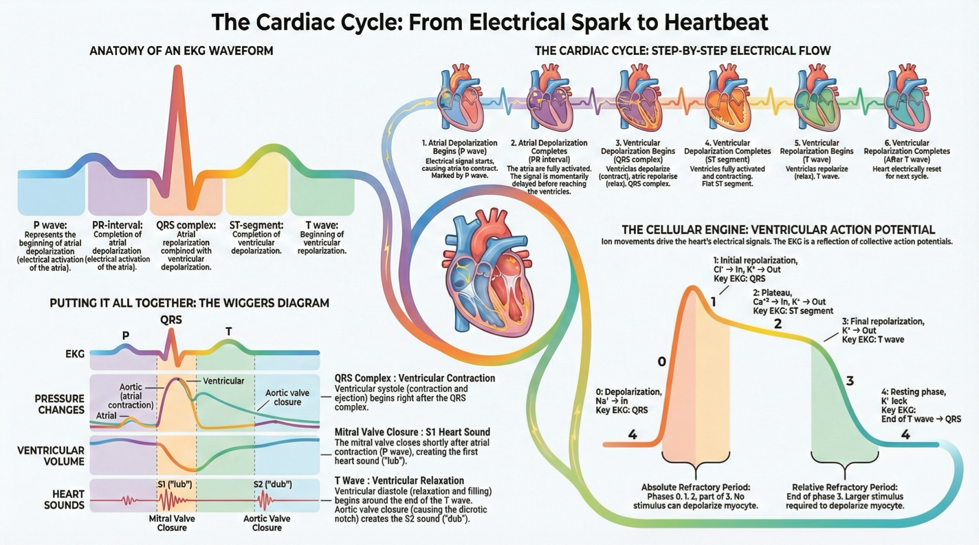
P wave: Atrial depolarization begins
PR interval: Atrial depolarization complete
QRS complex: Atrial repolarization + ventricular depolarization
ST segment: Ventricular depolarization complete
T wave: Ventricular repolarization begins
Correlation with Cardiac Action Potential
Match phases of ventricular action potential to EKG components:
Phase 0: QRS complex
Phase 2: ST segment
Phase 3: T wave
Ventricular Action Potential
Key events and EKG correlation:
Phase 0: Depolarization (Na+ in) → QRS
Phase 1: Initial repolarization (Cl- in, K+ out) → QRS
Phase 2: Plateau (Ca2+ in) → ST segment
Phase 3: Final repolarization (K+ out) → T wave
Phase 4: Resting phase (K+ leak) → End of T wave, start of QRS
Absolute refractory period: No stimulus can depolarize myocyte.
Relative refractory period: Larger stimulus required for depolarization.
Electrical Activity in Cardiac Cycle
Electrical events trace:
Atrial Depolarization: P wave
Ventricular Depolarization: QRS
Atrial Repolarization: Within QRS complex
Ventricular Repolarization: T wave
Integrating EKG with Cardiac Cycle
Electrical events precede mechanical events briefly.
Key mechanical events:
Isovol. Contraction
Rapid Ejection
Reduced Ejection
Isovol. Relaxation
Diastole: Includes rapid filling, reduced filling, atrial systole
Consider impacts of cardiac dysrhythmias on heart's mechanical functions and physiologic consequences.